173
Revista da Sociedade Brasileira de Medicina Tropical 44(2):173-176, mar-abr, 2011
INTRODUCTION
1. Laboratório de Zoonoses Bacterianas, Instituto Oswaldo Cruz, Fundação Oswaldo Cruz, Rio de Janeiro, RJ.
Address to: Dra. Deyse Christina Vallim. Lab. Zoonoses Bacterianas/IOC/FIOCRUZ. Av. Brasil 4365, Pavilhão Rocha Lima/sala 315, Manguinhos, 21040-900 Rio de Janeiro, RJ, Brasil.
Phone: 55 21 2270-6565 e-mail: [email protected] Received in 27/10/2010 Accepted in 15/12/2010
Antimicrobial susceptibilities of
Listeria monocytogenes human strains
isolated from 1970 to 2008 in Brazil
Suscetibilidade antimicrobiana de cepas humanas de
Listeria monocytogenes
isoladas no período
de 1970 a 2008 no Brasil
Cristhiane Moura Falavina dos Reis
1, André Victor Barbosa
1, Leonardo Alves Rusak
1, Deyse Christina Vallim
1and Ernesto Hofer
1ABSTACT
Introduction: Listeria monocytogenes is the causative agent of listeriosis, a foodborne illness that afects mainly pregnant women, the elderly and immunocompromised patients. he primary treatment is a combination of ampicillin with an aminoglycoside, in addition to a second-choice drug represented by chloramphenicol, erythromycin, tetracycline and rifampicin. he aim of this study was to analyze the antimicrobial susceptibility proile of strains isolated from human sources in the last four decades. Methods: Sixty-eight strains were selected from the culture collection of the Laboratory of Bacterial Zoonoses/LABZOO/FIOCRUZ isolated in diferent regions of Brazil from 1970 to 2008 and primarily isolated from cerebrospinal luid and blood culture. Susceptibility tests to antimicrobials drugs were evaluated using the criteria established by Soussy using the Kirby-Bauer method and E-Test strips were used to determine the minimum inhibitory concentration (MIC). Results: Among the strains tested, serovar L4b (60.3%) was the most prevalent, followed by serovar 1/2a (20.6%), 1/2b (13.2%) and the more uncommon serovars 1/2c, 3b and 4ab (5.9%). All strains were susceptible to ampicillin, cephalothin, erythromycin, gentamicin, teicoplanin and vancomycin. Only one strain (1.5%) showed resistance to rifampin, and two (3%) were resistant to trimethoprim-sulfamethoxazole.MICs with values up to 2μg/ml reinforce the need for microbiological surveillance. Conclusions: he study demonstrated low prevalence of strains resistant to the antimicrobial drugs indicated in the treatment of human listeriosis. Monitoring antimicrobial resistance proile is still very important to determine adequate treatment, especially in immunocompromised patients.
Keywords: Listeria monocytogenes. Antimicrobial susceptibilities. Listeriosis.
RESUMO
Introdução: Listeria monocytogenes é o agente etiológico da listeriose, doença de origem alimentar que acomete principalmente grávidas, pacientes imunodeprimidos e idosos. O tratamento primário é a associação de ampicilina a um aminoglicosídeo além de outros, em segunda escolha, representados por cloranfenicol, eritromicina, tetraciclina e rifampicina. O presente estudo teve como objetivo analisar o peril de susceptibilidade aos antimicrobianos de amostras de origem humana isoladas nas últimas quatro décadas. Métodos: Foram selecionadas 68 cepas provenientes de casos clínicos humanos ocorridos em diferentes regiões do país no período de 1970-2008. A susceptibilidade aos antimicrobianos testados foi determinada através dos critérios estabelecidos por Soussy pelo método de Kirby-Bauer e a concentração mínima inibitória realizada através do E-Test. Resultados: A amostragem constituiu-se de 68 cepas, isoladas principalmente de líquido cefalorraquidiano, e hemocultura no período, pertencentes ao Laboratório de Zoonoses Bacterianas/LABZOO/Fiocruz. O sorovar L4b (60,3%) foi o mais prevalente, seguido do sorovar 1/2a (20,6%), 1/2b (13,2%) e aqueles mais raros representados por 1/2c, 3b e 4ab (5,9%). Todas as cepas foram sensíveis à ampicilina, cefalotina, eritromicina, gentamicina, teicoplanina e vancomicina. Apenas uma cepa (1,5%) apresentou resistência à rifampicina, enquanto duas (3%) foram resistentes à associação de sulfametoxazol-trimetoprim.
Conclusões: Apesar de o estudo ter demonstrado uma baixa prevalência de amostras resistentes aos antimicrobianos indicados na terapêutica da listeriose humana, o sistema de monitoramento do peril de resistência antimicrobiana é de extrema importância para a orientação do tratamento adequado, principalmente nas infecções em pacientes imunocomprometidos.
Palavras-chaves: Listeria monocytogenes. Susceptibilidade antimicrobiana. Listeriose.
Listeria monocytogenes is a gram positive, facultative anaerobe, intracellular bacterium and the etiologic agent of human and animal listeriosis. The disease affects primarily pregnant women, newborns and patients with degenerative diseases and/or immunocompromised patients, is clinically manifested as meningitis and septicemia, has a high mortality rate, between 20 and 30% of cases, and causes neurological sequelae in some cases1-3.
Members of the genus Listeria are widely distributed in nature and can be detected in the environment (soil, vegetables, silage and water) and in the intestinal tract of humans and animals2. he
species is a signiicant food-borne pathogen4.
Listeria monocytogenes presents uniform antimicrobial susceptibility, including drugs commonly used for treating human listeriosis, such as ampicillin or in association with an aminoglycoside (e.g. gentamicin), and other second-choice antimicrobials represented by chloramphenicol, erythromycin, tetracycline and rifampicin4-6. However, clinical strains resistant
to chloramphenicol, erythromycin, streptomycin, tetracycline, vancomycin and trimethoprim have been recently described4. he widespread distribution of
epidemiologically serotypes of L. monocytogenes and their resistance to commonly used antibiotics indicate a potential public health risk. Given this situation, it is assumed that the system for monitoring antimicrobial resistance proile is extremely important to determine the appropriate treatment of human listeriosis. herefore, the main goal of this study was to analyze the proile of antimicrobial resistance in strains isolated from humans in diferent regions of Brazil during the last four decades.
METHODS
Bacterial strains
Sixty-eight strains isolated from 1970 to 2008 were selected, including human clinical cases occurring in diferent regions of the country. he samples belong to the collection of Bacteriological
Article/Artigo
174
RESULTS
Reis CMF et al - Antimicrobial susceptibilities of Listeria monocytogenes human strains
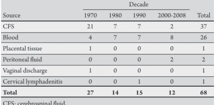
TABLE 1 - Distribution of the strains of Listeria monocytogenes analyzed, according to source of origin and decade of isolation.
Decade
Source 1970 1980 1990 2000-2008 Total
CFS 21 7 7 2 37
Blood 4 7 7 8 26
Placental tissue 1 0 0 0 1
Peritoneal luid 0 0 0 2 2
Vaginal discharge 1 0 0 0 1
Cervical lymphadenitis 0 0 1 0 1
Total 27 14 15 12 68
CFS: cerebrospinal luid.
TABLE 2 - List of primers used in PCR.
Primers Forward primer Reverse primer Product Speciicity
D1a CGATATTATCTACTTGTCA TGCTCCAAAGCAGGGCAT 214bp division I
D2b GCGGAGAAAGCTATCGCA TGTCAAACATAGGGCTA 140bp division II
23S rRNAc GGGGAACCCACTATCTTAGTC GGGCCTTCCAGACCGCTCA 239bp Listeria genus
Hlyd GCCTGCAAGTCCTAAGACGCCAATC CTGCAACTGCTCTTAGTAACAGC 706bp Listeria monocytogenes
a - D1: Division I consists of serotypes 1/2b, 3b, 4b, 4d, and 4e b - D2: Division II consists of serotypes 1/2a, 1/2c, 3a, and 3c c - 23S rRNA genes: marker of genus,
d - Hly: marker of the species Listeria monocytogenes
Culture Collection Laboratory, Bacterial Zoonoses of the Oswaldo
Cruz Institute/LABZOO/IOC/FIOCRUZ (Table 1), were
maintained in tryptose agar semi-solid at 4°C throughout the study period and stored at -20°C in BHI plus 20% glycerol.
Phenotypic identiication was performed in accordance with methods described by Rocourt & Seeliger7. For the identiication
of serogroups/serovars, the technique of slide agglutination test was used, with poly and monovalent somatic and lagellar antisera prepared by LABZOO, according to the technical guidance of Seeliger & Höhne8.
Genotypic analysis by PCR
he extraction of bacterial chromosomal DNA was performed using the Blood & Tissue Dneasy Kit (Qiagen), in accordance with the manufacturer's speciications.
To determine the strains detected, primers targeting the 23S rRNA genes (marker of genus), hly (marker of the species
L. monocytogenes) and the markers D1 and D2 to were used conirm the identiication of serogroups/serovars, according to literature9,10
(Table 2).
he ampliication reactions were performed in volumes of 25µl with a primer concentration of 50pmol/µl, 1U Taq polymerase, 0.2mM of each deoxynucleotide triphosphate, 2.5mM MgCl2 and 50ng of DNA. For the PCR, the PX2 thermal cycler equipment (hermo Fisher Scientiic Inc. Waltham, MA, USA) was used under
the following conditions (D1 + D2 primers): an initial step of 95°C for 3min followed by 25 cycles at 95°C/30s, 56°C/30s, 72°C/1min and a inal extension at 72°C for 10min. For ampliication with primers 23S rRNA + hly 95°C/5min followed by 40 cycles of 95°C/1min, 62°C/1min and 72°C/1min, followed by a inal extension at 72°C for 8min. All PCR products were determined by gel electrophoresis on 1% agarose 0.5 X TBE bufer and visualized under UV light ater staining with ethidium bromide. As molecular weight markers, the 2-log DNA ladders were used (New England BioLabs Inc.).
Antimicrobial susceptibility
Antimicrobial resistance was analyzed using the disk difusion method, in accordance with the CLSI11, and was performed
with standard discs (Oxoid) indicated for infections caused by Gram-positive bacteria: ampicillin (10mg), cephalothin (30μg), chloramphenicol (30μg), erythromycin (15μg), gentamicin (10mg), norloxacin (10mg), rifampicin (5μg), sulfamethoxazole/ trimethoprim (25μg), teicoplanin (30μg), tetracycline (30μg) and vancomycin (30μg). To maintain quality control of performance and reliability of the results, the standard strains of Escherichia coli ATCC 25922 and Staphylococcus aureus ATCC 25923 were used.
he size of the inhibition zone was determined according to CLSI guidelines, 2009, for Staphylococcus spp11. Ampicillin and
vancomycin were determined using the criteria established for
Listeria spp. by Soussy et al12. According to their behavior before the
use of antibiotics, the strains were classiied as sensitive, intermediate and resistant.
Determination of minimum inhibitory concentration Ater examination of the susceptibility by disk difusion method in agar, 43 strains were randomly selected to determine the minimum inhibitory concentration (MIC) to ampicillin (0.016-256µg/ml), tetracycline (0.016-256µg/ml) and rifampicin (0.016-256µg/ ml) by the E-test method, in accordance with the manufacturer's instructions (AB Biodisk). he MIC values were deined as the lowest concentration of antibiotic able to inhibit growth and the rate of change of MIC50 (where 50% of bacteria were inhibited) and MIC90 was calculated to specify the antimicrobial activity.
Of the 68 strains analyzed, 37 (53%) were isolated from cerebrospinal luid (CSF), 26 (41%) were isolated from blood and the remaining 6% were isolated from one of the following samples: placental tissue, vaginal secretion, cervical lymphadenitis and
175
Rev Soc Bras Med Trop 44(2):173-176, mar-abr, 2011
DISCUSSION
TABLE 3 - Distribution of serotypes in 68 strains of Listeria monocytogenes isolated from 1970 to 2000.
Decade
Serotypes 1970 1980 1990 2000 Number Percentage
1/2a 10 4 0 0 14 20.6
1/2b 1 0 2 6 9 13.2
1/2c 1 0 0 1 2 2.9
3b 0 0 0 1 1 1.5
4b 14 10 13 4 41 60.3
4ab 1 0 0 0 1 1.5
Number (%) 27 (35.5) 14 (20.6) 15 (22.1) 12 (17.6) 68 100.0
TABLE 4 - Antimicrobial susceptibility of 43 strains of Listeria monocytogenes by the E-test. Concentration Susceptibility Resistance Range MIC50 MIC90 (µ/ml) breakpoints breakpoints (µ/ml) (µ/ml) (µ/ml)
Ampicillin 0.016-256 ≤ 4 >16 0.25-4 1.0 2.0
Tetracycline 0.016-256 ≤ 4 >8 0.25-4 1.0 2.0
Rifampicin 0.016-256 ≤0.5 >16 0.016-0.94 0.047 0.25 MIC: minimum inhibitory concentration.
of the serotypes isolated are shown in Table 3. Currently, there is no criterion recommended by the CLSI for the interpretation of
Listeria susceptibility, except for penicillin and ampicillin breakpoint, hence the breakpoints recommended for the interpretive criteria for
Staphylococcus spp. were applied. All 68 strains analyzed were also susceptible to ampicillin, gentamicin, erythromycin, cephalothin, teicoplanin and vancomycin. Over the last four decades, a slight variation in the number of strains showing resistance to certain antimicrobials has been observed. In the 70s, only one strain of the serovar 1/2a (1.5%) was resistant to rifampicin isolated from CSF, and two serovar 4b (3%) samples isolated during the 1990s from blood were resistant to the association of trimethoprim-sulfamethoxazole. In contrast, in the 1980s and from 2000 to 2008, no resistance observed has been observed. A total of
29 (42.6%) strains have shown intermediate resistance proile for antimicrobials: chloramphenicol (7.4%), norloxacin (27.9%), tetracycline (5.9%) and rifampicin (1.5%), distributed over the last four decades.
All 43 strains tested against antimicrobial agents (rifampicin, ampicillin and tetracycline) using the E-test were sensitive. Rifampicin had the lowest MIC90
(0.25µg/ml), indicating its efective activity against Listeria. he values for ampicillin and tetracycline ranged from 0.25 to 4µg/ml and showed a level of MIC90 of 2µg/ml (Table 4).
Among the 13 serotypes of L. monocytogenes in the literature, serovar 4b is primarily responsible for most of the outbreaks in humans5,13 while the serovar 1/2a prevails in food and in some regions
of the world where it is more common in human cases14-16 .
In relation to serovars of L. monocytogenes identiied in this study, a higher incidence of serovar 4b (60.3%) was observed, which is in agreement with research by Hofer et al17 and reports dating back to
the 1970s. he frequency of serovar 4b was also demonstrated by Hofer et al18 in renal transplant recipients from the same hospital
in São Paulo. In the same state, Lemes-Marques et al19 observed
the incidence of the same serovar in clinical isolates from 1990 to 2005. Hofer et al20, performed phenotypic analysis of strains of
L. monocytogenes isolated from clinical material from 1969 to 2000 in diferent regions of the country, noting the higher incidence of serotype 4b, followed by 1/2a, in agreement with the results obtained in this study. In the aforementioned study20, the prevalence of serotype 4b
in CSF samples compared to blood isolates was also evident, which is consistent with the results obtained in this work, particularly for the 1970s. It is important to emphasize that all the strains tested were susceptible to ampicillin, which incidentally is the principal drug of choice for the treatment of listeriosis. It association with gentamicin has also been indicated and used successfully in the treatment of listeriosis, a situation supported by this study, since all strains were susceptible to gentamicin. he discrete level of rifampicin resistance in this study, another drug of choice for treatment is in agreement with the indings of Hofer & Oliveira21, and Pore-Gluchowska &
Markiewicz6, who reported resistance in clinical strains from diferent
parts of the world. It appears that virtually the same profile has been identiied over the years and in diferent countries. In relation to tetracycline, this research highlighted the extreme sensitivity of the 68 strains to this drug, contrasting with the emergence of clinical strains resistant to tetracycline, related to the gene tetM5,22,23.
No resistance to the association of trimethoprim-sulfamethoxazole was observed, which is important considering its nomination as an alternative in the treatment of listeriosis, primarily in patients with intolerance to penicillin5,24. he same result was obtained by
Lemes-Marques et al19, although reports in the literature demonstrate
resistance to trimethoprim25, as well as the combination of
trimethoprim-sulfamethoxazole26,27. MICs with values up to 2μg/
ml reinforce the need for microbiological surveillance.
In short, these results are compatible with most tests performed in various parts of the world, including Brazil, which showed a lower prevalence of strains resistant to antimicrobial therapy, indicated in cases of human listeriosis. However, the widespread use of antimicrobials in veterinary medicine, agriculture and particularly in animal food production could represent selective pressure on Listeria spp. In the environment in the future, allowing the acquisition of resistance mechanisms. herefore, to evaluate the progression of resistance, it is essential to establish a program of continuous monitoring of antimicrobial susceptibility of isolates of
L. monocytogenes and Listeria spp. isolated from human, animal, food and environmental sources.
ACKNOWLEDGMENTS
he authors declare that there is no conlict of interest.
CONFLICT OF INTEREST
FINANCIAL SUPPORT
REFERENCES
he authors would like to thank Evaldo Soares da Silva for his technical collaboration.
IOC/FIOCRUZ, CNPq (Proc. 301545/2006-5).
176
2. Hofer E, Hofer CB. Listeriose. In: Coura JR, editor. Dinâmica das Doenças Infecciosas e Parasitárias. Vol II. Rio de Janeiro: Guanabara Koogan; 2005. p. 1539-1545.
3. Cruz CD, Martinez MB, Destro MT. Listeria monocytogenes: um agente infeccioso ainda pouco conhecido no Brasil. Alim Nutr 2008; 19:195-206.
4. Aureli P, Ferrine AM, Mannoni V, Hodzic S, Wedell-Weergaard C, Oliva B. Susceptibility of Listeria monocytogenes isolated from food in Italy to antibiotics. Int J Food Microbiol 2003; 83:325-330.
5. Charpentier E, Courvalin P. Minireview. Antibiotic Resistence in Listeria spp. Antimicrob Agents Chemother 1999; 43: 2103-2108.
6. Poros-Gluchowska J, Markiewicz Z. Antimicrobial resistance of Listeria monocytogenes. Acta Microbiol Pol 2003; 52:113-129.
7. Rocourt J, Schretenbrunner A, Seeliger HPR. Diferénciation biochimique des groupes génomiques des Listeria monocytogenes (sensu lato). Ann Microbiol (Inst Pasteur) 1983; 134A:65-71.
8. Seeliger HPR, Höhne K. Serotyping of L. monocytogenes and related species. In: Bergan T, Norris JR, editors. Methods in microbiology. Vol 13. London: Academic Press; 1979. p. 31-49.
9. Hudson JA, Lake RJ, Savill MG, Scholes P, Mccormick RE. Rapid detection of Listeria monocytogenes in ham samples using immunomagnetic separation followed by polymerase chain reaction. J Appl Microbiol 2001; 90:614-621. 10. Boruck MK, Douglas R, Call DR. Listeria monocytogenes serotype identiication
by PCR. J Clin Microbiol 2003; 41:5537-5540.
11. Clinical and Laboratory Standards Institute. Performance Standards for Antimicrobial Disk and Dilution Susceptibility Tests for Bacteria Isolated From Animals. Approved Standard. 3rd ed. Wayne (PA): Clinical and Laboratory
Standards Institute; 2009. M31-A3.28.
12. Soussy CJ, Ckuzel R, Courvalin P. Comité de I’ Antibiogramme de la Société Française de Microbiologie.Deinition and Determination of in vitro Antibiotic Susceptibility Breakpoints for Bacteria in France. Eur J Clin Microbiol Infect Dis 1994; 13:238-246.
13. Gasanov U, Hughes D, Hansbro PM. Methods for isolation and identiication of Listeria spp. and Listeria monocytogenes: a review. FEMS Microbiol Rev 2005; 29:851-875.
14. Jay JM. Prevalence of Listeria spp. in meat and poultry products. Food Control 1996; 7:209-214.
15. Schlech WF. Overview of listeriosis. Food Control 1996; 7:183-186. 16. Gilbreth SE, Call JE, Wallace FM, Scot VN, Chen Y, Luchansky JB. Relatedness
of Listeria monocytogenes isolates recovered from selected ready-to-eat foods and listeriosis patients in the United States. Appl Environ Microbiol 2005; 71:8115-8122.
17. Hofer E, Pessoa GVA, Melles CEA. Listeriose humana. Prevalência dos sorotipos de Listeria monocytogenes isolados no Brasil. Rev Inst Adolfo Lutz 1984; 44:125-131.
18. Hofer CB, Melles CEA, Hofer E. Listeria monocytogenes in renal transplant recipients. Rev Inst Med TropSao Paulo 1999; 41:375-377.
19. Lemes-Marques EG, Cruz CD, Destro MT. Pheno - and genotypic characterization of Listeria monocytogenes clinical isolates from the southwestern region of the State of São Paulo, Brazil. Braz J Microbiol 2007; 38:287-292.
20. Hofer E, Reis CMF, Hofer CB. Serovars of Listeria monocytogenes and related species isolated from human clinical specimens. Rev Soc Bras Med Trop 2006; 39:32-37.
21. Hofer E, Oliveira LMA. Sensibilidade antimicrobiana em amostras de Listeria
isoladas de diferentes fontes e regiões do Brasil. Rev Microbiol 1988; 19:109-112.
22. Morvan A, Moubareck C, Leclereq A, Hervé-Bazin M, Bremont S, Lecuit M, et al. Antimicrobial resistance of Listeria monocytogenes human strains isolated in France. Antimicrob Agentes Chemother 2010; 54:2728-2731.
23. Poyart-Salmeron C, Trien-Cuot P, Carlier C, Macgowan A, McLauchlin J, Courvalin P. Genetic basis of tetracycline resistance in clinical isolates of
Listeria monocytogenes. Antimicrob Agents Chemother 1992; 36:463-466. 24. White DG, Zhao S, Simjee S, Wagner DD, Mcdemot PF.Antimicrobial resistance
of foodborne pathogens. Microbes and Infection 2002; 4:405-412.
25. Charpentier E, Gerbaud G, Jacquet C, Rocourt J, Courvalin P. Incidence of antibiotic resistance in Listeria species. J Infect Dis 1995; 172:277-281. 26. Hofer E, Nascimento RS, Oliveira MA. Meningite por L. monocytogenes. Relato
de casos em pacientes do Distrito Federal. Rev Soc Bras Med Trop 1998; 31:173-177.
27. Catão RMR, Vigolvino WA, Andrade WT, Hofer E. Meningite por Listeria monocytogenes em Campina Grande - Paraíba, Brasil: relato de um caso. Rev Bras Anal Clin 2003; 35:81-83.