Cop
yright
© ABE&M t
odos os dir
eit
os r
eser
vados
.
Cop
yright
© ABE&M t
odos os dir
eit
os r
eser
vados
.
The burden of osteoporosis in Brazil
O ônus da osteoporose no Brasil
Bruna Coelho Galvão Marinho1,2, Luiza Paulino Guerra1, Juliana Beaudette Drummond3, Barbara C. Silva4, Maria Marta Sarquis Soares5
ABSTRACT
Osteoporotic fractures impose severe physical, psychosocial, and inancial burden both to the patient and the society. Studies on the prevalence of osteoporosis and fragility fractures in Brazil show a wide variation, due to differences in sample size, the population studied, and me-thodologies. Few studies have been conducted in Brazil about the cost-effectiveness analyses of different intervention options aimed at the diagnosis and treatment of osteoporosis. Investiga-tion and treatment strategies based on cost-effectiveness and scientiic evidence are essential in the preparation of public health policies with the ultimate goal of reducing the incidence of fractures and, consequently, the direct and indirect costs associated with them. This article re-views the Brazilian burden of osteoporosis in terms of the prevalence and fractures attributable to the disease, the costs related to the investigation and management, as well as the impact of osteoporosis on the population as a whole and on affected individuals. Arq Bras Endocrinol Metab. 2014;58(5):434-43
Keywords
Expenses; osteoporosis; Brazil
RESUMO
Fraturas osteoporóticas impõem graves entraves físicos, psicossociais e inanceiros, tanto para o paciente quanto para a sociedade. Estudos sobre a prevalência de osteoporose e fraturas por fragilidade no Brasil mostram uma grande variação, em decorrência das diferenças no tamanho das amostras, da população estudada e da metodologia empregada. Poucos estudos têm sido realizados no Brasil sobre a análise de custo-efetividade das diferentes opções de intervenção que visam ao diagnóstico e ao tratamento da osteoporose. Estratégias de investigação e de tratamento com base na relação custo-eicácia e evidências cientíicas são essenciais para a elaboração de políticas de saúde pública, com o objetivo inal de reduzir a incidência de fratu-ras e, consequentemente, os custos diretos e indiretos associados a elas. Este artigo faz uma revisão sobre o ônus da osteoporose no Brasil em termos de prevalência e fraturas atribuí-veis à doença, de custos relacionados com a investigação, tratamento da osteoporose, bem como seu impacto na população como um todo e em indivíduos afetados. Arq Bras Endocrinol Metab. 2014;58(5):434-43
Descritores
Gastos; osteoporoses; Brasil
1 Hospital Felício Rocho, Belo
Horizonte, MG, Brazil
2 Faculdade de Medicina,
Universidade José do Rosário Vellano (Unifenas), Campus Belo Horizonte, Alfenas, MG, Brazil
3 Laboratório Hermes Pardini,
Belo Horizonte, MG, Brazil
4 INCT, Medicina Molecular,
Faculdade de Medicina, Universidade Federal de Minas Gerais (UFMG), Belo Horizonte, MG, Brazil
5 Faculdade de Medicina, UFMG,
Belo Horizonte, MG, Brazil
Correspondence to: Maria Marta Sarquis Soares Av. Alfredo Balena, 190 30130-100 – Belo Horizonte, MG, Brazil
Received on Jan/1/2014 Accepted on May/12/2014
DOI: 10.1590/0004-2730000003203
INTRODUCTION
O
steoporosis is a major health concern with up to 9 million new osteoporotic fractures expected an nually worldwide (1,2). Osteoporotic fractures impose severe physical, psychosocial, and inancial burden both to the patient and the society. They may be accompanied by pain, bone deformities, fear, distress, dificulty in per forming daily activities, loss of independence, and insti tutionalization (3). More important, fragility fracturesCop
yright
© ABE&M t
odos os dir
eit
os r
eser
vados
.
Osteoporosis is a “silent” disorder until it leads to one or more fractures (7). Since the treatment of osteo porosis can reduce the fracture risk, the early detection of osteoporosis by the measurement of bone mineral density (BMD) by dualenergy Xray absorptiometry (DXA) should be targeted in clinical practice (8). In fact, according to the 2002 Brazilian Consensus of Os teoporosis, BMD measurements should be performed in a number of settings – and as a rule, for all women over 65 years of age and men over 70 years (9).
This article reviews the Brazilian burden of osteo porosis in terms of the prevalence of osteoporosis and fractures attributable to the disease, the costs related to its management and its impact on the population as a whole and on affected individuals.
MATERIALS AND METHODS
We searched for English and Portugueselanguage ar ticles, in human subjects, available in full electronic me dia in MedLine (PubMed) and the database of the Latin American and Caribbean Literature on Health Sciences (LILACS) published within all dates. The search terms used were “osteoporosis” and “Brazil” in addition to one of the following: “costs or expenditure”, “burden”, “prevalence”, “fracture”, “quality of life”, or “impact”. Relevant articles were reviewed in detail. Pertinent data concerning the demographic proile of the Brazilian population were also used. Furthermore, author’s work published as an abstract regarding the direct costs re lated to the biochemical workup in the management of osteoporosis was also reviewed (10).
DEMOGRAPHIC PROFILE OF THE BRAZILIAN
POPULATION
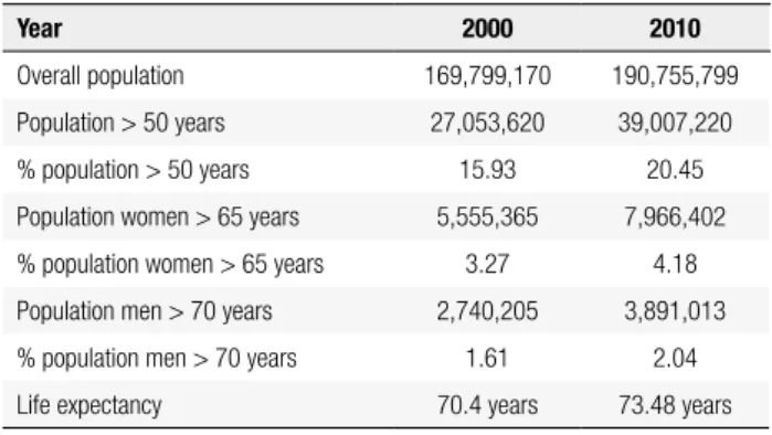
According to the 2010 census, conducted by the Ins tituto Brasileiro de Geograia e Estatística (IBGE), the Brazilian population in 2010 was 190,755,799 and ta ble 1 shows our demographic proile in 2000 and 2010. The percentage of elderly individuals increased in 2010 relative to 2000 (11) and consequently the prevalence of diseases such as osteoporosis is expected to rise. In 2010, there were more than 20 million Brazilians over 65 years of age, and the projection for this segment of the population is to exceed 50 million by 2050 (11,12). Moreover, 20.45% of the population was older than 50 years in 2010. Women over 65 years of age accounted for 4.18% of the population, while men over 70 years of age corresponded to 2.04% (Table 1) (11,12).
Table 1. Demographic characteristics of the Brazilian population in 2000 and 2010
Year 2000 2010
Overall population 169,799,170 190,755,799
Population > 50 years 27,053,620 39,007,220
% population > 50 years 15.93 20.45
Population women > 65 years 5,555,365 7,966,402
% population women > 65 years 3.27 4.18
Population men > 70 years 2,740,205 3,891,013
% population men > 70 years 1.61 2.04
Life expectancy 70.4 years 73.48 years
Source: Instituto Brasileiro de Geograia e Estatística (IBGE) (11).
PREVALENCE OF OSTEOPOROSIS IN BRAZIL
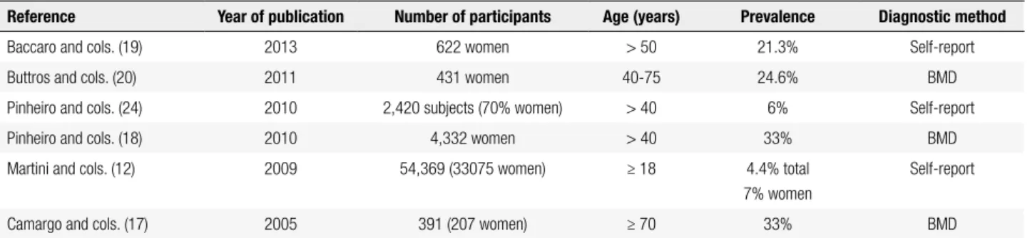
In Latin America, the estimated prevalence of spine os teoporosis in women aged 50 years and more ranges from 12.1% to 17.6%, while that of femoral neck os teoporosis ranges from 7.9% to 22% (13). Brazil is a country of extensive race mixing and heterogeneous re gional distribution, which implies different risk factors for osteoporosis and fractures (14). Moreover, access to BMD testing, which is essential for the detection of osteoporosis and intervention before fracture occurs, is still sparse in Brazil. The poor instrument availabili ty, high cost to patients and restrictive indications for BMD testing constitute the major barriers for the access to such testing in Brazil. Despite these limitations, few studies have evaluated the prevalence of osteopenia and osteoporosis in Brazil, which shows a wide variation, due to differences in sample size, eligibility criteria, and methodologies, as showed in table 2 (15). Overall, the prevalence of osteoporosis in Brazilian studies ranges from 6% to 33% depending on the population and other variables evaluated (1618).
Cop
yright
© ABE&M t
odos os dir
eit
os r
eser
vados
.
able in Latin America and Brazil. A review of Latin American studies showed that the incidence of osteopo rotic hip fractures ranged from 40 to 362 per 100,000 persons of 50 years or older, with a femaletomale ratio of 3:1. The majority of the studies showed lower inci dence of hip fractures in the Latin American population compared with the population of the United States, Canada, and Europe. This discrepancy was likely due to differences in the studied population, the adopted deinition of case, and methodological issues(13).
The Latin American Vertebral Osteoporosis Study (LAVOS) evaluated a randomized sample of 1,922 women aged 50 years and older from Argentina, Brazil, Colombia, Mexico, and Puerto Rico (22). The preva lence of vertebral fractures as assessed by lateral Xrays of the lumbar and thoracic spine was 11.18%, and was similar in the ive countries. Among women aged 50 to 59 years, the prevalence was 6.9%, which increased to 27.8% for those aged 80 years or older.
Studies exclusively conducted in Brazil show that the incidence of osteoporotic fractures varied with the population and the fracture site investigated. Overall, the ageadjusted annual incidence of fractures varied from 5.59 to 13 per 10,000 in women, and from 12.4 to 27.7 per 10,000 in men (16). The lowest incidences of fractures in the above ranges, both in women and men, correspond to the incidence of hip fractures in Sobral, a city located in the Northeast Region of Brazil (3ºS/40ºE) (23).
In BRAZOS, 2,420 Brazilian individuals from the ive geographic regions of the country were evaluated. The prevalence of selfreported fragility fractures de ined as that resulting of any fall from standing height or less in subjects > 40 years old, was 12.8% in men and 15.1% in women. Statistically signiicant differences among Brazilian regions, according to gender or social class, were not observed(24).
Table 2. Prevalence of osteoporosis based on different studies in the Brazilian population
Reference Year of publication Number of participants Age (years) Prevalence Diagnostic method
Baccaro and cols. (19) 2013 622 women > 50 21.3% Self-report
Buttros and cols. (20) 2011 431 women 40-75 24.6% BMD
Pinheiro and cols. (24) 2010 2,420 subjects (70% women) > 40 6% Self-report
Pinheiro and cols. (18) 2010 4,332 women > 40 33% BMD
Martini and cols. (12) 2009 54,369 (33075 women) ≥ 18 4.4% total
7% women
Self-report
Camargo and cols. (17) 2005 391 (207 women) ≥ 70 33% BMD
The overall prevalence of osteoporosis appears to be lower when women and men are included in the evalua tion. The Brazilian Osteoporosis Study (BRAZOS), which evaluated a representative sample of 2,420 Bra zilian individuals (women, 70%) > 40 years old, from different regions and economic classes revealed that the selfreported prevalence of osteoporosis was only 6% (16). Camargo and cols. assessed the BMD of 301 in dividuals (207 women) older than 70 years of age from different clinical centers in the city of São Paulo. The prevalence of osteoporosis ranged from 6.4% to 16.1% in men, and from 22.2% to 33.2% in women in the dif ferent centers studied (17).
Another crosssectional study conducted in São Pau lo evaluated 2,143 subjects ≥ 60 years old interviewed in the years of 2000 and 2006 (21). The prevalence of osteoporosis, assessed by selfreport, was greater among subjects with private health insurance coverage than in those without private insurance, regardless of sex and year of evaluation. This inding may represent a wider access to DXA tests and better understanding of the disease among those with private health insurance.
Finally, in a crosssectional study based on data from the VIGITEL system (Risk Factor Surveillance and Pro tection against Chronic Diseases through Telephone Survey) conducted in 2006, 54,369 Brazilian indivi duals (33,075 women) > 18 years of age were interviewed. The prevalence of osteoporosis as assessed by self report was 4.4% in the entire population, 7% in women and 1.3% in men (12). This lower prevalence of osteoporosis as compared to the previous studies reported here may be explained by the low age of subjects included.
INCIDENCE AND PREVALENCE OF OSTEOPOROTIC
FRACTURES
Cop
yright
© ABE&M t
odos os dir
eit
os r
eser
vados
.
Siqueira and cols. evaluated the prevalence of self reported fractures and its association with sociodemo graphic variables and medical diagnosis of osteoporosis in 3,100 individuals from Pelotas, a Southern Brazilian city (56.6% women) (25). The lifetime prevalence of any type of fracture was 37.5% among men and 21.3% among women (P < 0.001). While in men most fractures were caused by sports practice and happened in leisure time, most fractures in women were caused by falls and occurred inside the home. The prevalence of fractures throughout life was almost twice as higher (28.3%) than that observed in the BRAZOS study (14.4%) (24,25). It is important to emphasize that individuals aged 20 years or more were included in Siqueira’s study, as well as traumarelated and nontraumatic fractures.
In the SAPOS study, the prevalence of selfreported osteoporotic fractures was 11.5%, with a mean age of 65.5 ± 10 years at the time of the event. Among the 497 women with fractures, vertebral fractures were re ported in 6%, nonvertebral fractures in 86% (including the humerus, distal forearm, metacarpus, ribs, and hip) and femoral fractures in 8% of the cases (18).
A crosssectional study conducted in Chapecó, a Southern Brazilian city, explored the prevalence of as ymptomatic vertebral fractures by radiographs, in a pop ulation of 186 postmenopausal women over 60 years (26). Almost half of the women studied (48.9%) had at least one vertebral fracture not associated with a prior history of fracture. The higher prevalence of vertebral fractures in this study was mainly attributed to the crite ria used for the analysis and deinition of vertebral frac tures, the population studied (all subjects were white), the high frequency of risk factors such as glucocorticoid use, low dietary calcium intake, and alcohol abuse, and the latitude of the city (below the equator) (26).
In the city of São Paulo, Lopes and cols. evaluated 1,007 elderly subjects (600 women) using BMD test ing of hip and lumbar spine (27). The prevalence of osteoporotic fractures assessed by selfreport was 13.2% and the major sites affected were distal forearm (6.0%), humerus (2.3%), femur (1.3%), and ribs (1.1%). Wo men had greater prevalence of fractures (17.5%) com pared with men (6.9%) (27).
In the city of Campinas, Baccaro and cols. evalu ated 622 women aged 50 years or older and the overall prevalence of selfreported bone fragility fractures was 10.8%, whereas the prevalence of femoral/hip fractures was 1.8%. In the multiple regression analysis, a higher prevalence of fragility fractures was associated with a
longer time since the menopause and the presence of osteoporosis (19).
IMPACT OF FRAGILITY FRACTURES ON THE
INDIVIDUAL
Osteoporosis poses a signiicant negative impact on the quality of life of patients, particularly after fragility frac tures, which is supported by the majority of the studies reviewed below.
Osteoporotic fractures, commonly of the hip and spine, often result in secondary complications, such as functional impairment, increased hospital stays that may result in further health problems, increased medi cal costs, and increased dependence on others for living assistance (28). In addition to the evident physical and functional consequences, such as kyphosis, restriction of movement, and pain, the individual with osteoporo sis may sustain a psychosocial impact. Many patients, in the early stages of the disease, express marked anxiety, especially regarding the possibility of future fractures and physical deformity. As the disease progresses, de pression may be aggravated for those who sustain hip fractures or multiple vertebral fractures. Osteoporosis can lead to dependence, functional disability, and low ered selfesteem (29). Among Brazilian women over 45 years of age, 84% are concerned about having osteo porosis, and 13% of them have sustained at least one fragility fracture after the age of 40. Among women who had fractures, 52% reported worsened quality of life following the fracture (29).
The BRAZOS study showed a strong association between poor quality of life and the presence of low impact fractures, both in men and women older than 40 years of age, emphasizing that patients with osteo porosis and fractures have a higher incidence of chronic pain, decreased physical capacity, reduction in social activities, decreased perception of well being, and de pressed mood than individuals without fractures (16).
In a study that evaluated 56 elderly patients ≥ 60 years old with a lowtrauma hip fracture, from the Bra zilian city of São Paulo, there was an increase in the inability to walk and in the use of a supporting device. The hip fracture also led to a signiicant reduction in the functional ability to perform basic and instrumental activities of daily living (30).
Cop
yright
© ABE&M t
odos os dir
eit
os r
eser
vados
.
with or without vertebral fractures, the quality of life was assessed by the European quality of life question naire (Quality of Life Questionnaire of the European Foundation for Osteoporosis – QUALEFFO41) (31). The presence of asymptomatic vertebral fractures iden tiied on thoracic and lumbar spine radiographs was as sociated with a reduced quality of life regardless of age, BMI, and level of physical activity (31).
Another study assessed the prevalence and the as sociation between the number of vertebral fractures and quality of life in 126 postmenopausal Brazili an osteoporotic women (mean age 65.7 years). The prevalence of vertebral fractures identiied on thoracic and lumbar spine radiographs was 34.1%. While the QUALEFFO41 questionnaire showed no difference in scores between women with and without vertebral fractures, there was a direct correlation between the quality of life score and the number of vertebral frac tures(32).
Fortes and cols. evaluated the morbidity and mor tality deriving from proximal femoral fractures in in dividuals over 60 years of age who were admitted to two hospitals in the city of São Paulo (33). A marked decrease was noted in the indicators of functional dis ability evaluated using the Health Assessment Ques tionnaire(HAQ), after six months of the fracture event. The factors that correlated with poorer functional abili ty were HAQ score prior to fracture, institutionaliza tion after fracture and age. Six months after the event, 11.6% of the patients had become completely depen dent and 9.3% were institutionalized (33).
IMPACT OF OSTEOPOROSIS AND FRAGILITY
FRACTURES ON SOCIETY
Mortality
Hip fracture is one of the most feared consequences of osteoporosis. Hip fractures are associated with high post fracture disability, increased mortality, and high healthcare expenditures (34). The mortality rate from hip fractures in developed countries is around 25% in the irst year after the event (4,5). Mortality rates dur ing the hospitalization period range from 1.02% to 10% across countries (13).
In Brazil, it is estimated that 15% to 30% of patients with hip fractures die within the irst year following the event, frequently as a result of fracture complications such as infections, venous thrombosis, and pressure ul
cers, or comorbid conditions, especially cardiovascular diseases. The predominant factors related to increased risk of death are male gender, old age, impaired func tional ability prior to the fracture, greater number of comorbidities, sarcopenia and a fragile phenotype (15).
A study analyzed the proile of the Brazilian pub lic health care system (SUS) admissions due to osteo porotic hip fracture in patients over 60 years, in the years 2006 to 2008 in different regions of Brazil. The mortality rate due to femoral fracture was higher in fe males then in males (3.5 versus 1.9 per 1,000 elderly, respectively, in 2006, with similar rates in 2007 and 2008), which is in disagreement with other Brazilian studies. The proportion of outcomes of death increased with advancing age in the three analyzed years and the Southeast region had the highest percentage of deaths for elderly patients hospitalized with hip fracture (35).
A prospective study published in 2009 investigated the mortality rate in the irst year after hip fractures, as well as the factors associated with mortality in Bra zilian patients. In total, 246 individuals older than 60 years were followed for one year after hospitalization for hip fractures. Eightysix patients died (35%), with most of those deaths (74.4%) occurring after hospital discharge. Of the 67 men, 29 died (43.3%), and of the 179 women, 57 died (31.8%) within the irst year af ter fracture. Functional status prior to the fracture, age, male gender, and high surgical risk increased mortality risk, while antibiotics use and physical activity after the surgery reduced the risk (34).
A similar study conducted in Rio de Janeiro, showed a 21.5% overall mortality in the irst year after hip frac tures. Most of those deaths (55.1%) also occurred after hospital discharge, and were chiely associated with car diovascular events or infections (36).
Finally, a Brazilian study involving 56 elderly in dividuals from São Paulo showed a mortality rate of 23.2% over the six months following a hip fracture. The most frequent causes of death were infectious (46.1%), cardiovascular (46.1%) or indeterminate (7.8%) (33).
Cop
yright
© ABE&M t
odos os dir
eit
os r
eser
vados
.
Direct costs
The costs to society can be divided into direct and indi rect, and are related both to the prevention and treat ment of osteoporosis and rehabilitation following the fracture. These costs vary widely between countries, not only due to the varying incidence rates of the dis ease, but also because of the different degrees of em phasis placed on prevention, hospitalization, and treat ment (38).
Direct costs related to the management of osteoporosis: the biochemical workup
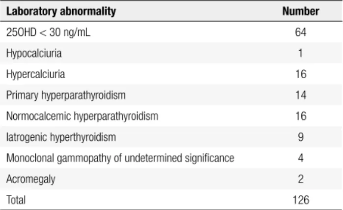
During the investigation of a low BMD, a biochemical workup aimed primarily at the detection of secondary causes of osteoporosis is indicated. The extent of the workup may vary according to the recommendation of each clinical institution or reference Service. Vari ous disorders affecting bone mass are common and of easy diagnosis and treatment. Some tests are key in the investigation of secondary factors that interfere with bone mass, such as serum levels of calcium, parathyroid hormone (PTH), 25hydroxy vitamin D (25OHD), thyroidstimulating hormone (TSH), and 24h urine cal cium. While patients with a clinical suspicion of sec ondary conditions should have extended diagnostic workup, the costs of ordering more complex tests for all patients with osteoporosis have not been estimated (10). Several studies have demonstrated the importance of laboratory studies in patients with osteoporosis (39 42). Our group evaluated the frequency and costeffec tiveness of the tests ordered for postmenopausal women who had been diagnosed as having osteoporosis based on BMD by DXA (10). The study was conducted in an outpatient clinic of general endocrinology in Belo Horizonte, Brazil, and in total, 185 medical records of postmenopausal women were reviewed. Patients with kidney or liver failure, or known to have a condition or a medication history that could cause bone loss were excluded. Of the 185 patients evaluated, 108 exhibited one or more abnormal tests (126 laboratory abnormali ties) indicative of disorders that could contribute to de creased bone mass or compromise the eficacy of the osteoporosis treatment (Table 3) (10).
In this study, serum calcium was the most frequently ordered test (100% of the patients) (Table 4); however, calcium concentrations were outside the reference range in only 9% of the patients (Table 4). The 25OHD test, which was ordered for 82% of the patients, was altered
Table 3. Laboratory abnormalities found in the workup for primary osteoporosis at the General Endocrinology Service (n = 185)
Laboratory abnormality Number
25OHD < 30 ng/mL 64
Hypocalciuria 1
Hypercalciuria 16
Primary hyperparathyroidism 14
Normocalcemic hyperparathyroidism 16
Iatrogenic hyperthyroidism 9
Monoclonal gammopathy of undetermined signiicance 4
Acromegaly 2
Total 126
Source: Marinho BCG, Soares MMS. Custo-efetividade da investigação laboratorial da
osteoporose. In: IOF Regionals Brazil 2012. 1st Latin America Osteoporosis Meeting, 2012, São
Paulo. Arch Osteoporos. 2012;7:S191.
Table 4. Frequency of ordered tests and its abnormalities found by the General Endocrinology Service for osteoporosis workup (n = 185)
Laboratory tests Number (%) of tests ordered abnormal testsNumber (%) of
Serum calcium 185 (100%) 16 (9%)
TSH 175 (95%) 22 (13%)
Complete blood cell count 173 (94%) 8 (5%)
PTH 152 (82%) 54 (36%)
25OHD 152 (82%) 64 (42%)
Phosphate 140 (76%) 4 (3%)
24-h urine calcium 124 (67%) 23 (19%)
Protein electrophoresis 44 (24%) 6 (14%)
Cortisol 16 (9%) 0
Antigliadin antibody 12 (6%) 0
IGF-1 (somatomedin) 5 (3%) 2 (40%)
Source: Marinho BCG, Soares MMS. Custo-efetividade da investigação laboratorial da
osteoporose. In: IOF Regionals Brazil 2012. 1st Latin America Osteoporosis Meeting, 2012, São
Paulo. Arch Osteoporos. 2012;7:S191.
Cop
yright
© ABE&M t
odos os dir
eit
os r
eser
vados
.
Direct costs related to the management of osteoporosis: treatment
It is estimated that SUS accounts for the provision of healthcare to 75.5% of the Brazilian citizens, and medications represent a sizable fraction of the public spending with health (47). The expenses of Brazil’s Ministry of Health with drugs acting on bone struc ture and mineralization correspond to approximately 10.9% of the total spending with highcost medications (48). In the São Paulo public health system, in 1998, the mean annual cost for postmenopausal osteoporo sis treatment in ambulatory patients amounted to 775 dollars per patient, and outofthepocket payments by the patients corresponded to 9% of the monthly family income (49). In Minas Gerais State, Brandao and cols. examined a historical cohort of 72,265 women, mean age 64.8 ± 9.8 years old, who used highcost medi cations supplied by the SUS to treat postmenopausal osteoporosis in the period 2000 to 2006 (50). The study demonstrated that the mean monthly percapita
expenditurein the irst year of treatment was 51 USD, and this gradually increased with increasing age from 50 years. The average monthly expenditure was 3.9% greater in women aged 50 to 59 years than in women < 49 years old. Similarly, the monthly expenditure was 55.8% greater in women who had sustained a fragil ity fracture as compared to those without fractures. A total of 6,429 (8.9%) patients died during the study period. The average monthly per capita expenditure was 2.2% higher in women who died than in those who were alive at the end of the study (p = 0.02). The most commonly used drug at the beginning of the treatment was alendronate (57%), followed by calcitonin (24.6%) and raloxifene (15.5%). The type of antiosteoporosis therapy used was the variable with the highest impact on the average monthly per capita expenditure, with tamoxifen and calcitonin having the greatest impact on the mean monthly spending, using alendronate as the standard drug (50).
Hip fractures typically demand inhospital care, while hospitalization is less frequent in vertebral, wrist, and other fractures (15). A recent study evaluated the proile of SUS hospitalizations for the treatment of femoral osteoporotic fracture in elderly patients in Brazil between 2006 and 2008 (35). Over this 3year period, 1% of the senior citizens hospitalized had a femoral fracture as the primary diagnosis. The total spending with hospital stays for femoral fractures in the elder fore, it is likely that these diseases were not found in
the present study because of the population sample size (small in relation to the prevalence of these two dis eases, especially Cushing syndrome) and the reduced number of screening tests performed (antigliadin anti body and cortisol levels were measured in 6%, and 9% of the patients, respectively).
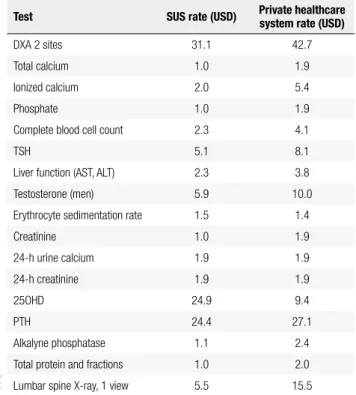
In order to assess the costeffectiveness of different testing strategies for the diagnosis of secondary causes of osteoporosis, we further evaluated a subgroup of 62 patients with no treatment for osteoporosis who under went all the tests of a basic workup routine, which were: serum calcium, 24h urine calcium, PTH, 25OHD, and TSH. This workup was selected based on different recommendations of several Societies (7,40,42,43,46). The strategy of ordering PTH, 24h urine calcium and 25OHD for all patients, and TSH for those in use of le vothyroxine, resulted in 100% of diagnoses at a cost of 69.7 U.S. dollars (USD) per patient screened and 84.7 USD per diagnosis. The testing strategy that proposes ordering PTH and 24h urine calcium measurements for all patients, 25OHD for those with an abnormal PTH and/or urinary calcium, and TSH for the levo thyroxine users identiies 84.31% of the disorders at a lower cost, of 55.6 USD per patient and 80.2 USD per diagnosis (10). The current cost of different diagnostic tests for osteoporosis in Brazil is depicted in table 5.
Table 5. Costs in U.S. dollar (USD) of diagnostic tests for osteoporosis
Test SUS rate (USD) Private healthcare system rate (USD)
DXA 2 sites 31.1 42.7
Total calcium 1.0 1.9
Ionized calcium 2.0 5.4
Phosphate 1.0 1.9
Complete blood cell count 2.3 4.1
TSH 5.1 8.1
Liver function (AST, ALT) 2.3 3.8
Testosterone (men) 5.9 10.0
Erythrocyte sedimentation rate 1.5 1.4
Creatinine 1.0 1.9
24-h urine calcium 1.9 1.9
24-h creatinine 1.9 1.9
25OHD 24.9 9.4
PTH 24.4 27.1
Alkalyne phosphatase 1.1 2.4
Total protein and fractions 1.0 2.0
Cop
yright
© ABE&M t
odos os dir
eit
os r
eser
vados
.
ly population in Brazil, including intensive care unit stays, prostheses and ortheses expenses, added up to 17,437,273.50 USD in 2006, 21,067,819.40 USD in 2007, and 27,358,429.50 USD in 2008, which cor responded to 2% of the total spending with hospitali zation for the elderly (35). The mean cost of treating a femoral fracture in two hospitals that maintained an agreement with the SUS ranged between 994.32 USD and 4,215.79 USD. This was a comprehensive analysis and included direct costs of staff (medical, nurse and physiotherapy services) as well as hospital resources (emergency room, operating room, intensive care unit, medications, prostheses, laboratory and imaging exams). This great disparity is due to the fact that cost analysis methodologies vary between hospitals (51). These ig ures contrast with the mean cost of hospitalization for osteoporotic femur fractures in individuals older than 50 years of age in the Brazilian private healthcare sys tem, which amounted to 10,104.00 USD according to a study conducted between 2003 and 2004 (52). This number is closer to direct hospitalrelated costs re ported in other countries, which suggests that the costs calculated by the hospitals with an agreement with the SUS are lower than the actual cost of the treatment for acute femur fracture. Femoral fractures were ob served in 4.99% of the 129,611 cases of osteoporosis managed within the private healthcare system. The an nual economic burden of these fractures for the health insurance companies was estimated in approximately 6 million USD (15,52). However, these results do not provide consistent data on the total costs over the me dium and long term (15).
Regarding pharmacoeconomic analyses, a Brazilian study performed a systematic review of the strategies used in Brazil and worldwide, focusing on the treat ment of osteoporosis in postmenopause. The use of bisphosphonates was the most extensively evaluated strategy, and produced the best costeffectiveness. Hor mone therapy, calcium and vitamin D supplementation, strontium ranelate, raloxifene, teriparatide, and deno sumab were also evaluated, with varying outcomes. Given the particularities of the Brazilian context, it was impossible to extrapolate any of the external results to our population, which restricted the applicability of such results to the decisionmaking process in public health policies (38).
Costeffectiveness analyses of different interven tion options aimed at the diagnosis and treatment of osteoporosis, with the ultimate goal of reducing the in
cidence of fractures and, consequently, the direct and indirect costs associated with them, are paramount in the preparation of public health policies. Few studies have been conducted in Brazil in this regard. In 2003, Silva and cols., using the decision tree tool, calculated in 34,800.00 USD the cost per fracture that could be spared if the BMD/alendronate intervention were ap plied to women over 65 years of age, and in 4,315.00 USD if only the association of calcium and vitamin D were used (53). Both estimates far exceeded the mean cost to treat femoral fractures (750.00 USD) estimated from DATASUS 2001 data, which led the authors to question the indication to implement any prevention and treatment strategy, given the limited resources. The analysis that those authors undertook was pre liminary and shows methodological shortcomings, yet it reinforces the need for enhanced knowledge of cost analysis strategies in our setting. In 2008, Araújo and cols. evaluated the costeffectiveness of the osteoporosis treatment with zoledronic acid in the Brazilian private healthcare system, in a hypothetical cohort of women over 65 years of age using the Markov model (54). The use of zoledronic acid has proved to be costeffective in the prevention of proximal femur fracture in that spe ciic setting; however, these data cannot be extrapolated to the public system or to other sites of osteoporotic fractures. Additionally, the use of new tools enabling the calculation of individual risks of fracture based on clini cal estimators, such as FRAX Brazil (Fractures Risk As sessment) and SAPORI (São Paulo Osteoporosis Risk Index), could contribute to a more effective use of the available diagnostic and therapeutic resources (55).
Indirect costs
Cop
yright
© ABE&M t
odos os dir
eit
os r
eser
vados
.
CONCLUSIONS
Osteoporosis is a disabling disease associated with high rates of morbidity and mortality. The projected number of individuals with osteoporosis in Brazil will increase as the population ages, which may have serious economic impact on our society and on the quality of life of the affected individuals.
In spite of the heterogeneity of Brazilian studies, the overall prevalence of osteoporosis and fragility frac tures in our country is high. In addition to the different ways to assess osteoporosis (e.g. selfreport vs. DXA), the sociodemographic characteristics of our population also inluence the results found.
There are few Brazilian studies of costeffectiveness strategies used in the workup and treatment of osteopo rosis. Nonpharmacological strategies should always be considered, however without neglecting the proven ef icacy of the various pharmacological options available. Early detection and sensible use of antifracture medi cations are invaluable in reducing the morbidity and mortality deriving from fractures. More comprehensive, multicentric studies are needed to enable us to outline evaluation and treatment protocols for our population.
Public policies aiming at the education of the popu lation regarding the importance of osteoporosis pre vention, which should be initiated in childhood and adolescence, could minimize the future economic bur den of the disease.
Disclosure: no potential conlict of interest relevant to this article was reported.
REFERENCES
1. Johnell O, Kanis JA. An estimate of the worldwide prevalence and disability associated with osteoporotic fractures. Osteoporos Int. 2006;17(12):1726-33.
2. Kanis J. Assessment of osteoporosis at the primary health care level. WHO Scientiic Group Technical Report, on behalf of the World Health Organization Scientiic Group 2007. Available at: http://www.shef.ac.uk/FRAX/pdfs/WHO_Technical_Report.pdf. 3. Gold D. The clinical impact of vertebral fractures: quality of life in
women with osteoporosis. Bone. 1996;18(2 Suppl):185S-9S. 4. Browner WS, Pressman AR, Nevitt MC, Cummings SR.
Mortal-ity following fractures in older women. The study of osteoporotic fractures. Arch Intern Med. 1996;156(14):1521-5.
5. Hannan EL, Magaziner J, Wang JJ, Eastwood EA, Silberzweig SB, Gilbert M, et al. Mortality and locomotion 6 months after hospi-talization for hip fracture: risk factors and risk-adjusted hospital outcomes. JAMA. 2001;285(21): 2736-42.
6. U.S. Department of Health and Human Services, Bone Health and Osteoporosis: A Report of the Surgeon General. Rockville, MD: U.S. Department of Health and Human Services, Ofice of the
Surgeon General, 2004. Available at: http://www.surgeongeneral. gov/library.
7. NIH (National Institute of Health). Consensus Development Panel on Osteoporosis Prevention, Diagnosis, and Therapy. Osteo-porosis: prevention, diagnosis, and therapy. J Am Med Assoc. 2001;85:785-95.
8. Sampaio PRL, Bezerra AJC, Gomes L. A osteoporose e a mu-lher envelhecida: fatores de risco. Rev Bras Geriatr Gerontol. 2011;14(2):295-302.
9. Neto A, Soares A, Urbanetz A, Souza A, Ferrari A, Amaral B, et al. Brazilian consensus on osteoporosis. Rev Bras Reumatol. 2002;42(6): 344-54.
10. Marinho B, Soares M. Custo-efetividade da investigação labo-ratorial da osteoporose. Arch Osteoporos. 2012;7(Suppl):S191 (abstract).
11. Instituto Brasileiro de Geociências e Estatística (IBGE). Censo 2010. Distribuição da população por sexo, segundo os grupos de idade. Available at: http://www.ibge.gov.br/home/estatistica/ populacao/censo2000/.
12. Martini LA, Moura EC, Santos LC, Malta DC, Pinheiro Mde M. Prevalence of self-reported diagnosis of osteoporosis in Brazil, 2006. Rev Saude Publica. 2009;43 Suppl 2:107-16.
13. Morales-Torres J, Gutierrez-Urena S. Osteoporosis Commit-tee of Pan-American League of Associations for Rheumatology. The burden of osteoporosis in Latin America. Osteoporos Int. 2004;15(8):625-32.
14. Fontes T, Araújo L, Soares P. Osteoporosis in climacteric I: epidemiology, deinition, screening and diagnosis. Femina. 2012;40(2):109-16.
15. Pinheiro MM, Eis SR. Epidemiology of osteoporotic fractures in Brazil: what we have and what we need. Arq Bras Endocrinol Me-tabol. 2010;54(2):164-70.
16. Pinheiro MM, Ciconelli RM, Martini LA, Ferraz MB. Clinical risk factors for osteoporotic fractures in Brazilian women and men: the Brazilian Osteoporosis Study (BRAZOS). Osteoporos Int. 2009;20(3):399-408.
17. Camargo MB, Cendoroglo MS, Ramos LR, de Oliveira Latorre Mdo R, Saraiva GL, Lage A, et al. Bone mineral density and osteo-porosis among a predominantly Caucasian elderly population in the city of Sao Paulo, Brazil. Osteoporos Int. 2005;16(11):1451-60. 18. Pinheiro M, Reis Neto E, Yang J, Machado F, Omura F, Szejnfeld
J, et al. Risk factors for osteoporotic fractures and low bone density in pre and postmenopausal women. Rev Saude Publica. 2010;44(3):479-85.
19. Baccaro LF, de Souza Santos Machado V, Costa-Paiva L, Sousa MH, Osis MJ, Pinto-Neto AM. Factors associated with osteopo-rosis in Brazilian women: a population-based household survey. Arch Osteoporos. 2013;8(1-2):138.
20. Buttros Dde A, Nahas-Neto J, Nahas EA, Cangussu LM, Barral AB, Kawakami MS. [Risk factors for osteoporosis in postmeno-pausal women from southeast Brazilian]. Rev Bras Ginecol Ob-stet. 2011;33(6):295-302.
21. Hernandes ES, Lebrao ML, Duarte YA, Santos JL. [Health insur-ance coverage of the elderly and socioepidemiological character-istics associated]. Rev Saude Publica. 2012;46(6):1030-8. 22. Clark P, Cons-Molina F, Deleze M, Ragi S, Haddock L, Zanchetta JR,
et al. The prevalence of radiographic vertebral fractures in Latin American countries: the Latin American Vertebral Osteoporosis Study (LAVOS). Osteoporos Int. 2009;20(2):275-82.
23. Castro da Rocha FA, Ribeiro AR. Low incidence of hip fractures in an equatorial area. Osteoporos Int. 2003;14(6):496-9.
Cop
yright
© ABE&M t
odos os dir
eit
os r
eser
vados
.
from fractures in adult men and women--the Brazilian Osteopo-rosis Study (BRAZOS). Rev Bras Reumatol. 2010;50(2):113-27. 25. Siqueira FV, Facchini LA, Hallal PC. The burden of fractures in
Bra-zil: a population-based study. Bone. 2005;37(2):261-6.
26. Oliveira PP, Marinheiro LP, Wender MC, Roisenberg F, Lacativa PG. [Prevalence of vertebral fractures and risk factors in women over 60 years of age in Chapeco, Santa Catarina State, Brazil]. Cad Saude Publica. 2010;26(9):1777-87.
27. Lopes JB, Figueiredo CP, Caparbo VF, Takayama L, Menezes PR, Scazufca M, et al. Osteoporotic fractures in the Brazilian commu-nity-dwelling elderly: prevalence and risk factors. J Clin Densi-tom. 2011;14(3):359-66.
28. Bulletin of the World Health Organization. Round Table Discus-sion. 1999;77(5):423-35. Available at: http://www.ncbi.nlm.nih. gov/pmc/articles/PMC2557664/pdf/10361763.pdf.
29. Pesquisa Firme Forte Osteoporose, 2012. Available at: http:// www.sejairmeforte.com.br.
30. Garcia R, Leme MD, Garcez-Leme LE. Evolution of Brazilian el-derly with hip fracture secondary to a fall. Clinics (Sao Paulo). 2006;61(6):539-44.
31. Lopes JB, Fung LK, Cha CC, Gabriel GM, Takayama L, Figueiredo CP, et al. The impact of asymptomatic vertebral fractures on qual-ity of life in older communqual-ity-dwelling women: the Sao Paulo Ageing & Health Study. Clinics (Sao Paulo). 2012;67(12):1401-6. 32. de Oliveira Ferreira N, da Silva RB, Arthuso M, Pinto-Neto AM,
Caserta N, Costa-Paiva L. Prevalence of vertebral fractures and quality of life in a sample of postmenopausal Brazilian women with osteoporosis. Arch Osteoporos. 2012;7(1-2):101-6.
33. Fortes EM, Raffaelli MP, Bracco OL, Takata ET, Reis FB, Santili C, et al. [High morbid-mortability and reduced level of osteoporosis diagnosis among elderly people who had hip fractures in Sao Paulo City]. Arq Bras Endocrinol Metabol. 2008;52(7):1106-14. 34. Pereira SR, Puts MT, Portela MC, Sayeg MA. The impact of
pre-fracture and hip pre-fracture characteristics on mortality in older per-sons in Brazil. Clin Orthop Relat Res. 2010;468(7):1869-83. 35. Bortolon P, Andrade C, Andrade C. O peril das internações do
SUS para fratura osteoporótica de fêmur em idosos no Bra-sil: uma descrição do triênio 2006-2008. Cad Saude Publica. 2011;27(4):733-42.
36. Vidal EI, Coeli CM, Pinheiro RS, Camargo KR Jr. Mortality within 1 year after hip fracture surgical repair in the elderly according to postoperative period: a probabilistic record linkage study in Brazil. Osteoporos Int. 2006;17(10):1569-76.
37. Pinheiro MM, Castro CM, Szejnfeld VL. Low femoral bone min-eral density and quantitative ultrasound are risk factors for new osteoporotic fracture and total and cardiovascular mortality: a 5-year population-based study of Brazilian elderly women. J Gerontol A Biol Sci Med Sci. 2006;61(2):196-203.
38. Brandao CM, Machado GP, Acurcio Fde A. Pharmacoeconomic analysis of strategies to treat postmenopausal osteoporosis: a systematic review. Rev Bras Reumatol. 2012;52(6):924-37. 39. Johnson BE, Lucasey B, Robinson RG, Lukert BP. Contributing
di-agnoses in osteoporosis. The value of a complete medical evalu-ation. Arch Intern Med. 1989;149(5): 1069-72.
40. Cerda Gabaroi D, Peris P, Monegal A, Albaladejo C, Marti-nez MA, Muxi A, et al. Search for hidden secondary causes
in postmenopausal women with osteoporosis. Menopause. 2010;17(1):135-9.
41. Cooper A, Brew S, de Lusignan S. The effectiveness of blood tests in detecting secondary osteoporosis or mimicking conditions in postmenopausal women. Br J Gen Pract. 2002;52(477):311-3. 42. Tannenbaum C, Clark J, Schwartzman K, Wallenstein S, Lapinski
R, Meier D, et al. Yield of laboratory testing to identify secondary contributors to osteoporosis in otherwise healthy women. J Clin Endocrinol Metab. 2002;87(10):4431-7.
43. Rajeswaran C, Spencer J, Barth JH, Orme SM. Utility of biochemi-cal screening in the context of evaluating patients with a presump-tive diagnosis of osteoporosis. Clin Rheumatol. 2007;26(3):362-5. 44. Stewart P. The adrenal cortex. In: Williams Textbook of Endocrinol-ogy. Melmed S, Polonsky KS, Larsen PR, Kronenberg HM, Editor. 2002, Saunders: Philadelphia. p. 491-551.
45. Silva T, Furnaletto T. Diagnóstico de doença celíaca em adultos. Rev Assoc Med Bras. 2010;56(1):122-6.
46. Hodgson SF, Watts NB, Bilezikian JP, Clarke BL, Gray TK, Har-ris DW, et al. American Association of Clinical Endocrinologists medical guidelines for clinical practice for the prevention and treatment of postmenopausal osteoporosis: 2001 edition, with selected updates for 2003. Endocr Pract. 2003;9(6): 544-64. 47. Bahia L, Costa A, Fernandes C, Luiz RR, Cavalcanti MdLT.
Seg-mentation of the demand of the plans and private insurances of health: an analysis of the information of PNAD/98. Ciênc Saúde Coletiva. 2002;7(4):671-86.
48. Brandão CMR, Júnior AAG, Cherchiglia ML, Andrade EIG, Almei-da AM, Silva GDd, et al. Gastos do Ministério Almei-da Saúde do Brasil com medicamentos de alto custo: uma análise centrada no paci-ente. Value in Health. 2011;14(5):S71-7.
49. Kowalski SC, Sjenzfeld VL, Ferraz MB. Resource utilization in postmenopausal osteoporosis without incident fractures. J Rheu-matol. 2004;31(5):938-42.
50. Brandao CM, Ferre F, Machado GP, Guerra AA Jr, Andrade EI, Cherchiglia ML, et al. [Public spending on drugs for the treat-ment of osteoporosis in post-menopause]. Rev Saude Publica. 2013;47(2):390-402.
51. Bracco O, Fortes E, Raffaelli M, Araújo D, Santili C, Castro M. Custo hospitalar para tratamento da fratura aguda do fêmur por osteoporose em dois hospitais-escola conveniados ao Sistema Único de Saúde. J Bras Econ Saúde. 2009;1(1):3-10.
52. Araujo DV, Oliveira JH, Bracco OL. [Cost of osteoporotic hip frac-ture in the Brazilian private health care system]. Arq Bras Endo-crinol Metabol. 2005;49(6):897-901.
53. Silva LK. [Technology assessment in health care: bone densitom-etry and alternative therapies in post-menopausal osteoporosis]. Cad Saude Publica. 2003;19(4):987-1003.
54. Araújo D, Bahia L, Souza C, Fernandes R, Navarro J, Bueno R. Análise de custo-efetividade do ácido zoledrônico na prevenção da fratura osteoporótica proximal de fêmur no cenário do Siste-ma Suplementar de Saúde Brasileiro. Rev Bras Geriatr Gerontol. 2008;11(3):357-68.