Cop
yright
© ABE&M t
odos os dir
eit
os r
eser
vados
.
Arq Bras Endocrinol Metab. 2011;55/4
288
Cerebrovascular complications of
diabetic ketoacidosis in children
Complicações cerebrovasculares da cetoacidose diabética em crianças
Luis Felipe Mendonça de Siqueira1
SUMMARY
Neurological deterioration in children with diabetic ketoacidosis (DKA) is commonly caused by ce-rebral edema. However, subtle cece-rebral injuries including strokes should also be suspected, since children with hyperglycemia and DKA are prone to thrombosis. In this paper,a case involving a 2 month-old patient that presented cerebral edema and stroke as complications of DKA is reported. In the discussion, the literature on neurological complications of DKA in children is briefly reviewed, emphasizing the prothrombotic tendency of these patients. Arq Bras Endocrinol Metab. 2011;55(4):288-90
SUMÁRIO
Alterações neurológicas em crianças com cetoacidose diabética (CAD) são comuns, sobretudo em decorrência de edema cerebral. Contudo, lesões cerebrais agudas, como acidente vascular cerebral (AVC), também devem ser investigadas, já que as crianças com hiperglicemia e cetoa-cidose têm maior chance de apresentar essa complicação. Neste relato, descreve-se a história de um paciente de 2 meses de idade que apresentou edema cerebral e AVC como complicações de um quadro de cetoacidose diabética. Durante a discussão, será feita uma breve revisão da literatura sobre as complicações neurológicas da CAD nos pacientes pediátricos enfatizando sua tendência pró-trombótica. Arq Bras Endocrinol Metab. 2011;55(4):288-90
clinical case report
1 Hospital das Clínicas, Universidade
Federal de Minas Gerais (UFMG); Department of Pediatrics, Faculty of Medicine, UFMG, Belo Horizonte, MG, Brazil
Correspondence to:
Luis Felipe Mendonça de Siqueira Av. Professor Moraes, 532/82 30150-370 − Belo Horizonte, MG, Brazil
Received on Jul/24/2010 Accepted on Apr/26/2011
INTRODUCTION
C
hildren with new onset type 1 diabetes mellitus(T1DM) frequently have diabetic ketoacidosis (DKA) as their initial presentation, a disorder that is as-sociated with signiicant morbidity and mortality. In this context, neurological complications, including cerebral edema and other subtle cerebral injuries have long been recognized as the most frequent serious complications of DKA in children.
Indeed, acute injuries during brain maturation can result not only in neurological deicits but may also directly interfere with skills acquisition and psycholo-gical development of children with T1DM. This paper presents the case of a 2-month old infant who showed edema of the central nervous system and ischemic/he-morrhagic stroke as complications of DKA.
CASE REPORT
A previously healthy 68 day-old male infant was admit-ted in the emergency room with dehydration and
som-nolence. The symptoms had started about one hour be-fore, with vomiting, abdominal pain and deep, gasping breathing. Despite his altered level of consciousness, he was afebrile, normotensive and had normal parameters in general and neurological examination. After his irst evaluation, he presented a 5-minute episode of right--sided focal seizure. Laboratory investigations showed ketonuria, acidosis (pH 7.13, bicarbonate 7 mmol/L) and hyperglycemia (plasma glucose 480 mg/dL). His brain computed tomography (CT) scan showed global cerebral edema.
Cop
yright
© ABE&M t
odos os dir
eit
os r
eser
vados
.
289
Arq Bras Endocrinol Metab. 2011;55/4
On the third day, left hemiparesis was noted and a new brain CT was performed, showing bilateral asym-metrical parieto-occipital areas of hypodensity and hyperdensity, compatible with ischemic/hemorrhagic infarction of slight preponderance on the right side. Screening for prothrombotic conditions showed nega-tive results. After 12 days of hospitalization to select the right dose and timing of insulin therapy, he was relea-sed in good clinical and neurological recovery.
Over the next months of follow-up, phenobarbital was changed to valproic acid (44 mg/kg/day), and physical therapy, occupational therapy and speech the-rapy interventions started promptly for appropriate ha-bilitation and rehaha-bilitation of the patient.
At six months of age, he had axial hypotonia and was not able to sit on his own. He also had soft left hemiparesis with left patellar hyperrelexia, although he presented good cognitive development, with normal auditory and visual reactivity. His head circumference, weight and length were between the 25th and 50th
per-centile in the NCHS growth charts. His sleep electro-encephalogram showed an organized background, with no epileptiform discharges.
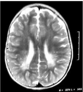
At the age of one year, he had normal language and social skills, was able to stand alone but could not walk by himself. He still had discrete left hemiparesis, but his axial tonus and patellar relexes were normal. His cra-nial magnetic resonance imaging was compatible with the diagnosis of old ischemic/hemorrhagic bilateral parieto-occipital stroke (Figures 1 and 2).
Currently, the patient is 1.6 years old and on two doses of NPH insulin (0.32 IU/kg/day). His most re-cent HbA1c level measurement was 7.2%. He still has a very discrete distal left leg weakness, but already walks very well, climbs stairs, speaks several words and has, therefore, normal psychomotor development.
Diagnosis of a genetic syndrome associated with neonatal diabetes was considered improbable, since the child presented good weight gain, absence of di-morphisms, good recovery from neurological deicits, progressive acquisition of developmental milestones, and his epileptic syndrome was classiied as focal symptomatic epilepsy due to stroke, discarding diag-nosis of progressive encephalopathy, and pointing to secondary neurological complications of DKA.
DISCUSSION
Cerebral edema is a major cause of serious complica-tions in children with DKA. The irst large population--based prospective study on this subject was published in 2001, and estimated risk of cerebral edema was 6.8 per 1,000 cases of DKA. Cerebral edema was also as-sociated with signiicant mortality (24%) and morbidity (35% of survivors) (1).
In addition, other causes of neurological deterio-ration have been described in association with DKA. In a retrospective study with 69 pediatric patients with neurological complications, CT scans and post-mortem Figure 2. T2 weighted image − Almost symmetrical areas of hyperintensity affecting bilaterally the semioval center and the periventricular parieto-occipital white matter.
Figure 1. T1 weighted image − Discrete areas of hyposignal affecting bilaterally the periventricular parieto-occipital white matter (cavitations).
Cop
yright
© ABE&M t
odos os dir
eit
os r
eser
vados
.
290 Arq Bras Endocrinol Metab. 2011;55/4
Diabetic ketoacidosis-associated stroke
studies showed that approximately 20% of the patients were found to have localized basilar edema, hemorrha-ge, thrombosis or infection (2).Some case reports on cerebral and systemic infarctions associated with T1DM have also been published, all of them emphasizing the low probability of occurrence of the disorder, and the importance of early diagnosis, investigation and treat-ment for patients that present new onset T1DM and stroke with DKA (3-5).
The causes of the hypercoagulable state and endo-thelial dysfunction in T1DM patients are still unclear. However, evidence of increased thrombin–antithrombin complex, plasma factor VII coagulant activity, and D--dimers, besides increased intima-media thickness of the carotid artery were found in children with T1DM (6,7).
Several other haemostatic changes that could lead to a thrombotic tendency during DKA have also been identiied, including evidence of endothelial injury, activation of platelets and relative hypoibrinolysis. It was also reported that children with severe DKA deve-lop decreased protein C activity and elevated levels of von Willebrand factor (vWF) antigen and vWF activity, both before and during DKA treatment.
Besides, both hyperglycemia and DKA were repor-ted to be accompanied by a proinlammatory state with elevated levels of cytokines, which also predispose to an acquired procoagulant state in in vitro and in vivo
studies. Fortunately, after DKA treatment, platelet ac-tivity returns to normal and all the other measures of hemostasis improve (8-10).
This data supports the concept that cerebral throm-bosis can occur unrelated to clinical cerebral edema, and that cerebrovascular dysregulation occurs at several sites during DKA and its treatment. Thus, it is likely that cerebrovascular accidents in children with DKA are diverse in their pathophysiology. In the case analyzed here, it is assumed that the hypercoagulable state was responsible for the stroke(8-10).
In summary, although the major cause of neurolo-gical deterioration in patients with DKA is cerebral ede-ma, practitioners must also consider stroke in
differen-tial diagnosis, especially in patients with focal indings, because of their prothrombotic tendency. DKA patients require close monitoring of their neurological status for at least 48 h after presentation, even if metabolic deran-gements have normalized. However, studies involving a multi-specialty approach are still needed to develop strategies to improve the outcomes for children with cerebrovascular events caused by DKA (11).
Disclosure: no potential conlict of interest relevant to this article was reported.
REFERENCES
1. Edge JA, Hawkins MM, Winter DL, Dunger DB. The risk and outco-me of cerebral oedema developing during diabetic ketoacidosis. Arch Dis Child. 2001;85:16-22.
2. Rosenbloom AL. Intracerebral crises during treatment of diabetic ketoacidosis. Diabetes Care. 1990;13:22-33.
3. Lee HS, Hwang JS. Cerebral infarction associated with tran-sient visual loss in child with diabetic ketoacidosis. Diabet Med. 2011;28(5):516-8
4. Reverter JL, Reverter JC, Tassies D, Rius F, Monteagudo J, Rubi-és-Prat J, et al. Thrombomodulin and induced tissue factor ex-pression on monocytes as markers of diabetic microangiopathy: a prospective study on hemostasis and lipoproteins in insulin--dependent diabetes mellitus. Am J Hematol. 1997;56:93-9. 5. Roe TF, Crawford TO, Huff KR, Costin G, Kaufman FR, Nelson MD
Jr. Brain infarction in children with diabetic ketoacidosis. J Diabe-tes Complications. 1996;10(2):100-8.
6. Giusti C, Schiaffini R, Brufani C, et al. Coagulation pathways and diabetic retinopathy: abnormal modulation in a selected group of insulin dependent diabetic patients. Br J Ophthalmol. 2000;84(12):591-5.
7. Ileri NS, Buyukasik Y, Karaahmetoglu S, Ozatli D, Sayinalp N, Ozcebe OI, et al. Evaluation of the haemostatic system during ketoacidotic deterioration of diabetes mellitus. Haemostasis. 1999;29:318-25.
8. Carl CF, Hoffman WH, Passmore GG, Truemper EJ, Lightsey AL, Cornwell PE, et al. Diabetic ketoacidosis promotes a prothrombo-tic state. Endo Res. 2003;29:73-82.
9. Hoffman WH, Burek CL, Waller JL, Fisher LE, Khichi M, Mellick LB. Cytokine response to diabetic ketoacidosis and its treatment. Clin Immunol. 2003;108:175-81.
10. Van der Poll T, Buller HR, ten Cate H, Wortel CH, Bauer KA, van Deventer SJ, et al. Activation of coagulation after administra-tion of tumor necrosis factor to normal subjects. N Engl J Med. 1990;322:1622-7.