Infection proile of patients undergoing autologous bone
marrow transplantation in a Brazilian institution
Peril de infecção em pacientes submetidos a transplante autólogo de medula
óssea em uma instituição brasileira
Kelli Borges Santos
I, Abrahão Elias Hallack Neto
II, Girlene Alves Silva
III, Angelo Atalla
IV, Marcus Matta Abreu
V, Luiz Cláudio Ribeiro
VIUniversity Hospital, Universidade Federal de Juiz de Fora (UFJF), Juiz de Fora, Minas Gerais, Brazil
ABSTRACT
CONTEXT AND OBJECTIVE: Hematopoietic stem cell transplantation (HSCT) has been widely used for treating oncological and hematological diseases. Although HSCT has helped to improve patient survival, the risk of developing infection during hospitalization is an important cause of morbidity and mortality. This study aimed to analyze the infection proile during hospitalization and the associated risk factors among patients undergoing autologous HSCT at the University Hospital, Universidade Federal de Juiz de Fora. DESIGN AND SETTING: This was a cross-sectional study on patients undergoing autologous HSCT at a public university hospital.
METHODS: Patients with febrile neutropenia between 2004 and 2009 were retrospectively evaluated re-garding their infection proile and associated risk factors.
RESULTS: Infection occurred in 57.2% of 112 patients with febrile neutropenia. The main source of infec-tion was the central venous catheter (25.9%). Infecinfec-tion was chiely due to Gram-positive bacteria, although Gram-negative-related infections were more severe and caused a higher death rate. Sex, age, skin color, nutritional status and underlying disease were not associated with the development of infection. Patients with severe mucositis (Grades III and IV) had a higher infection rate (P < 0.001). Patients who developed pulmonary complications during hospitalization had higher infection rates (P = 0.002). Infection was the main cause of death (57.1%) in the study sample.
CONCLUSION: Strategies aimed at reducing infection-related mortality rates among patients undergoing autologous HSCT are necessary.
RESUMO
CONTEXTO E OBJETIVO: O transplante de células-tronco hematopoiéticas (TCTH) vem sendo ampla-mente utilizado no tratamento das doenças onco-hematológicas. Embora o TCTH tenha colaborado para a melhora na sobrevida dos pacientes, o risco de desenvolver infecção no período de internação é uma importante causa de morbi-mortalidade. O presente estudo teve como objetivo analisar o peril das in-fecções no período de internação e os fatores de risco associados entre os pacientes submetidos ao TCTH autólogo, no Hospital Universitário da Universidade Federal de Juiz de Fora.
TIPO DE ESTUDO E LOCAL: Trata-se de um estudo transversal sobre pacientes submetidos a transplante autólogo, em um hospital público universitário.
MÉTODOS: Foram analisados retrospectivamente os pacientes que apresentaram neutropenia febril no período de 2004 a 2009, com relação ao peril infeccioso e os fatores de risco associados.
RESULTADOS: A infecção foi determinada em 57,2% dos 112 pacientes com neutropenia febril. A prin-cipal fonte de infecção foi o cateter venoso central (25,9%). A infecção ocorreu prinprin-cipalmente devido a bactérias Gram-positivas, apesar de as infecções causadas por bactérias Gram-negativas terem sido mais graves e causado maior taxa de morte. Sexo, idade, cor da pele, estado nutricional e doença de base não estiveram associados com o desenvolvimento da infecção. Pacientes com mucosite grave (graus III e IV) apresentaram maior taxa de infecção (P < 0.001). Os pacientes que desenvolveram complicações pulmo-nares durante a internação apresentaram maiores taxas de infecção (P = 0,002). A infecção foi a principal causa do óbito (57,1%) na amostra estudada.
CONCLUSÃO: São necessárias estratégias voltadas para a redução da taxa de mortalidade relacionada com infecção entre pacientes submetidos ao TCTH autólogo.
IMSc. Assistant Professor, School of Nursing,
Universidade Federal de Juiz de Fora (UFJF), Juiz de Fora, Minas Gerais, Brazil.
IIPhD. Adjunct Professor, Department of Clinical
Medicine, Universidade Federal de Juiz de Fora (UFJF), Juiz de Fora, Minas Gerais, Brazil.
IIIPhD. Adjunct Professor, School of Nursing,
Universidade Federal de Juiz de Fora (UFJF), Juiz de Fora, Minas Gerais, Brazil.
IVMSc. Adjunct Professor, Department of Clinical
Medicine, Universidade Federal de Juiz de Fora (UFJF), Juiz de Fora, Minas Gerais, Brazil.
VMD. Specialist in Thoracic Surgery and Professor
of Surgery, Faculdade de Ciências Médicas de Juiz de Fora (FCMS/JF), Juiz de Fora, Minas Gerais, Brazil.
VIPhD. Demographer and Associate Professor,
Department of Statistics, Universidade Federal de Juiz de Fora (UFJF), Juiz de Fora, Minas Gerais, Brazil.
KEY WORDS:
Hematopoietic stem cell transplantation. Transplantation, autologous.
Infection. Risk factors. Infection control.
PALAVRAS-CHAVE:
Transplante de células-tronco hematopoéticas. Transplante autólogo.
INTRODUCTION
Hematopoietic stem cell transplantation (HSCT) has been widely used for treating oncological and hematological diseases. Although its use has increased patient survival, the risk of infec-tion is an important cause of morbidity and mortality among those undergoing this therapeutic approach.1
In spite of lower infection rates with autologous bone marrow transplantation (BMT), in comparison with allogeneic BMT, infec-tion is the second most frequent cause of death among patients under-going autologous BMT, and is second only to disease relapse.2,3
Patient who have undergone autologous BMT are at higher risk of infection during the neutropenia period, and for up to 30 days ater bone marrow grating. he neutropenia period and breach of the mucocutaneous barrier are the main risks for devel-opment of infections.4,5
Hospital-acquired infection rates among HSCT patients depend on disease severity, drugs used and invasive procedures performed.6 Furthermore, the more severe the neutropenia is, the more frequent and aggressive the infections are.4
OBJECTIVE
Taking into account the susceptibility of autologous BMT patients to infection during the neutropenia period, we assessed the infectious events occurring during hospitalization and their associated risk factors.
METHODS
One hundred and iteen patients underwent autologous BMT at the University Hospital, Universidade Federal de Juiz de Fora (UFJF), between 2004 and 2009. Of these, 112 patients developed febrile neutropenia, and were analyzed. his study was approved by the research ethics committee of the University Hospital, UFJF.
All patients received antiviral prophylaxis consisting of acy-clovir 240 mg/m2/day, divided into four doses, until they received the grat, and also received granulocyte colony-stimulating factor (G-CSF), 5 mg/kg/day, in accordance with our local protocol.
hree blood samples were obtained for blood cultures: one from each line of the central venous catheter and one from peripheral blood. Empirical treatment with cefepime (4 g/day, divided into two doses) was instituted for every neutropenic patient with fever.
Febrile neutropenia was considered to be present when-ever a patient with a neutrophil count below 500/mm3, or below 1,000/mm3 but predicted to fall to below 500/mm3,7 developed a single axillary temperature reading ≥ 38 °C, or two readings ≥ 37.5 °C within a 12-hour period. Cultures of samples obtained from diferent sites were performed whenever a speciic infec-tious site was suspected.
Ater three days of treatment, the patients were reassessed and, if the fever persisted, a new blood culture was performed. he empirical therapeutic regimen was adjusted in accordance
with the recommendations of the Centers for Disease Control and Prevention (CDC).8
When blood cultures and cultures from the catheter tip yielded the same organism, with a higher number of colony-forming units (CFU) in the latter, and when the catheter tip culture grew more than 15 CFU, the infection was considered to be catheter-related.
For univariate analysis, the chi-square or Fisher exact test was used to determine the association between infection and risk factors (sex, age, underlying disease, duration of neutropenia, drugs used in the conditioning regimen, presence of mucositis, pulmonary compli-cation, diarrhea and sinusoidal obstructive syndrome). Logistic regres-sion was used for multivariate analyses, with the incluregres-sion of variables that presented P values < 0.05 on univariate analysis. he Statistical Package for the Social Sciences (SPSS) 13.0 sotware was used for cal-culations, and P values < 0.05 were considered statistically signiicant.
RESULTS
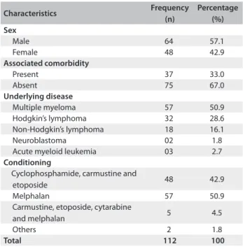
Out of the 115 patients who underwent autologous BMT, 112 developed febrile neutropenia (97.39%). he median age of this latter group was 43 years (range: 8-69 years). Table 1 shows the characteristics of the study population.
he neutropenia period ranged from four to 25 days (median: nine days). Multiple myeloma patients remained neutropenic for seven days, on average, while those with lymphoma remained neutropenic for 12 days, on average (P < 0.001). Although the diference between the groups was signiicant, the duration of neutropenia was not associated with occurrences of infection (P = 0.323). here was no impact on the number of infused cells on occurrences of infection (P = 0.129).
Six out of 60 patients with positive blood culture had more than one microorganism. Table 2 shows the microorganisms
Characteristics Frequency
(n)
Percentage (%) Sex
Male 64 57.1
Female 48 42.9
Associated comorbidity
Present 37 33.0
Absent 75 67.0
Underlying disease
Multiple myeloma 57 50.9
Hodgkin’s lymphoma 32 28.6
Non-Hodgkin’s lymphoma 18 16.1
Neuroblastoma 02 1.8
Acute myeloid leukemia 03 2.7
Conditioning
Cyclophosphamide, carmustine and
etoposide 48 42.9
Melphalan 57 50.9
Carmustine, etoposide, cytarabine
and melphalan 5 4.5
Others 2 1.8
Total 112 100
Microorganism Number of cases in irst blood culture %
Number of cases in second blood culture %
Total number
of cases % total Gram-positive bacteria
Coagulase-negative Staphylococcus 23 20.0 3 2.6 23 20.0
Staphylococcus aureus 9 7.8 5 4.3 11 9.5
Streptococcus alpha haemolyticus 1 0.9 - - 1 0.9
Total 32 27.8 8 6.9 34 29.5
Gram-negative bacteria
Enterobacteriaceae family
Klebsiella pneumoniae 13 11.3 - - 13 11.3
Escherichia coli 5 4.3 1 0.9 6 5.2
Enterobacter cloacae 5 4.3 - - 5 4.3
Serratia marcescens 1 0.9 - - 1 0.9
Non-fermenting
Acinetobacter baumanii 2 1.7 - - 2 1.7
Pseudomonas aeruginosa 3 2.6 - - 3 2.6
Non-identiied non-fermenting
gram-negative bacteria - - 1 0.9 1 0.9
Total 29 25.2 2 1.7 31 26.9
Fungi
Candida parapilosis 1 0.9 1 0.9 1 0.9
Total 1 0.9 1 0.9 1 0.9
Total (all microorganisms) 62 53.9 11 10.4 66 57.3
Table 2. Frequency of microorganisms isolated from blood cultures
Characteristics Frequency (n) Percentage (%)
Catheter-related infection 29 34.1
Oral candidiasis 24 28.2
Pneumonia 10 11.7
Bacteremia 05 5.8
Urinary infection 05 5.8
Skin infection 04 4.7
Intestinal infection 04 4.7
Systemic fungal infection 02 2.3
Vaginal candidiasis 02 2.3
Total 85 100
Table 3. Sources of infection
yielded in the irst and second blood cultures. One hundred and four diferent microorganisms were isolated, and the most fre-quent ones were: coagulase-negative Staphylococcus (24.3%), Staphylococcus aureus (13%), Klebsiella pneumoniae (12.1%), Escherichia coli (9.7%), Pseudomonas aeruginosa (4.3%) and Enterobacter cloacae (4.3%). here were also isolates of fungi (Candida parapilosis, 0.9%; Candida albicans, 5.2%; and Fusarium sp., 0.9%) and intestinal parasites (Cryptosporidium spp., 1.7%; Isospora sp, 0.9%; and Strongyloides stercoralis, 2.6%).
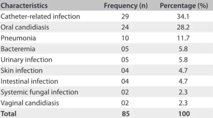
Eighty-ive instances of infection were identiied in 63 patients (57.2% of the study population), of whom 22 (34.9%) had a sec-ond source of infection. Table 3 shows the sources of infections identiied in the study population.
he catheter was the main infection source identiied in the study population, and accounted for the infections of 29 patients (25.9% of the study population). Catheter-related infec-tion was caused by Gram-positive bacteria in 48.3% of the cases (P < 0.001).
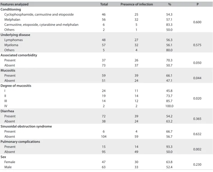
An analysis was made between diferent risk factors and occurrences of infection. Two cases were taken to be missing data, since it was not possible to determine the occurrence of infection. Table 4 shows the results from this analysis.
Among the 63 patients for whom some kind of microorgan-ism was identiied as the cause of their infection, eight (12.6%) had fungal infection (Candida albicans), 30 (47.6%) had Gram-positive bacteria and 25 (39.6%) had Gram-negative bacteria. Viral infection due to cytomegalovirus was identiied by means of the polymerase chain reaction(PCR) in only one patient.
Oral candidiasis was associated with higher grades (III and IV) of mucositis (P < 0.001). Two patients developed fungemia: one due to Candida parapilosis and one due to Fusarium spp. Fungi (Cryptosporidium spp. and Isospora spp.) were isolated from the stools of two patients.
Mycobacterium tuberculosis grew in a culture from secretions obtained from a cutaneous abscess. Out of the 104 microorgan-isms isolated, nine (8.6%) were cefepime-resistant. Although three (21.4%) of the Staphylococcus aureus isolates were methicillin-resis-tant (MRSA), they were uniformly sensitive to vancomycin. Blood cultures from six patients presented oxacillin-resistant coagulase-negative Staphylococcus.
Infection-associated factors
Mucositis (P = 0.04) and pulmonary complications (P = 0.002) during hospitalization were the only variables with a statistically signiicant impact on the occurrence of infection, on univariate analysis. Patients with higher grades (III and IV) of mucositis developed infections more frequently (P = 0.02).
Patients with associated comorbidities, such as diabetes melli-tus and systemic arterial hypertension, were more likely to develop infection, although this was not statistically signiicant (P = 0.05).
Both variables that showed statistical signiicance in relation to infection on univariate analysis (mucositis and pulmonary complications) were included in the logistic regression models.
he results from this model are shown in Table 5. Only pulmo-nary complications remained an independent risk on multivari-ate analysis (P = 0.015), with an odds ratio of 13. Although the conidence interval of the odds ratio for the variable of pulmo-nary complications was large, this was due to the low frequency of events. Nonetheless, 14 out of the 15 individuals who had pulmo-nary complications developed infections, which shows that there was a strong association between these variables. Even though the P value related to mucositis was slightly above 5%, stratiied analysis showed that among individuals who did not have pul-monary complications, the infection rate was signiicantly higher among patients who developed mucositis (P = 0.032).
Features analyzed Total Presence of infection % P
Conditioning
Cyclophosphamide, carmustine and etoposide 46 25 54.3
0.600
Melphalan 56 32 57.1
Carmustine, etoposide, cytarabine and melphalan 6 5 83.3
Others 2 1 50.0
Underlying disease
Lymphomas 48 27 56.3
0.575
Myeloma 57 32 56.1
Others 5 4 80.0
Associated comorbidity
Present 37 26 70.3
0.050
Absent 73 37 50.7
Mucositis
Present 59 39 66.1
0.044
Absent 51 24 47.1
Degree of mucositis
I 24 11 45.8
0.020
II 19 14 73.7
III 14 12 85.7
IV 2 2 100.0
Diarrhea
Present 72 39 54.2
0.365
Absent 38 24 63.2
Sinusoidal obstruction syndrome
Present 6 4 66.7
0.632
Absent 104 59 56.7
Pulmonary complications
Present 15 14 93.3
0.002
Absent 95 49 50.0
Sex
Female 47 30 63.8
0.230
Male 63 33 52.4
*Missing: two patients without data on infection.
Table 4. Factors associated with infection during hospitalization*
Table 5. Multivariate analysis with risk factors for infection*
Variables P-value Odds ratio 95% conidence interval
Lower Upper
Mucositis 0.054 2.21 0.98 4.94
Pulmonary complications 0.015 13.23 1.65 105.98
DISCUSSION
In this study, 57.2% of the population had an infection identiied during hospitalization. According to published data from a single Brazilian center, the infection rate among a pedi-atric population undergoing allogeneic and autologous BMT was 58.5%.9 In a multicenter study undertaken in southern and southeastern Brazil, the infection rate was 55%.2 Although these values are similar to ours, the comparison is hampered because we studied patients undergoing autologous BMT alone.
Studies in other countries on patients undergoing autolo-gous BMT have indicated infection rates ranging from 27.6% to 48.2%,10,11 i.e. lower than the rate that we found. Although not statistically signiicant, there was a diference in the occur-rence of infection in relation to the irst years covered by this study. his might be accounted for by the little experience of managing autologous BMT patients that existed in those early days, which may have led to infection rates that were higher than those in the literature.
In disagreement with the literature,5,12,13 age was not asso-ciated with the risk of infection in our study. his inding was possibly due to the small number of children in our sample.
In agreement with the indings of Poutsiaka et al.,14 the length of the neutropenia period was not signiicantly associ-ated with higher rates of infectious complications in our study. he same was observed in the study by Ninin et al.,15 in which patients undergoing autologous and allogeneic BMT had simi-lar infection rates.
Although not statistically signiicant, multiple myeloma patients had the lowest infection rates, a inding that is similar to the data reported by Meyer et al.16 According to Gil et al.,10 acute myeloid leukemia patients undergoing autologous BMT have the highest risk of infection, since they remain neutro-penic for longer periods. In our study, there was no association between the conditioning regimen and infection.
he most common infection source was the cathe-ter (34.1%). Our data were very close to those of Nucci and Maiolino,2 who reported a catheter-related infection rate of 38% in their patients. he associated factors may include: han-dling frequency, insertion site, contamination during inser-tion, skin colonization around the insertion site, contamina-tion of the conneccontamina-tion device between the infusion system and vascular access, contamination of the infusion luid, contami-nation of solutions used to ensure catheter permeability and presence of distant infectious sites with hematogenic spread of infection.4,8
We also made this observation, such that patients with mucositis of grades III and IV had higher infection rates.
Infection-related pulmonary complications have been considered to be important determiners of morbidity and
mortality. According to Meyer et al.,16 pneumonia was the sec-ond most frequent cause of infection in these patients, occur-ring in 7.8% of the cases. According to Dettenkofer et al.,6 pneumonia is the most frequent infection in patients undergo-ing autologous BMT.
hree patients had oxacillin-resistant Staphylococcus aureus. his microorganism increases the infection-related mortality rate of grated patients, and has been isolated with higher fre-quency, in diferent places, over the past few years.17 No vanco-mycin-resistant microorganism was isolated in our study.
In accordance with the Brazilian and foreign literature, Gram-positive bacteria were the most frequent isolates in our study. Nonetheless, a signiicant increase in the number of infec-tions due to Gram-negative bacteria has been reported over the past few years.1,7,12,18-21 his may be accounted for by greater use of prophylactic antimicrobials, chiely quinolones.22
According to Laws et al.,19 Gram-negative bacterial infec-tions are more severe and cause higher death rates, compared with Gram-positive bacterial infections. his may be explained by their greater virulence and resistance, and the latter is also due to their double cell membrane, which blocks the entry of some drugs.23
he most frequent isolate in our study was coagulase-neg-ative Staphylococcus, in agreement with indings from diferent BMT centers.6,15,16,19,24,25 Until recently, these bacteria were con-sidered contaminants, devoid of serious clinical importance.26 However, over the past few years, they have been acknowledged as important infectious agents. Catheters are the main means of entry for these microorganisms.26
Fungi were the second most frequently identified micro-organisms in the infections we found. The main isolate was Candida albicans, and its presence was associated with mucosi-tis. In autologous BMT, the likelihood of systemic candidiasis is very low, and occurs chiely ater the neutropenia period has resolved, or in the absence of mucositis.27
Similarly to indings from other centers,2,8,11,14 infection was the main cause of death in our patients during hospitalization. It seemed to contribute towards longer hospital stay, although this association did not present statistical signiicance (P = 0.081).
CONCLUSIONS
REFERENCES
1. Naoum FA, Martins LTV, Castro NS, Barros JC, Chiattone CS. Peril
microbiológico dos pacientes nos primeiros trinta dias pós transplante
de medula óssea do Serviço de Transplantes da Santa Casa de São
Paulo [Microbiological proile of patients in the irst thirty days post
bone marrow transplantation of the Transplantation Service in Santa
Casa, São Paulo]. Rev Bras Hematol Hemoter. 2002;24(2):91-6.
2. Nucci M, Maiolino A. Infecções em transplante de medula óssea.
[Infection in bone marrow transplant recipients]. Medicina (Ribeirão
Preto). 2000;33(3):278-93.
3. Bueno ND, Saboya R, Martins MC, et al. O transplante de medula óssea
na leucemia mielóide aguda: análise de 80 pacientes transplantados
no complexo do Hospital das Clínicas da Faculdade de Medicina da
Universidade de São Paulo [The allogeneic and autologous bone
marrow transplantation in acute myeloid leukemia: analysis of 80
patients – Bone Marrow Transplantation Service – Hospital das
Clínicas of the Medical School, University of São Paulo]. Rev Bras
Hematol Hemoter. 2004;26(2):84-92.
4. Dykewicz CA; Centers for Disease Control and Prevention (U.S.);
Infectious Diseases Society of America; American Society of Blood and
Marrow Transplantation. Summary of the Guidelines for Preventing
Opportunistic Infections among Hematopoietic Stem Cell Transplant
Recipients. Clin Infect Dis. 2001;33(2):139-44.
5. Mackall C, Fry T, Gress R, et al. Background to hematopoietic cell
transplantation, including post transplant immune recovery. Bone
Marrow Transplant. 2009;44(8):457-62.
6. Dettenkofer M, Wenzler-Röttele S, Babikir R, et al. Surveillance of
nosocomial sepsis and pneumonia in patients with a bone marrow
or peripheral blood stem cell transplant: a multicenter project. Clin
Infect Dis. 2005;40(7):926-31.
7. Hughes WT, Armstrong D, Bodey GP, et al. 2002 guidelines for the
use of antimicrobial agents in neutropenic patients with cancer. Clin
Infect Dis. 2002;34(6):730-51.
8. Centers for Disease Control and Prevention; Infectious Diseases
Society of America; American Society of Blood and Marrow
Transplantation. Guidelines for preventing opportunistic infections
among hematopoietic stem cell transplant recipients. MMWR
Recomm Rep. 2000;49(RR-10):1-125, CE1-7.
9. Castro Júnior CG, Gregianin LJ, Brunetto AL. Análise clínica e
epidemiológica do transplante de medula óssea em um serviço de
oncologia pediátrica [Clinical and epidemiological analysis of bone
marrow transplantation in a pediatric oncology unit]. J Pediatr (Rio J).
2003;79(5):413-22.
10. Gil L, Styczynski J, Komarnicki M. Infectious complication in 314
patients after high-dose therapy and autologous hematopoietic
stem cell transplantation: risk factors analysis and outcome. Infection.
2007;35(6):421-7.
11. Reich G, Mapara MY, Reichardt P, Dörken B, Maschmeyer G. Infections
complications after high-dose chemotherapy and autologous stem
cell transplantation: comparison between patients with lymphoma
or multiple myeloma and patients with solid tumors. Bone Marrow
Transplant. 2001;27(5):525-9.
12. Bailey LC, Reilly AF, Rheingold SR. Infections in pediatric patients with
hematologic malignancies. Semin Hematol. 2009;46(3):313-24.
13. Mendes AVA, Sapolnik R, Mendonça N. Novas diretrizes na abordagem
clínica da neutropenia febril e da sepse em oncologia pediátrica
[New guidelines for the clinical management of febrile neutropenia
and sepsis in pediatric oncology patients]. J Pediatr (Rio J.). 2007;
83(2, supl.): S54-S63.
14. Poutsiaka DD, Price LL, Ucuzian A, et al. Blood stream infection
after hematopoietic stem cell transplantation is associated with
increased mortality. Bone Marrow Transplant. 2007;40(1):63-70.
15. Ninin E, Milpied N, Moreau P, et al. Longitudinal study of bacterial, viral,
and fungal infections in adult recipients of bone marrow transplants.
Clin Infect Dis. 2001;33(1):41-7.
16. Meyer E, Beyersmann J, Bertz H, et al. Risk factor analysis of blood
stream infection and pneumonia in neutropenic patients after
peripheral blood stem-cell transplantation. Bone Marrow Transplant.
2007;39(3):173-8.
17. Shaw BE, Boswell T, Byrne JL, Yates C, Russell NH. Clinical impact of
MRSA in a stem cell transplant unit: analysis before, during and after
an MRSA outbreak. Bone Marrow Transplant. 2007;39(10):623-9.
18. Oliveira AL, de Souza M, Carvalho-Dias VM, et al. Epidemiology
of bacteremia and factors associated with multi-drug-resistant
gram-negative bacteremia in hematopoietic stem cell transplant
recipients. Bone Marrow Transplant. 2007;39(12):775-81.
19. Laws HJ, Kobbe G, Dilloo D, et al. Surveillance of nosocomial infections
in paediatric recipients of bone marrow or peripheral blood stem cell
transplantation during neutropenia, compared with adult recipients.
J Hosp Infect. 2006;62(1):80-8.
20. Moura MEB, Campelo SMA, Brito FCP, et al. Infecção hospitalar: estudo
de prevalência em um hospital público de ensino [Nosocomial
infection: study of prevalence at a public teaching hospital]. Rev Bras
Enferm. 2007;60(4):416-21.
21. Walsh T.J. Advances and challenges in infectious diseases supportive
care of patients with hematologic malignancies, hematopoietic stem
cell transplantation, and severe aplastic anemia. Semin Hematol.
2009;46(3):191-7.
22. Cattaneo C, Quaresmini G, Casari S, et al. Recent changes in bacterial
epidemiology and the emergence of luoroquinolone-resistant
Escherichia coli among patients with haematological malignancies:
results of a prospective study on 823 patients at a single institution. J
Antimicrob Chemother. 2008;61(3):721-8.
23. Alterthun F. Morfologia e estrutura da célula bacteriana. In: Trabulsi LR,
Alterthun F, editors. Microbiologia. São Paulo: Atheneu; 2008. p. 1-19.
24. Çelebi H, Akan H, Akçağlayan, Ustün C, Arat M. Febrile neutropenia in
allogeneic and autologous peripheral blood stem cell transplantation
and conventional chemotherapy for malignancies. Bone Marrow
Transplant. 2000;26(2):211-4.
chemotherapy and following autologous stem cell transplantation.
Bone Marrow Tranplant. . 2001;28(12):1129-34.
26. Bueris V, Moreira CG, Teixeira LM, Santos KRN, Trabulsi LR.
Staphylococcus epidermidis e outras espécies de Staphylococcus,
micrococcus e Rothia (Stomatococcus). In: Trabulsi LR, Alterthum F,
editors. Microbiologia. São Paulo: Atheneu; 2008. p. 183-7.
27. Marr KA, Bow E, Chiller T, et al. Fungal infection prevention after
hematopoietic cell transplantation. Bone Marrow Transplant.
2009;44(8):483-7.
Sources of funding: None Conlict of interest: None
Date of irst submission: January 10, 2011 Last received: July 13, 2011
Accepted: July 20, 2011
Address for correspondence:
Kelli Borges Santos
Joaquim Carneiro Filho, 45/501
Cascatinha — Juiz de Fora (MG) — Brasil
CEP 36033320