Surgical treatment of congenital
lung malformations in pediatric patients*
Tratamento cirúrgico das malformações pulmonares congênitas em pacientes pediátricos
Hylas Paiva da Costa Ferreira, Gilberto Bueno Fischer, José Carlos Felicetti, José de Jesus Peixoto Camargo, Cristiano Feijó Andrade
Abstract
Objective: To determine the main congenital lung malformations treated and the principal diagnostic methods employed, as well as the indications for surgical treatment and the results obtained, at a referral facility for pediatric thoracic surgery. Methods: We reviewed the medical charts of 52 patients anatomopathologically diagnosed with congenital lung malformations and who had been submitted to pulmonary resection between January of 1997 and December of 2006. Exclusion criteria were age ≥ 12 years and incomplete clinical data. The final sample comprised 35 patients. Results: In this sample, the mean age was 31 months, and there was a predominance of males (n = 21). The anatomopathological findings were cystic adenomatoid malformation (n = 14), congenital lobar emphysema (n = 13), pulmonary sequestration (n = 8) and arteriovenous malformation (n = 1). The most common type of lung resection was left lower lobectomy (in 25.71%) followed by different types of segmentectomy (in 22.85%), left upper lobectomy (in 22.85%), right upper lobectomy (in 14.28%), right lower lobectomy (in 8.57%) and middle lobectomy (in 5.71%). Of the 35 patients, 34 (97.14%) were submitted to closed pleural drainage, with a mean duration of thoracic drainage of 3.9 days. Ten patients (28.5%) presented with postoperative complications. There were no deaths in our sample. Conclusions: Pulmonary resection for the treatment of congenital lung malformations is a safe procedure, presenting low morbidity and no mortality at a referral facility for pediatric thoracic surgery.
Keywords: Cystic adenomatoid malformation of lung, congenital; Bronchopulmonary sequestration; Pneumonectomy; Pulmonary surgical procedures; Pulmonary emphysema.
Resumo
Objetivo: Determinar as principais malformações congênitas pulmonares e os principais métodos diagnósticos utilizados, assim como as indicações de tratamento cirúrgico e os seus resultados em um serviço de referência de cirurgia torácica pediátrica. Métodos: Foram revisados 52 prontuários de pacientes com diagnóstico anatomopa-tológico de malformações congênitas pulmonares e que foram submetidos à ressecção pulmonar entre janeiro de 1997 e dezembro de 2006. Os critérios de exclusão foram idade ≥ 12 anos e dados clínicos incompletos. A amostra final foi composta de 35 pacientes. Resultados: Nesta amostra, a média de idade foi de 31 meses, com predo-minância do sexo masculino (n = 21). Os achados anatomopatológicos foram malformação adenomatoide cística (n = 14), enfisema lobar congênito (n = 13), sequestro pulmonar (n = 8), e malformação arteriovenosa (n = 1). A ressecção mais comum foi a lobectomia inferior esquerda (25,71%), seguida por diferentes tipos de segmentec-tomia (22,85%), lobecsegmentec-tomia superior esquerda (22,85%), lobecsegmentec-tomia superior direita (14,28%), lobecsegmentec-tomia inferior direita (8,57%) e lobectomia média (5,71%). Dos 35 pacientes, 34 (97,14%) foram submetidos à drenagem pleural fechada, com tempo médio de permanência do dreno torácico de 3,9 dias. Dez pacientes (28,5%) apresentaram complicações pós-operatórias. Não houve óbitos nesta série. Conclusões: A ressecção pulmonar para o tratamento das malformações pulmonares é um procedimento seguro, apresentando baixa morbidade e nenhuma mortalidade em um serviço de referência para doenças pulmonares.
Descritores: Malformação adenomatoide cística congênita do pulmão; Sequestro broncopulmonar; Pneumonectomia; Procedimentos cirúrgicos pulmonares; Enfisema pulmonar.
* Study carried out at the Santo Antônio Children’s Hospital, Porto Alegre, Brazil.
Correspondence to: Cristiano Feijó Andrade. Serviço de Cirurgia Torácica Pediátrica, Rua Annes Dias, 285, Centro, CEP 90020-090, Porto Alegre, RS, Brasil.
Tel 55 51 3214-8674. E-mail: cristianofa@cirurgiatoracica.net
Financial support: Cristiano Feijó Andrade is a recipient of a Research Productivity Scholarship (Level II) from the Conselho Nacional de Desenvolvimento Científico e Tecnológico (CNPq, National Council for Scientific and Technological Development).
Methods
We retrospectively analyzed the charts of all patients anatomopathologically diagnosed with congenital lung malformations and submitted to pulmonary resection at the Santa Casa Sisters of Mercy Hospital Complex, located in the city of Porto Alegre, Brazil, between January of 1997 and December of 2006. Charts of patients aged 12 years or older were excluded, as were charts from which data were missing. The present study was approved by the Research Ethics Committee of the Santa Casa Sisters of Mercy Hospital (Protocol no. 1436/06).
We reviewed 52 charts of patients anato-mopathologically diagnosed with congenital lung malformations, 39 of whom were less than 12 years of age. Four cases (2 cases of pulmo-nary hypoplasia and 2 cases of arteriovenous fistula) were excluded due to the fact that their charts were incomplete. Since the cases of bron-chogenic cysts presented as extrapulmonary or mediastinal cysts, they were not included in the data collection. The patients were divided into groups by malformation as follows: congenital lobar emphysema; extralobar and intralobar sequestration; arteriovenous malformation; and CAM. The following parameters were analyzed: age; gender; signs and symptoms; imaging tests employed; location of the lesions; surgical procedures performed; and postoperative compli-cations. The patients diagnosed with CAM were divided into types I to IV, in accordance with the anatomopathological classification devised by Stocker.(3,10,17)
Data were entered into a Microsoft Excel spreadsheet. Mean and median ages were calcu-lated, as were mean length of hospital stay and the percentage and location of each disease. Student’s t-test was used in order to identify significant differences (p < 0.05) in the dura-tion of sucdura-tion pleural drainage and water-seal pleural drainage.
Results
In this sample of 35 patients, the mean age at the time of surgery was 31 months (median, 17 months; range: 1-120 months) and there was a predominance of males (60%). Of those 35 patients, 31 were White (88.6%), 3 were Black (8.6%) and 1 was of indigenous ethnicity (2.8%).
Introduction
Congenital lung malformations form a spectrum of rare diseases, occurring from the development of the primitive intestine and its differentiation into a respiratory system onward.(1,2) Although it is difficult to determine
the incidence of such malformations worldwide, the advent of prenatal ultrasound has led to an increase in the number of diagnoses,(3) the
esti-mated annual incidence rates now ranging from 30 to 42 cases per 100,000 population.(4)
The spectrum of congenital lung malfor-mations includes cystic adenomatoid malformation (CAM), pulmonary sequestration, congenital lobar emphysema and bronchogenic cysts, the last being intrapulmonary or extrapul-monary.(5) Although they represent alterations in
the pulmonary vessels, rather than in the lungs themselves, arteriovenous malformations are also included in the list of lung malformations.
Congenital lung malformations can be diag-nosed at any age.(4,6,7) Their clinical presentation
can range from respiratory failure after birth to imaging alterations in asymptomatic adult patients.(1,4,6) The understanding of such
malfor-mations is of utmost importance, since their early recognition in neonates can mean success in the treatment of neonatal respiratory failure and other complications.(7-10)
The diagnosis of these lesions usually requires various imaging tests, such as prenatal/postnatal Doppler ultrasound, prenatal magnetic reso-nance imaging and chest CT, for proper surgical planning.(1,3,6-9) Pulmonary resection is the
prin-cipal treatment for various forms of congenital lung malformations; resections should be as conservative as possible, lobectomy being the most common procedure.(1,3,8-11) Resections can
be performed by thoracoscopy or by thora-cotomy, depending on the experience of the facility.(12-16)
There have been few studies evaluating the surgical treatment of lung malformations in children in Brazil.(4) Therefore, we sought to
required mechanical ventilation for more than 24 h (range: 1-5 days).
Discussion
Although pulmonary malformations have been known to medicine for more than three centuries, there is still no consensus on how or when to treat them.(4) With the advances
in imaging testing, pulmonary malforma-tions began to be diagnosed more frequently in the prenatal period or on the first days of life, allowing an increase in the prevention of complications in the neonatal period. Even with the routine use of prenatal ultrasound,(3,6)
the differential diagnosis of various pulmonary malformations is often difficult, requiring the use of additional tests, such a prenatal magnetic resonance imaging or neonatal CT, for diagnostic confirmation.(3) In our sample, few patients
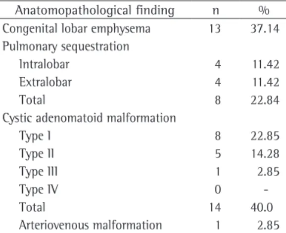
were found to have been prenatally diagnosed with congenital malformation, probably due to the fact that most patients were residents of The anatomopathological findings in the
35 patients analyzed are described in Table 1. One patient had type I CAM accompanied by extralobar pulmonary sequestration.
Of the 35 patients studied, 9 (25.71%) had no preoperative symptoms. Of those 9 patients, 2 (5.71%) had type I CAM, 2 (5.71%) had type II CAM, 2 (5.71%) had intralobar pulmonary sequestration and 3 (8.57%) had extralobar pulmonary sequestration. All patients with congenital lobar emphysema had preoperative symptoms, the most common of which was labored breathing (in 9 cases—69.23%).
All of the patients had been submitted to some type of imaging test for the preoperative diagnosis: 28 (80%) had been submitted to chest CT; 35 (100%) had undergone routine chest X-ray; and only 4 (11.42%) had been prenatally diagnosed by ultrasound (3 cases of CAM and 1 case of extralobar pulmonary sequestration). The locations of the lesions are described in Table 2, and the types of resections are shown in Table 3.
Of the patients studied, 17 (48.57%) had a history of treatment for bacterial pneumonia, 14 (40.00%) had been submitted to diagnostic bronchoscopy, and only 2 (5.71%) had under-gone preoperative pulmonary function tests. The mean length of hospital stay was 11 days (range: 3-30 days), whereas the mean length of postoperative hospital stay was 8 days (range: 2-21 days).
Ten patients (28.5%) presented with post-operative complications, the most common of which was pulmonary infection (in 7), followed by pneumothorax after the removal of the drainage tubes (in 2) and pulmonary atelectasis (in 1). There were no deaths in our sample.
Thirty-four patients (97.14%) had been submitted to closed pleural drainage: water-seal drainage in 18 (51.42%); and continuous suction drainage in 16 (45.71%). There was no significant difference between these two groups in terms of duration of drainage (p > 0.05). In 28 patients (80%), drainage was performed with one chest tube, whereas two chest tubes were used in 6 (17.14%). The mean dura-tion of thoracic drainage was 3.9 days (range: 1-9 days). Only 1 patient diagnosed with extralobar sequestration was not submitted to thoracic drainage. In our sample, 8 patients had
Table 1 - Anatomopathological findings in 35 patients with congenital lung malformations.a
Anatomopathological finding n % Congenital lobar emphysema 13 37.14 Pulmonary sequestration
Intralobar 4 11.42
Extralobar 4 11.42
Total 8 22.84
Cystic adenomatoid malformation
Type I 8 22.85
Type II 5 14.28
Type III 1 2.85
Type IV 0
-Total 14 40.0
Arteriovenous malformation 1 2.85
aThere was 1 patient with two lung malformations
(extralobar pulmonary sequestration and type I cystic adenomatoid malformation), which were resected simultaneously.
Table 2 - Location of lung malformations (based on preoperative imaging studies).
Location n (%)
Left lower lobe 7 (20)
Left upper lobe 13 (37.14) Right lower lobe 6 (17.14) Right upper lobe 7 (20)
mately until 8 years of age.(2-4) In asymptomatic
patients, the mean age for surgery increases,(1,11)
and surgical treatment is indicated for those who begin to have symptoms.
Video-assisted thoracoscopy has been widely used in the resection of pulmonary sequestra-tions in adults, presenting good results due to safe resection of the lesion, with lower rates of complications and early discharge from hospital.
(15,16) Regarding pediatric patients, video-assisted
thoracoscopy can be performed in children of all ages, being similar to the procedures performed in adults in terms of safety and efficacy.(18)
In our sample, the most common type of resection was upper lobe lobectomy, espe-cially among the patients with congenital lobar emphysema.(1,11) Patients with CAM or pulmonary
sequestration predominantly had lower lobe involvement, as observed in various studies,(5,9,10)
whereas those with congenital lobe emphysema had a greater number of lesions in the upper lobes. Although not all of the patients in our study had been submitted to bronchoscopy, we recommend that bronchoscopy be routinely used in the preoperative evaluation, since this proce-dure, in addition to allowing the collection of endobronchial samples, can provide important information for surgical strategy by means of the visualization of the anatomy of the tracheo-bronchial tree.(8)
Combined pulmonary malformations are seen in up to 50% of cases.(2,6,8) In addition,
pulmo-nary malformations can occur concomitantly with malformations in other organs, the most common being pectus excavatum, esophageal malformations and cardiac malformations.(7-9)
Although we observed such concomitance in our sample, its incidence was much less lower than that reported in the literature.(2,6-9)
The most common postoperative compli-cations and the causes of reintervention in patients with pulmonary malformations who are submitted to pulmonary resection are pleural empyema, pneumonia, hemothorax, pleural effusion and prolonged air leak.(1,7,9,13) Among the
postoperative complications seen in the present study, pneumonia was the most common, which corroborates what has been reported in the liter-ature.(4) The duration of thoracic drainage varies
from study to study. In our study, it was longer than the mean duration of 1.2 days reported by another group of authors.(18) None of our
other areas and their mothers had not received adequate prenatal care. Patients with pulmonary malformations will eventually have symptoms, typically before the age of 13 years.(4)
In the presence of symptoms, surgical treat-ment of pulmonary malformations should be performed as early as possible, seeking to preserve as much normal lung parenchyma as possible.
(7,10) In asymptomatic lesions, it remains
contro-versial as to whether or not pulmonary resection is necessary, and its indication is based on the risk of recurrent infections and possible pleurop-ulmonary complications, as well as on the risk of malignancy in cases of CAM ( rhabdomyosarcoma and bronchioloalveolar carcinoma).(1,3,4,6,8,10) In
such cases, pulmonary resections are safe and have a low rate of complications.(1,7,8)
The resection of choice in cases of CAM and in cases of intralobar sequestration is lobec-tomy or segmenteclobec-tomy, when possible,(3-5,8,12,13)
whereas sequestrectomy is indicated for cases of extralobar sequestration(3,4,7-9,12,13) and, very
rarely, pneumonectomy will be indicated for selected cases of CAM.(3)
There is controversy as to whether the treat-ment of congenital lobar emphysema should be surgical or conservative.(3,11) When patients are
symptomatic, surgery is indicated immediately after diagnosis, and lobectomy is the resection of choice,(1,11) whereas segmental resection is to
be used only in cases of localized disease.(4) This
type of early indication solves the problem, and patients show no reduction in pulmonary func-tion since there is compensatory growth of the lung until the end of early childhood,
approxi-Table 3 - Resections performed in 35 patients with congenital lung malformations.
Type of resection n (%)
Left lower lobectomy 5 (14.28) Left upper lobectomy 8 (22.85) Right lower lobectomy 3 (8.57)
Middle lobectomy 2 (5.71)
Right upper lobectomy 5 (14.28) Anterior LUL segmentectomy 3 (8.57)
Left basal pyramid 2 (5.71)
Apical RLL segmentectomy 1 (2.75) Anterior RUL segmentectomy 1 (2.75) Apical RUL segmentectomy 1 (2.75)
Sequestrectomy 4 (11.42)
evaluation and management. Eur J Cardiothorac Surg. 2005;27(1):45-52.
9. Van Raemdonck D, De Boeck K, Devlieger H, Demedts M, Moerman P, Coosemans W, et al. Pulmonary sequestration: a comparison between pediatric and adult patients. Eur J Cardiothorac Surg. 2001;19(4):388-95. 10. Waszak P, Claris O, Lapillonne A, Picaud JC, Basson E,
Chappuis JP, et al. Cystic adenomatoid malformation of the lung: neonatal management of 21 cases. Pediatr Surg Int. 1999;15(5-6):326-31.
11. Mei-Zahav M, Konen O, Manson D, Langer JC. Is congenital lobar emphysema a surgical disease? J Pediatr Surg. 2006;41(6):1058-61.
12. Tanaka T, Ueda K, Sakano H, Hayashi M, Li TS, Zempo N. Video-assisted thoracoscopic surgery for intralobar pulmonary sequestration. Surgery. 2003;133(2):216-8. 13. Gezer S, Taştepe I, Sirmali M, Findik G, Türüt H, Kaya
S, et al. Pulmonary sequestration: a single-institutional series composed of 27 cases. J Thorac Cardiovasc Surg. 2007;133(4):955-9.
14. Suda T, Hasegawa S, Negi K, Hattori Y. Video-assisted thoracoscopic surgery for extralobar pulmonary sequestration. J Thorac Cardiovasc Surg. 2006;132(3):707-8.
15. Vu LT, Farmer DL, Nobuhara KK, Miniati D, Lee H. Thoracoscopic versus open resection for congenital cystic adenomatoid malformations of the lung. J Pediatr Surg. 2008;43(1):35-9.
16. Kestenholz PB, Schneiter D, Hillinger S, Lardinois D, Weder W. Thoracoscopic treatment of pulmonary sequestration. Eur J Cardiothorac Surg. 2006;29(5):815-8.
17. Tawil MI, Pilling DW. Congenital cystic adenomatoid malformation: is there a difference between the antenatally and postnatally diagnosed cases? Pediatr Radiol. 2005;35(1):79-84.
18. Koontz CS, Oliva V, Gow KW, Wulkan ML. Video-assisted thoracoscopic surgical excision of cystic lung disease in children. J Pediatr Surg. 2005;40(5):835-7.
patients underwent reintervention or died, a finding that is in agreement with those of most other studies.(1,10)
Pulmonary resection for the treatment of congenital lung malformations is a safe proce-dure, presenting low morbidity and no mortality at a referral facility for pediatric thoracic surgery.
References
1. Khosa JK, Leong SL, Borzi PA. Congenital cystic adenomatoid malformation of the lung: indications and timing of surgery. Pediatr Surg Int. 2004;20(7):505-8. 2. McLean SE, Pfeifer JD, Siegel MJ, Jensen ER, Schuler
PM, Hirsch R, et al. Congenital cystic adenomatoid malformation connected to an extralobar pulmonary sequestration in the contralateral chest: common origin? J Pediatr Surg. 2004;39(8):e13-7.
3. Azizkhan RG, Crombleholme TM. Congenital cystic lung disease: contemporary antenatal and postnatal management. Pediatr Surg Int. 2008;24(6):643-57. 4. Costa Júnior Ada S, Perfeito JA, Forte V. Surgical treatment
of 60 patients with pulmonary malformations: what have we learned? J Bras Pneumol. 2008;34(9):661-6. 5. Hashemzadeh S, Aslanabadi S, Jafari Rouhi AH, Azhough
R, Kaleibar NA. Congenital malformations of the lung. Indian J Pediatr. 2007;74(2):192-4.
6. Sapin E, Lejeune V V, Barbet JP, Carricaburu E, Lewin F, Baron JM, et al. Congenital adenomatoid disease of the lung: prenatal diagnosis and perinatal management. Pediatr Surg Int. 1997;12(2/3):126-9.
7. Halkic N, Cuénoud PF, Corthésy ME, Ksontini R, Boumghar M. Pulmonary sequestration: a review of 26 cases. Eur J Cardiothorac Surg. 1998;14(2):127-33. 8. Shanmugam G, MacArthur K, Pollock JC. Congenital
About the authors
Hylas Paiva da Costa Ferreira
Assistant Professor of Emergency Medicine. Universidade Federal do Rio Grande do Norte – UFRN, Federal University of Rio Grande do Norte – Natal, Brazil.
Gilberto Bueno Fischer
Full Professor of Pediatrics. Universidade Federal de Ciências da Saúde de Porto Alegre – UFCSPA, Federal University of Health Sciences of Porto Alegre – Porto Alegre, Brazil.
José Carlos Felicetti
Assistant Professor of Thoracic Surgery. Universidade Federal de Ciências da Saúde de Porto Alegre – UFCSPA, Federal University of Health Sciences of Porto Alegre – Porto Alegre, Brazil.
José de Jesus Peixoto Camargo
Assistant Professor of Thoracic Surgery. Universidade Federal de Ciências da Saúde de Porto Alegre – UFCSPA, Federal University of Health Sciences of Porto Alegre – Porto Alegre, Brazil.
Cristiano Feijó Andrade