249
1. PhD; Associate professor, Department of Pediatrics, UniversidadeFede-ral de Minas Gerais (UFMG), Belo Horizonte, MG, Brazil.
2. PhD; Assistant professor, Department of Ophthalmology, Universidade Federal de Minas Gerais (UFMG), Belo Horizonte, MG, Brazil. 3. Assistant professor, Department of Pediatrics, Universidade Federal de
Minas Gerais (UFMG). Pediatric neurologist, Hospital das Clínicas, UFMG, Belo Horizonte, MG, Brazil.
4. MD, Universidade Federal de Minas Gerais (UFMG), Belo Horizonte, MG, Brazil.
Manuscript received Apr 15 2003, accepted for publication Oct 15 2003.
Marcus Gunn Phenomenon:
differential diagnosis
of palpebral ptoses in children
Marcia R. F. Torres1, Nassin Calixto Jr.2, Luiz R. Oliveira3, Sílvia A. Steiner4, Amarilis M. Iscold4
0021-7557/04/80-03/249 Jornal de Pediatria
Copyright © 2004 by Sociedade Brasileira de Pediatria
C
ASER
EPORTIntroduction
The eyelids are mobile protective covers for the eyes formed by superimposed plates, within which are located certain muscles. The upper eyelid levator muscle, connected to the third cranial nerve (oculomotor nerve), is responsible for opening the eyelids, with the Müller and frontalis muscles giving support. The orbicularis oculi muscle, connected to the seventh cranial nerve (facial nerve), performs eye closure, an important function for visual dynamics.1
Ptosis is a condition of the eyelids that results from an abnormally low upper eyelid position. It is agreed that the normal position of the free border of the eyelid is, on average, 2 mm below the superior corneal limbus; any additional obscuring of the cornea is considered pathological, in this manner the upper eyelid already covers 1 to 2 mm of the cornea.1 Ptosis can result from a number of different diseases that can be grouped into four categories: aponeurotic, mechanical, myogenic e neurogenic. Aponeurotic causes include senile ptosis, post-ocular surgery ptosis, blepharochalasia, Graves disease and pregnancy. Among mechanical causes we can cite scar-related ptosis and ptosis associated with tumors. Myasthenia gravis and familial myopathic progressive ptosis are examples of myogenic ptoses. The most common neurogenic forms are Horner syndrome, third cranial nerve palsy (ophthalmoplegia) and synkinetic ptosis.2,3
Synkinesis is defined as the occurrence of simultaneous movements or a coordinated sequence of movements of muscles innervated by different nerves or by different branches of the same nerve.4
Abstract
Objective: The aim of this paper is to review existing literature on the subject and to report on and discuss a case of Marcus Gunn Phenomenon.
Description: A five year-old female, otherwise a healthy patient, while still a few months old, was seen by a pediatrician who detected a disorder of the right eye, initially believed to be strabismus, at a follow-up childcare consultation. Several ophthalmologists failed to establish a precise diagnosis. After a pediatric ophthalmologist had examined the child at four years of age, a diagnosis of Marcus Gunn Phenomenon, otherwise known as jaw-winking phenomenon, was confirmed. Apart from this anomaly, physical, ophthalmological, and neurological examinations were normal. Since ptosis was mild and no association with strabismus, amblyopia or other conditions was established, no surgical procedures were necessary until now.
Comments: This report is an alert to pediatricians regarding the presence of this largely unknown phenomenon, making it possible for pediatricians to identify the phenomenon, refer the patient to an ophthalmologist, and establish differential diagnosis from other, more severe forms of ptosis, requiring more aggressive treatment.
250 Jornal de Pediatria - Vol. 80, No.3, 2004 Marcus Gunn Phenomenon Torres MRF et alii
In 1883, Marcus Gunn first described synkinesis that progresses with congenital eyelid ptosis in a peculiar manner, associated with jaw movement. Since then his finding has been known as Marcus Gunn Phenomenon.New cases have been described in scientific literature, but this affliction remains little known, particularly among pediatricians.
The Marcus Gunn Phenomenon (jaw-winking phenomenon) is characterized by eyelid ptosis, of varying degrees and generally unilateral, which is reduced, or even transformed into eyelid retraction when the jaw is moved.2 In general it occurs sporadically although cases of dominant irregular autosomal heredity have been described.5 The phenomenon is responsible for 2 to 13% of congenital ptoses. Two recent studies found a prevalence of 5%.5,6
Case history
A five year-old female, otherwise a healthy patient, visited the pediatrician at the Hospital das Clínicas, Universidade Federal de Minas Gerais (UFMG) while still a few months old. The clinical examination was normal with the exception of an ocular disorder initially believed to be strabismus. This hypothesis was discarded by the ophthalmologist. The mother reported that, when the child stared her eyelid seemed to open more and her head inclined to one side. The ophthalmologist then directed her to return when the child was seven months old. At this point, after an ophthalmological examination, including fundoscopy, it was concluded that the child did not exhibit strabismus or any other ophthalmological abnormality. Nevertheless, as the child grew the ocular disorder became more obvious, causing family anxiety. At three years of age, a third and
later a fourth opinion were sought and astigmatism was detected. During this period she was examined by several different pediatricians, because of clinical events and her ptosis was not valued at any point. At four years of age, on the insistence of the pediatrician who had looked after the child since birth, she was referred to the ophthalmologist at the Hospital das Clínicas at UFMG, who performed a meticulous and complete ophthalmological examination.
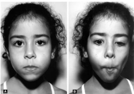
During the physical examination, mild right eyelid ptosis could be seen. Upper right eyelid retraction was also observed, triggered by chewing and the lateral movements of the mandible (Figure 1). The patch test, which consists of the occlusion of one eye and observation of the type and magnitude of eye movement caused by this occlusion, detected orthotropy. The refraction test demonstrated physiological childhood hyperopia. Biomicroscopy, an examination that uses a stereomicroscope along with a slit-lamp allowing the details of both normal and pathological structures that escape other techniques to be viewed, found no relevant abnormalities and neither did the fundoscopy. No abnormalities were detected in the left eye.
According to the clinical case described, a hypothesis of Marcus Gunn Phenomenon was suggested. Orthoptic examinations were requested to assess the presence of strabismus. Mild exophoria with foveal fusion was found. Since in this case ptosis is mild and does not prejudice visual acuity and there is no strabismus, no surgical intervention was indicated, just periodic follow-up appointments.
A neurological examination was performed by the neurologist at the Hospital das Clínicas at UFMG and was normal. Computerized tomography of the skull was also performed and did not detect any abnormalities.
Jornal de Pediatria - Vol. 80, No.3, 2004 251 Marcus Gunn Phenomenon Torres MRF et alii
Discussion
In Marcus Gunn Phenomenon, elevation and even retraction of the affected eyelid is triggered by chewing, suction, lateral mandible movement, smiling, sternocleidomastoid contraction, protruding the tongue, the Valsalva maneuver and even by simply breathing in.2 In the case described above, mild ptosis was observed and the presence of eyelid retraction when chewing, opening the mouth or sticking out the tongue.7
Etiopathogenesis of the phenomenon is not well defined, being attributed to an anomalous connection between the nerves and the levator and external pterygoid muscles of the eyelid. In this scenario, the eyelid levator muscle is innervated by motor ramifications of the trigeminal nerve and by the oculomotor nerve.3 Recently, a case was described of a child with Marcus Gunn Phenomenon associated with synkinesis of the extra-ocular, trigeminal and abducent muscles.7,8 Some authors have described cases in which there were lesions to the oculomotor nerve and subsequent innervation of the eyelid by a ramification of the fifth cranial nerve. Such reports constitute the rare, acquired form of the phenomenon.9
Some authors claim that the ptosis improves with time, but there is no scientific proof that this really does take place.9 It is believed that, as time passes, the affected individual will come to recognize which movements are responsible for the synkinesis and learn to control or avoid them and so minimize or mask the syndrome. In the case in question, the childs eyelid ptosis is becoming more obvious and causing relationship problems with other children.
It should be highlighted that patients who are suspected to have Marcus Gunn Phenomenon should be quickly referred to ophthalmologists and neurologists because of the chance of other associated conditions and secondary complications. Published literature describes the following possible associated conditions: aponeurosis, unequal focusing, strabismus, elevator palsy, paralysis of the rectus superior, congenital nystagmus and congenital fibrosis syndrome5,10,11 (Table 1).
The degree of ptosis can vary and is considered mild when the eyelid covers 1-2 mm more of the cornea than the 1-2 mm already covered in a normal state, moderate when it is up to 3 mm, and severe when equal or greater than 4 mm.12 Careful analysis of the degree of ptosis, its etiology and levator muscle function define whether surgery is indicated, and also which surgical approach would be most appropriate. When ptosis is moderate and esthetically acceptable, surgery is not recommended. Amblyopia and vertical strabismus, if present, demand surgical eyelid repair. Patients with severe ptosis and levator muscle dysfunction benefit from frontalis muscle elevation, associated with myectomy of the upper eyelid levator. If ptosis is accentuated, but levator muscle function is preserved, then a simple shortening of the eyelid levator muscle aponeurosis can be performed.12,13 In a retrospective analysis of the results of surgical correction of eyelid ptosis associated with Marcus Gunn Phenomenon it was found that the degree of ptosis is not, in general, adequately assessed during conventional ophthalmological examinations. These authors concluded that surgical results are better when the degree of ptosis is evaluated preoperatively using the technique of immobilizing the jaw and temporarily occluding the ipsilateral eye.14
Furthermore, it should be emphasized that the esthetic aspect and the possibility of developing emotional problems or difficulties justifies psychological evaluation and, if necessary, additional treatment.
Other synkinetic abnormalities which may be associated with eyelid ptosis are Inverted Marcus Gunn Phenomenon and Marin-Amat syndrome.15,16 Both conditions progress with ptosis that becomes more obvious when the mandible is moved. Initially they were believed to be the same disease, but recent studies have found differences between them. The first is a congenital condition in which the eyelid levator muscle is inhibited. Marin-Amat syndrome is acquired, occurring after facial paralysis and in this case orbicularis oculi and levator muscle function is not compromised.9,15,16
Neurological and ophthalmological examination is fundamental to the differentiation of the various types of ptosis. Sudden manifestation of eyelidptosis, for example, is suggestive of myasthenia gravis. An association between ptosis and homolateral ipslateral myosis implies a possibility of Horners syndrome.9
This case report was carried out with the objective of alerting pediatricians to the existence of the Marcus Gunn Phenomenon, which remains little known. It is usual to recommend a routine ophthalmological examination as the child reaches three to four years of age and when a certain degree of cooperation can be expected. The pediatrician is in frequent contact with the child from birth throughout the period of rapid growth development and, t h e r e f o r e , h a s a n e s s e n t i a l r o l e i n d e t e c t i n g ophthalmological abnormalities, such as strabismus, blindness, tumors and others, such as the Marcus Gunn Phenomenon described above.
Findings n. of cases
Aponeurosis 42
Strabismus 41
Unequal focusing 18
Elevator palsy 18
Paralysis of the rectus superior 16 Congenital nystagmus 02
Duane syndrome 02
Congenital fibrosis syndrome 03
Cleft palate 01
Lobular capillary hemangioma 01 Corneal dystrophy 01
252 Jornal de Pediatria - Vol. 80, No.3, 2004
References
1. Moribe I, Cruz AAV, Habib JT. Anomalias palpebrais. In: Rodrigues MLV, editor. Oftalmologia Clínica. Rio de Janeiro: Cultura Médica; 1992. p. 306-07.
2. Disorders of the eyelids. In: Kanski JJ, editor. Clinical Ophthalmology. 3rded. Butterworth-Heinemann Medical; 1997. p. 1-26.
3. Chaves PS, Hoyt, WF. Neuro-oftalmologia. In: Vanghan DG, Asbury T, Riordan P, editor. Oftalmologia Geral. 4a ed. São
Paulo: Atheneu; 1998. p. 84.
4. McLeod AR, Glaser JS. Deglution-Trochlear synkinesis. Arch Ophthalmol. 1974;92:171-2.
5. Pratt SP, Beyer CK, Johnson CC. The Marcus Gunn phenomenon. Ophthalmology. 1984;91:27-30.
6. Lucci LMD, Portelinha W, SantAnna AEBPP. Ptose palpebral: estudo de 390 casos. Arquivos Brasileiros de Oftalmologia, volume 60, fascículo 5. Disponível em: http:// www.abonet.com.br/abo/abo605.htm. Acessado em 20 de fevereiro de 2003.
7. Freedman HL, Kushner BJ. Congenital ocular aberrant innervation new concepts. J Pediatr Ophtalmol Strabismus. 1997;34(1): 10-6.
8. Kodsi S. Marcus Gunn jaw winking with trigemino-abducens synkinesis. AAPOS. 2000;4(5):316-7.
9. Odehnal M, Malec J. New views on aberrant innervation of oculomotor muscles. Cesk Slov Oftalmol. 2002;58(5):307-14. 10. Pieh C, Goebel HH, Engle EC, Gottlob I. Congenital fibrosis syndrome associated with central nervous system abnormalities. Graefes Arch Clin Exp Ophthalmol. 2003;241(7):546-53. Epub 2003 Jun 18.
11. Brodsky MC, Pollock SC, Buckley EG. Neural misdirection in congenital ocular fibrosis syndrome: implications and pathogenesis. J Pediatr Ophtalmol Strabismus. 1989;26(4): 159-61.
Marcus Gunn Phenomenon Torres MRF et alii
Corresponding author: Marcia Regina Fantoni Torres Rua Pirapetinga, 204/300
CEP 30220-150 - Belo Horizonte, MG, Brazil Tel.: +55 (31) 3227.7289/9983.0283 E-mail: marciaft@medicina.ufmg.br
12. Neto GH, Yamane, R. Doenças das pálpebras. In: Dantas AM, editor. Oftalmologia Pediátrica. Rio de Janeiro: Cultura Médica; 1995. p. 115.
13. Friedhofer H, Camargos CP, Ferreira MC. Implante suspensor palpebral de silicone para correção da blefaroptose severa. Revista da Sociedade Brasileira de Cirurgia Plástica. 1999;14(3): 7-20.
14. Khwarg SI, Tarbet KJ, Dortzback RK, Lucarelli MJ. Management of moderate-to-severe Marcus-Gunn jaw-wink ptosis. Ophtalmology. 1999;106(6):1191-6.
15. Wong JF, Theriault JF, Bouzouaya C, Codere F. Marcus Gunn jaw-winking phenomenon: a new supplemental test in the preoperative evaluation. Ophthal Plast Reconstr Surg. 2001; 17(6):412-8.
16. Rana PSV. The Marin-Amat syndrome: an unusual facial synkinesia. J Neurosurg Psychiatry. 1985;48:939-41. 17. Lubkin V. The Inverse Marcus Gunn Phenomenon: An