C A SE REPORT
248 J Vasc Bras. 2017 Jul-Set;16(3):248-251 http://dx.doi.org/10.1590/1677-5449.001917
Abnormal origin of posterior circumflex humeral artery and
subscapular artery: case report and review of the literature
Origem anômala das artérias circunflexa posterior do úmero e subescapular:
relato de caso e revisão de literatura
Rajani Singh1
*
Abstract
he subscapular, anterior circumlex, and posterior circumlex arteries arise from the third part of the axillary artery. During dissection of the right upper limb of the cadaver of a 70-year-old male, a common trunk was observed arising from the third part of the axillary artery which, after traveling for 0.5 cm, bifurcated into subscapular and posterior circumlex humeral arteries. he common trunk was crossed anteriorly by the radial nerve. he medial nerve was formed by medial and lateral roots on the medial side of the third part of the axillary artery, remaining medial to the brachial artery up to the cubital fossa and then following its usual course thereafter. Awareness of the vascular variations observed in the present case is important when conducting surgical procedures in the axilla, for radiologists interpreting angiographs, and for anatomy-pathologists studying rare indings.
Keywords: axillary artery; variation; radial nerve; pectoralis minor.
Resumo
As artérias subescapular, circunlexa anterior e circunlexa posterior se originam da terceira parte da artéria axilar. Durante a dissecção de membro superior direito de um cadáver humano com 70 anos de idade, do sexo masculino, um tronco comum foi observado originando-se da terceira parte da artéria axilar, após um percurso de 0,5 cm, bifurcando-se em artéria subescapular e artéria circunlexa posterior do húmero. O tronco comum era cruzado anteriormente pelo nervo radial. O nervo medial era formado por raízes medial e lateral, no lado medial da terceira parte da artéria axilar, permanecendo em posição medial à artéria braquial até a fossa cubital e seguindo seu curso usual a partir de então. Conhecimento das variações vasculares observadas neste caso é importante ao executar procedimentos cirúrgicos na axila, para radiologistas que interpretam radiograias, e também para anatomo-patologistas que estudam achados raros.
Palavras-chave: artéria axilar; variações; nervo radial; peitoral menor.
1All India Institute of Medical Siences Rishikesh – AIIMS, Department of Anatomy, Rishikesh, Uttrakhand, India. Financial support: None.
Conlicts of interest: No conlicts of interest declared concerning the publication of this article. Submitted: March 17, 2017. Accepted: June 02, 2017.
249 J Vasc Bras. 2017 Jul-Set;16(3):248-251
Rajani Singh
INTRODUCTION
The axillary artery is a continuation of the subclavian
artery and begins at the outer border of the irst rib.
The axillary artery is anatomically divided into three parts
with relation to the pectoralis minor muscle. The irst
part lies proximal, the second part deep, and the third
part distal to the pectoralis minor muscle.1 The irst
part of the artery gives rise to the superior thoracic
artery. The second part of the axillary artery gives off the lateral thoracic and thoracoacromial arteries. The subscapular, anterior, and posterior circumlex
humeral arteries originate from the third part of axillary
artery.2 Although variations in the branching pattern
of the axillary artery are common, a rare anatomical variation of the origin of the subscapular and posterior
circumlex humeral arteries from the axillary artery
combined with an anomalous relationship between the radial nerve and the common trunk and a variant formation and course of the median nerve all detected
simultaneously nevertheless merit reporting. Besides
its importance as an anatomical curiosity, knowledge
of these variations relating to variant conigurations of the axillary, subscapular, and posterior circumlex
humeral arteries and the radial and median nerves is clinically important for orthopedic and vascular surgeons to avoid complications during various
surgical procedures in the axilla. Additionally the
neural variations may be useful when conducting regional neurosurgery and for successful repair of
peripheral nerves. Hence the case is reported here.
CASE REPORT
During dissection of the right upper limb of the
cadaver of a 70-year-old male ixed in 10% formaline,
conducted at the department of anatomy, AIIMS Rishikesh, Uttrakhand, India, multiple variations were
observed in the axilla. The third part of the axillary
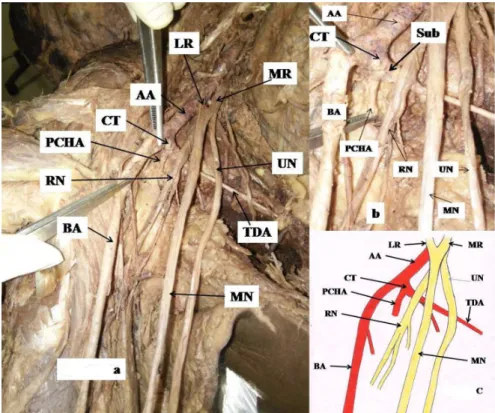
artery gave off an anomalous common trunk, which,
after traveling 0.5 cm, bifurcated into subscapular and posterior circumlex femoral arteries (Figure 1). The common trunk was crossed anteriorly by the radial
nerve. The lateral root from the lateral cord crossed
the axillary artery and fused with the medial root of the medial cord, forming the median nerve, lying
medial to the axillary artery. This remained medial
to the brachial artery throughout the arm and cubital
fossa. There was no abnormality in the left upper limb.
Figure 1. (a) showing a common trunk from the third part of the axillary artery and abnormal positions of the radial nerve and median
250 J Vasc Bras. 2017 Jul-Set;16(3):248-251 Abnormal posterior circumlex humeral artery
DISCUSSION
As described in standard anatomy text books, the
anterior circumlex, posterior circumlex humeral
arteries, and, subsequently, the subscapular artery normally emanate from the third part of the axillary
artery (Figure 2). A common trunk arising from the third part of the axillary artery and bifurcating into
posterior circumlex humeral and subscapular arteries has been reported by Gaur et al.,3 similar to the present
study, but our case is different from that described by
Gaur et al. as regards the anomalous coniguration of the radial nerve with respect to the common trunk.
Normally, the radial nerve lies deep to the 3rd part
of the axillary artery and branches. In the present
case, it crossed the common trunk anteriorly, from
medial to lateral. The radial nerve may compress the
common trunk, causing ischemic changes in the area
supplied by the posterior circumlex humeral and subscapular arteries. Moreover, the median nerve,
which usually remains lateral to the axillary artery,
is located medial to it. The median nerve continued
anomalously, medial to the brachial artery up to the
cubital fossa. This is the irst time that formation of
a common trunk and its bifurcation into posterior
circumlex and subscapular arteries in combination
with abnormal disposition of the radial nerve has
been detected and similar variant conigurations of
the median nerve have rarely been described in the
literature.
Bergman et al.4 reported emergence of anterior
circumlex humeral, posterior circumlex humeral,
subscapular, and profunda brachii arteries from a
common trunk arising from the third part of the axillary
artery. Saeed et al.5 reported the origin of a common
subscapular-circumlex humeral trunk from the third
part of axillary artery, trifurcating into subscapular,
anterior circumlex humeral, and posterior circumlex humeral arteries in 3.8% of cases. Ramesh et al.6
described the emergence of a common trunk from
the third part of the left axillary artery. This common trunk divided into subscapular, anterior circumlex humeral, posterior circumlex humeral, profunda brachii, and ulnar collateral arteries. Vijaya et al.7
reported a common trunk that emerged from the third part of the axillary artery and then divided into anterior
circumlex humeral, posterior circumlex humeral,
subscapular, radial collateral, middle collateral, and
superior ulnar collateral arteries. Here, the profunda brachii artery was absent. Astik and Dave8 found
a common trunk from the third part of the axillary
artery in 25% of a sample of 40 limbs. Of these, in 10% the common trunk gave origin to anterior circumlex humeral, posterior circumlex humeral,
subscapular, and profunda brachii arteries, whereas
in 15% of these cases anterior circumlex humeral, posterior circumlex humeral, and profunda brachii arteries originated from the common trunk.
Embryological background
The arteries of the limbs arise as a number of vessels contributing to a primitive capillary plexus, but eventually only one trunk persists – the subclavian artery – with the positions and relations of the seventh intersegmental artery, probably representing its lateral
branch. The main trunk to the upper limb, the axillary, forms later. The anomalous blood vessels observed
in the present case could be due to9 (i) choice of
unusual paths in the primitive vascular plexuses,
(ii) persistence of vessels normally obliterated, (iii) disappearance of vessels normally retained, (iv) incomplete development, and (v) fusions or absorption of parts that are usually distinct.
Awareness of variations in the branching pattern of the axillary artery is essential when performing bypasses between the axillary and subclavian arteries
in surgical treatment of subclavian artery occlusions.10
Aneurysms and axillary artery traumas may require
reconstructive interventions. The variations highlighted in the present case may present dificulties in this type of procedure. Aneurysms of the axillary artery and its branches are often observed in baseball pitchers.11
Repetitive positional compression of the axillary artery in athletes can cause focal intimal hyperplasia, aneurysm formation, segmental dissection, and branch
vessel aneurysms. These conditions favor thrombosis
251 J Vasc Bras. 2017 Jul-Set;16(3):248-251
Rajani Singh
and distal embolism and may need positional
arteriography for diagnosis.12 Variant coniguration
of branches of the axillary artery similar to the present case may increase positional compression of the common trunk, due to its variant relation to the radial nerve, and so increase propensity to the
aforementioned pathologies. The axillary arteries are
used as cannulation sites in cardiopulmonary bypass and thoracic and aortic procedures, and for insertion
of intra-aortic balloon pumps. They are also under consideration for use as inlow vessels in coronary artery surgery.13 Variant common trunks from the
axillary artery, as observed in the current study, can
be considered for cannulation. Radiological studies
can thus be performed before proceeding to the
aforementioned procedures. It has been shown that injection of oily suspension into deltoid muscle can
cause ischemic changes in the scapular and pectoral
region. It was presumed that these changes were
caused by action of medication on nerves or entrance
of substance into blood vessels causing embolism.14
In the present case, the radial nerve crossed the common trunk, which may compress the common trunk emanating from the axillary artery and cause ischemia of structures supplied by branches of the
common trunk. If one is unaware of this variation, the radial nerve could be injured during surgical procedures in the axilla.
Awareness of the variations observed in the present case may therefore be essential for vascular surgeons during surgical procedures in the axilla and for radiologists, to prevent misinterpretation of angiographs, and of interest to anatomists studying
rare variations.
REFERENCES
1. Standring S, editor. Gray’s Anatomy: the anatomical basis of clinical practice. 40th ed. London: Churchill-Livingstone Elsevier; 2008. p. 815.
2. Snell R. Clinical anatomy for medical students. 7th ed. London: Lippincott Williams and Wilkins; 2004. p. 475-7.
3. Gaur S, Katariya SK, Vaishnani H, et al. A cadaveric study of branching pattern of the axillary artery. Int J Biol Med Res. 2012;3(1):1388-91.
4. Bergman RA, Thompson SA, Afifi AK, Saadeh FA. Compendium of human anatomic variations. Munich: Urban and Schwarzenberg; 1988. p. 70-3.
5. Saeed M, Rufai AA, Elsayed SE, Sadiq MS. Variations in the subclavian-axillary arterial system. Saudi Med J. 2002;22(2):206-12. PMid:11938385.
6. Ramesh RT, Shetty P, Suresh R. Abnormal branching pattern of the axillary artery and its clinical significance. Int J Morphol. 2008;26:389-92.
7. Vijaya PS, Venkata RV, Satheesha N, Mohandas R, Sreenivasa RB, Narendra P. A rare variation in the branching pattern of the axillary artery. Indian J Plast Surg. 2006;39(2):222-3. http://dx.doi. org/10.4103/0970-0358.29559.
8. Rajesh A, Urvi D. Variations in branching pattern of the axillary artery: a study in 40 human cadavers. J Vasc Bras. 2012;11(1):12-7. http://dx.doi.org/10.1590/S1677-54492012000100003.
9. Arey LB. In development of the arteries, developmental anatomy. 6th ed. Philadelphia: W.B. Saunders’co; 1957. p. 375.
10. Bhat KMR, Gowda S, Potu BK, Rao MS. A unique branching pattern of the axillary artery in a South Indian male cadaver. Bratisl Med J. 2008;109(12):587-9. PMid:19348386.
11. Schneider K, Kasparyan NG, Altchek DW, Fantini GA, Weiland AJ. An aneurysm involving the axillary artery and its branch vessels in a major league baseball pitcher: a case report and review of the literature. Am J Sports Med. 1999;27(3):370-5. PMid:10352776. http://dx.doi.org/10.1177/03635465990270031801.
12. Duwayri YM, Emery VB, Driskill MR, et al. Positional compression of the axillary artery causing upper extremity thrombosis and embolism in the elite overhead throwing athlete. J Vasc Surg. 2011;53(5):1329-40. PMid:21276687. http://dx.doi.org/10.1016/j. jvs.2010.11.031.
13. Karambelkar RR, Shewale AD, Umarji BN. Variations in branching pattern of axillary artery and its clinical significance. Anatomica Karnataka. 2011;5(2):47-51.
14. Duque FL, Chagas CA. Intramuscular accident with drug injection in the deltoid muscle: local and distant lesions, review of 32 cases. J Vasc Bras. 2009;8(3):238-46. http://dx.doi.org/10.1590/ S1677-54492009000300009.
*
Correspondence
Rajani Singh All India Institute of Medical Siences Rishikesh – AIIMS, Department of Anatomy Veerbhadra marg Pashulok 249203, Rishikesh, Uttrakhand, India Tel.: +91 (94) 5319-3659 E-mail: [email protected]
Author information