Asse ssme nt o f fe tal risk asso ciate d
with e xpo sure to cance r che mo the rapy
during pre gnancy: a multice nte r study
1Departamento de Genética, Universidade Federal do Rio Grande do Sul,
Porto Alegre, RS, Brasil
2Sistema Nacional de Informações sobre Agentes Teratogênicos,
Serviço de Genética Médica, and 3Serviço de Hematologia,
Hospital de Clínicas de Porto Alegre, Porto Alegre, RS, Brasil
4Serviço de O ncologia, Hospital Nossa Senhora da Conceição, Porto Alegre, RS, Brasil 5Serviço de Hematologia, Hospital Universitário de Santa Maria, Santa Maria,
RS, Brasil R.M. Peres1,
M.T.V. Sanseverino2,
J.L.M. Guimarães4,
V. Coser5,
L. Giuliani5,
R.K. Moreira2,
T. O rnsten3
and L. Schüler-Faccini1,2
Abstract
The objective of the present study was to evaluate and quantify fetal risks involved in the administration of cancer chemotherapy during gestation, as well as to assess the long-term effects on the exposed children. In this retrospective, cohort study, we reviewed the records of women aged 15 to 45 years with a diagnosis of malignancy or benign tumors with malignant behavior at three reference services in the State of Rio Grande do Sul, Brazil, from 1990 to 1997. All patients with a diagnosis of pregnancy at any time during the course of the disease were selected, regardless of whether or not they received specific medication. Fetal outcomes of 14 pregnancies with chemo-therapy exposure were compared to that of 15 control pregnancies in which these drugs were not used. Long-term follow-up of the exposed children was carried out. Fisher’s exact test was used to compare the groups. Continuous variables were compared by the Wilcoxon-Mann-Whitney test. We found an increased rate of prematurity (6/8 vs 2/10;
RR: 3.75; CI: 1.02-13.8; P = 0.03) in the exposed group. There was a trend to an increased fetal death rate (4/12 vs 0/10; P = 0.07) in the group exposed to chemotherapy. No malformations were detected in any child, which can be related to our small sample size as well as to the fact that most exposures occurred after the first trimester of pregnancy. Other larger, controlled studies are needed to establish the actual risk related to cancer chemotherapy during pregnancy.
Co rre spo nde nce
L. Schüler-Faccini
SIAT, Serviço de Genética Médica Hospital de Clínicas de Porto Alegre Rua Ramiros Barcelos, 2350 3º andar
90035-003 Porto Alegre, RS Brasil
Fax: + 55-51-3316-8010 E-mail: lavinia.faccini@ ufrgs.br
Research supported by CNPq and PRO NEX/FINEP.
Received February 8, 2001 Accepted September 11, 2001
Ke y words
·Cancer ·Pregnancy ·Chemotherapy ·Teratogenicity ·Fetal outcome
Intro ductio n
Cancer is the second most common cause of death in women during their reproductive years (1,2). Therefore, one should not be surprised by the fact that this disease is diag-nosed in 0.7 to 1.0 out of a thousand preg-nancies (3). The most common maligpreg-nancies
associated with pregnancy are those that have an ascending incidence curve during the re-productive years, such as Hodgkin’s disease, melanoma, breast and cervical cancer (4).
management of malignancies during preg-nancy do not exist. It is universally recog-nized that a large number of malignant tu-mors, specifically breast cancer and hemato-logical malignancies, cannot have their treat-ment delayed until the end of pregnancy. Due to the high rate of tumor cell prolifera-tion, fetal well being may be compromised. A delay in treatment, in such cases, would certainly result in worsening of maternal prognosis (6,7). Likewise, the decision to terminate a pregnancy is complicated by ethi-cal, moral and religious issues.
Almost all cytotoxic agents are known to be teratogenic in animals, being associated with increased rates of malformations, fetal loss, intrauterine growth retardation, and other long-term effects on the progeny (8). In humans, however, the risk of teratogen-esis related to this class of drugs has not been well established. The literature consists mostly of case reports, which tend to overes-timate potential side effects of the pharma-cologic substances being studied because of reporting bias (9,10). The few case series and cohort studies available are of limited value due to their small sample sizes and, for the most part, lack of control groups (5,11-14). Due to limited data, quantifying the risk of fetal damage associated with exposure to this class of drugs becomes a complex task. The estimated risk for the exposed fetuses, according to these studies, varies from 7 to 75% for all major malformations (9). The lack of a precise risk estimation makes it difficult for physicians and patients to make clinical decisions.
Here, we conducted a multicenter, col-laborative study involving three oncology and hematology clinics in order to establish and quantify the risk associated with fetal exposure to antineoplastic agents.
Mate rial and Me thods
In this retrospective, cohort study, we reviewed the records of women aged 15 to
45 years with a diagnosis of malignancy or benign tumors with malignant behavior at three reference services in the State of Rio Grande do Sul, Brazil: University Hospital of Porto Alegre, Oncology Service at Nossa Senhora da Conceição Hospital, and Hema-tology Service of the University Hospital of Santa Maria. We reviewed the records from December 1990 to December 1997. The Eth-ics Committees of all three hospitals evalu-ated and approved this study.
We selected all patients with a diagnosis of pregnancy at any time during the course of the disease, regardless of whether or not they received specific medication. All the infor-mation about the disease, its evolution and therapeutics, such as histopathologic diag-nosis, staging, date of diagnosis and period of gestation during which patients received specific medication was obtained by review-ing the medical records. We also recorded a detailed obstetric history including date of conception and obstetric or clinical compli-cations. For pregnancies that resulted in live-born children, data such as Apgar scores, anthropometry, neonatal complications and pediatricians’ assessment of possible con-genital malformations were obtained from hospital records.
between the two groups. Fisher’s exact test was used to compare the rates of stillbirths, abortion, prematurity, low Apgar scores, neonatal complications, and congenital mal-formations. Prematurity was defined as de-livery occurring at a gestational age of less than 37 weeks. Continuous data such as maternal age, birth weight and gestational age at birth were compared by the Wilcoxon-Mann-Whitney test, a nonparametric test for independent samples. Birth weight was ad-justed for gestational age according to stan-dardized charts (15).
Long-term follow-up of the exposed chil-dren was carried out by assessment of child-hood development milestones. Clinical his-tory and physical examination data were collected by a dysmorphologist.
The statistical analysis was performed with the SPSS 8.0 program for Windows.
Re sults
Of the patients with a diagnosis of a malignant tumor made at the referring cen-ters, 860 were women of reproductive age. Of these, 29 (3.4%) were pregnant and were included in the present study. Fourteen of them received chemotherapy (exposed group) and 15 did not (non-exposed group).
In our sample of pregnant women, the mean maternal age at the time of diagnosis was 29.6 ± 6.6 years, and the median age was 28.5 years (range: 18 to 43). The specific type of malignancy found in each group is presented in Table 1. The proportion of he-matopoietic malignancies and solid tumors did not differ between the two groups.
No significant difference in mean mater-nal age was observed between groups (P = 0.77). History of smoking or alcohol intake during gestation was negative for all pa-tients. Regarding associated maternal dis-eases, only one case of systemic hyperten-sion and one case of congestive heart disease secondary to the use of doxorubicin was observed in the exposed group. These
find-ings, however, were not significant. A statis-tically significant increase in the rate of preterm births was observed in the exposed group. No case of major malformation was reported in either group (Table 2).
Once adjusted for gestational age, no case of low birth weight was observed. In addition, the mean birth weight did not differ between groups.
The specific chemotherapic drugs, as well as the gestational age of administration to the exposed group are presented in Tables 3 and 4.
Table 2. Comparison of fetal outcome betw een exposed and non-exposed patients.
Fetal outcome Exposed Non-exposed RR 95% CI P value
(N = 14) (N = 15)
N % N %
Livebirths 8/12 67 10/10 100 0.67 0.45-1.0 0.07
Preterm 6/8 75 2/10 20 3.75 1.02-13.8 0.03
M ajor malformations 0/8 0 0/10 0 - -
-Neonatal complications 3/8 38 1/10 10 3.75 0.48-29.52 0.20
Apgar score <7 1' 2/8 25 1/10 10 2.50 0.27-22.86 0.41
Spontaneous abortions 1/13 8 0/10 0 ? ? 0.60
Therapeutic abortions 1/13 8 5/15 33 0.23 0.03-1.7 0.12
Stillbirths 4/12 33 0/10 0 ? ? 0.07
Note: For comparison of the exposed and non-exposed groups, w e excluded thera-peutic abortions. In the non-exposed group, therathera-peutic abortions w ere performed very early, before 8-10 w eeks and the products of conception w ere not examined. ? = Not possible to calculate due to zero value.
Table 1. Neoplasms found in the exposed and non-exposed groups.
Neoplasm Exposed group Non-exposed group
N % N %
Breast 3 21 4 27
HD 2 14 6 40
CM L 3 21 1 7
ALL 3 21
-AM L 2 14
-NHL 1 7
-Uterine cervix - 2 13
Lung - 1 7
Stomach - 1 7
Total 14 100 15 100
Table 3. Exposed group: outcome (livebirths) and chemotherapy employed.
Case Neoplasm Drugs M other Baby
number (stage)
Trimester exposure Gestational Birth w eight Neonatal Age at Abnormal
(w eeks) age (g) complication follow -up development
1 HD Cisplatin + etoposide + 2 (26) 36 2,540 Jaundice, -
-cytarabine non-hemolytic
anemia
2 ALL Vincristine + idarubicin + 2 (24; 28) 36 2,490 No 2 years No
L-asparaginase
3 CM L Busulfan 2 (26-36) 36 2,600 No 11 years No
4 Breast (IV) Doxorubicin 3 (28) 31 2,070 RDS, BCP 6 years No
neonatal sepsis
5 CM L Hydroxyurea 2 (22) 39 3,800 No 4 years No
6 CM L Hydroxyurea 1 (?) 35 3,195 Jaundice 4 months No
Vincristine + doxorubicin 2 (25)
7* Breast (IV) 5-Fluorouracil + doxorubicin 1 and 2 (2; 6; 10; 34 2,170 No -
-+ cyclophosphamide 14; 18; 22; 26)
8 AM L Cytarabine + daunorubicin 2 (19) 39 3,000 No 9 years No
* The only case of preterm delivery due to w orsening of maternal state. RDS: respiratory distress syndrome; BCP: bronchopneumonia; HD: Hodgkin’s disease; ALL: acute lymphoblastic leukemia; CM L: chronic myeloid leukemia; AM L: acute myeloblastic leukemia.
Table 4. Exposed group: outcome (pregnancy losses) and chemotherapy employed.
Case number Neoplasm Drugs Trimester of Fetal death Necropsy Result
exposure time
(w eeks) (w eeks)
9 ALL Vincristine + epirubicin + 2 (19) 30 No
-prednisone
10 AM L Idarubicin + cytarabine Yes IUGR, oligohydramnios; no malformation
11 NHL (IV) Cisplatin + etoposide 2 (22) 26 Yes Fetal autolysis; no malformations
12 Breast (IV) Cyclophosphamide + 1 (5; 8) 25 Yes No malformations
methotrexate + 5-fluorouracil
13 (spontaneous ALL Vincristine + doxorubicin 1 (13) 17 No
-abortion)
14 (elective HD M OPP-ABVD 1 (3-7) 18 Yes No malformations; toxic degenerative
abortion) changes in liver and kidneys, placenta
w ith villus degeneration and vascular toxic degeneration
Due to the small numbers of patients on a particular regimen, we were not able to asso-ciate a specific chemotherapeutic scheme with a particular adverse outcome. We also tested the effect of monotherapy versus polytherapy on fetal outcome, but no differ-ence was observed in our sample [RR (CI): 2.0 (0.33-12.18), P = 0.4 for stillbirths; RR (CI): 1.25 (0.52-3.0), P = 0.58 for preterm delivery]. In the exposed group, among liveborns, only two exposures occurred dur-ing the period of organogenesis. One prema-ture birth was induced because of worsening of maternal disease and all others were due to obstetric complications.
Long-term follow-up did not detect any abnormality in the development of the six liveborns examined. We lost two long-term follow-ups due to a change in address.
The non-exposed group is characterized in Table 5.
Two preterm deliveries occurred (cases 16 and 17) due to premature rupture of mem-branes. Therapeutic abortion was induced in five patients. Among these cases of thera-peutic abortion, one patient was exposed to radiotherapy (mantle field), and another was submitted to surgery (radical modified mas-tectomy during the second trimester).
D iscussio n
Most cytotoxic agents exert their effect by interfering with some stage of DNA and RNA synthesis, by interrupting essential metabolic pathways, and by destroying mac-romolecules, not only of tumor tissue, but also of normal tissue. In the case of preg-nancy, both maternal and fetal tissue may be affected, resulting in systemic toxicity and teratogenicity (16).
Almost all chemotherapeutic and cyto-toxic agents are known to be highly terato-genic to animals. When progeny is exposed to such substances during organogenesis, major malformations as well as an increased risk of miscarriage are observed. Exposure
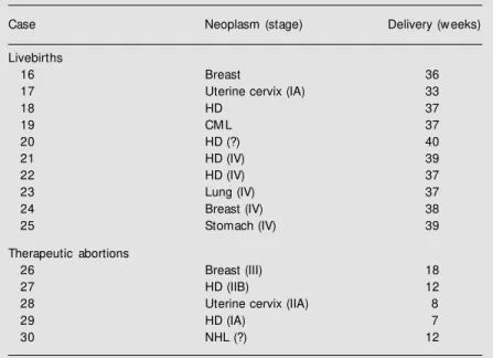
Table 5. Non-exposed pregnancies: outcome.
Case Neoplasm (stage) Delivery (w eeks)
Livebirths
16 Breast 36
17 Uterine cervix (IA) 33
18 HD 37
19 CM L 37
20 HD (?) 40
21 HD (IV) 39
22 HD (IV) 37
23 Lung (IV) 37
24 Breast (IV) 38
25 Stomach (IV) 39
Therapeutic abortions
26 Breast (III) 18
27 HD (IIB) 12
28 Uterine cervix (IIA) 8
29 HD (IA) 7
30 NHL (?) 12
A marked difference exists in the characteristics of maternal disease betw een the liveborn group and the therapeutic abortion group. In the former, mothers had malig-nancies in advanced stages, in w hich early treatment and pregnancy interruption w ould not result in clinical benefits, w hereas in the therapeutic abortion group, mothers had cancer at an early stage, in w hich pregnancy interruption and administra-tion of aggressive therapy w ould be beneficial. HD: Hodgkin’s disease; CM L: chronic myeloid leukemia; NHL: non-Hodgkin’s lymphoma.
after organogenesis is associated with in-creased rates of prematurity, stillbirths, and intrauterine growth retardation (8). How-ever, the doses used in animal experiments are much higher than those employed to treat humans. This, in addition to the fact that the entire process of teratogenesis varies from one species to another, makes extrapolation of animal data to humans a difficult task (17).
Case reviews found in the medical litera-ture indicate that administration of chemo-therapy during the first trimester of preg-nancy is associated with higher rates of abor-tion and malformaabor-tions, and administraabor-tion later in pregnancy is associated with a greater risk of prematurity, fetal death and intrauter-ine growth retardation (7,18-23). Table 6 presents case reviews published in the litera-ture about exposure during pregnancy.
congeni-tal malformations as compared to mono-chemotherapy (22-24).
Cohort studies published in the literature involve sample sizes ranging from 9 to 43 gestations (Table 7).
In only one of these studies, however, was a control group used to estimate the relative risk associated with fetal exposure to chemotherapeutic agents (14). This con-trol group, however, was composed of healthy pregnant women, and, as such, could not identify potential risks due to maternal
can-cer itself.
Contrary to what case report reviews in-dicate, the rates of miscarriage observed in these cohort studies do not seem to be el-evated, with the exception of the study by Zemlickis et al. (14). These findings, how-ever, can be explained by the fact that, in our study, as well as in other cohort studies, not all exposures occurred during the first tri-mester of gestation. When only exposures that took place during the first trimester are considered, abortion rates of 20% (or 1/5) (our study), 25% (or 1/4) (12) and 44% (or 4/ 9) (14) have been reported. Therefore, we may conclude that exposure to chemothera-peutic agents during the first trimester of gestation is possibly associated with an in-creased risk of early fetal losses.
A significantly higher rate of prematurity was observed in the exposed group in our study compared to the control group. The high rates of prematurity observed in the two other existing cohort studies, on the other hand, may be explained by frequent induc-tion of birth due to the worsening of mater-nal disease (11,14). In our study, however, only one birth was induced for this reason, which implies that chemotherapy itself could
Table 6. Pregnancy outcome for patients w ith cancer in pregnancy: literature review .
Reference N 1st trimester Preterm SA Stillbirth Birth LBW
exposure defects
(% )
Nicholson (18) 185 + + + + 7.5a +
Catanzarite and 47 + + + + - +
Ferguson II (21)
Doll et al. (22) 139 + * * * 17a, 6b *
Wiebe and Sipila (20) 121 + + + + + +
Ebert et al. (26) 217 + + + + 8.3a +
SA: spontaneous abortion; LBW: low birth w eight. * Not tested.
+: presence of exposure during the 1st trimester; -: absence of exposure during the 1st trimester.
apolychemotherapy; bmonochemotherapy.
Table 7. Pregnancy outcomes in cancer patients: cohort studies and the present study.
Reference N Number of Livebirths SA SB§ SGA Preterm Birth Long-term follow -up Development
1stexposures defects exposed group
Pizzuto et al. (11) 9 4 8 none 1/9 5/8 2/8 none yes Normal 2 months up to
15 years (6/9)
M ulvihill et al. (12) 32 4 23 2/25 1/25 none none 5/23 no
-Avilés et al. (13) 43 20 43 none none none none none yes Normal 3 months up to
19 years (43/43)
Zemlickis et al. (14)* 21 13 11 4/16 4/16 9/11 5/11 2/11 no
-Berry et al. (5) 24 1 24 none none 1/24 3/24 none no
-Present study* * 14 6 8 1/14 4/14 0/8 6/8 none yes Normal 4 months up to
11 years (6/8)
SA: spontaneous abortion; SB: stillbirth; SGA: small for gestational age.
* Used a control group (healthy w omen) for assessment of potential fetal hazards.
have a direct effect on prematurity rates. Most of our preterm babies were born at 36 weeks of pregnancy, which does not mean extreme prematurity. Even so, they are at higher risk of morbidity than full-term ba-bies. There was a trend to an increased rate of stillbirths in our study, which, however, was not significant. This lack of significance could be explained by the small sample size. Elevated rates of stillbirths were also re-ported in three cohort studies (11,12,14) and in several case series studies (18,20,21,25), representing an important effect of chemo-therapy. In our study, cytotoxic treatment was started earlier in women who had still-births compared to those women whose ba-bies were born alive. This is related to the fact that more aggressive tumors need earlier treatment.
We emphasize that our study, to our knowledge, is the first to use a control group of pregnant women with diagnosed malig-nancy who were not receiving chemotherapy of any kind. Although we used a group not exposed to chemotherapy as control, we ob-served more aggressive tumors in the ex-posed group compared to the non-exex-posed women. This could explain the higher inci-dence of stillbirths and preterm babies in the exposed women. Also, since there were more therapeutic abortions in the non-exposed group we cannot predict what the outcome would have been in these cases. Therefore, we cannot infer that the high rates of still-births as well as of prematurity reported in this study were only due to a direct effect of the cytotoxic therapy, and not also to com-plications secondary to maternal disease.
As opposed to the majority of published reports such as cohort studies and reviews of case reports, which use birth weights lower than 2,500 g as an indicator of low birth weight, in our investigation weight was cor-rected for gestational age. This fact partially explains the absence of children classified as having low birth weight reported here. It should be noted, however, that Zemlickis et
al. (14) also used this correction and found a significantly higher proportion of neonates who were small for gestational age. In addi-tion, complications secondary to maternal disease may have had adverse effects on fetal intrauterine growth, representing an im-portant bias in all studies which did not use a control group. Unfortunately, we cannot evaluate the real impact of chemotherapy on birth weight and its role in intrauterine growth restriction in the exposed group due to the increased number of stillbirths.
The study of Avilés et al. (13) was the only one which differed significantly with regard to fetal outcome when compared to other studies, including ours. In their cohort of 43 women treated for hematological ma-lignancies, the rates of prematurity, abor-tions and stillbirths were not different from those expected for the general population. This difference could be explained by the fact that only livebirths were included in this study.
We did not observe any malformation among the six liveborn babies who were exposed to chemotherapy during gestation. Two explanations for this observation are possible: 1) with the exception of three cases who were exposed to cyclophosphamide, mechlorethamine, and methotrexate, which are known to have a teratogenic potential, the other drugs used have not been incrimi-nated as definite teratogens in the literature (22,23); 2) the sample was too small to de-tect an increase in the rate of congenital malformations.
Also, these children were exposed to a highly heterogeneous group of cytotoxic agents. Most of the published literature on this topic, however, also includes a variety of chemotherapeutic regimens.
The long-term follow-up of the six chil-dren who were exposed to chemotherapy in
utero is described in Table 7. We did not
sub-ject. One of the possible long-term conse-quences of intrauterine chemotherapy is car-cinogenicity. There is only one case report of a child born to a mother who used cyclo-phosphamide throughout pregnancy. She developed a papillary carcinoma of the thy-roid at 11 years of age and a neuroblastoma at 14 years of age. She was also the only child exposed to chemotherapy during preg-nancy reported to have mental retardation (25). However, in order to establish the ac-tual long-term risk, especially that related to the development of carcinogenesis, a longer follow-up period than the one reported here would be necessary.
In our study, no significant difference was observed when the fetal outcome of women who had hematopoietic malignan-cies was compared to that of women who had solid tumors. Although some authors suggest that hematopoietic malignancies dur-ing pregnancy might be associated with an increased number of complications due to the drugs employed to treat them as well as to their more aggressive behavior (11,21), this subject remains controversial. In fact, Ebert et al. (26) recently compared the rates of abortion, stillbirths, and fetal malforma-tions among 217 literature cases, afflicted by several diseases, in which cytotoxic treat-ment was employed during pregnancy. Ac-cording to this study, rheumatic diseases formed a group which was associated with the highest rates of adverse effects in preg-nancy (37.5%), followed, in order of fre-quency, by miscellaneous diseases, gyneco-logical malignancies, and finally by lympho-mas and leukemias, which had the lowest
complication rates.
The results of our study indicate that cytotoxic agents could exert adverse effects when employed during gestation, causing an increased rate of prematurity and stillbirths. Due to the small sample size and to the fact that only six exposures occurred in the first trimester of gestation, the potential terato-genic effect of cytotoxic agents, when used during the organogenesis period, could not be assessed, although no case of congenital malformation was observed in our study. The normal development of the six children exposed in utero indicates a good prognosis for the liveborn children without malforma-tions. The period of follow-up ranged from 4 months to 11 years after birth.
The present data serve as additional evi-dence that a rigorous, multidisciplinary ap-proach is needed for pregnant women with malignant neoplasms. Additionally, our study supports the need for a meta-analysis on this subject. Other cohort studies with larger sample sizes are necessary to elucidate the true risk associated with administration of antineoplastic agents during pregnancy.
Ackno wle dgm e nts
We are grateful to Dr. Myla Moretti, Assistant Director of the Mother Risk Pro-gram, for technical support with the statisti-cal analysis and for translating the manu-script into English. We also thank the Direc-tors of the Ethics Committees of the Medical Services involved in this multicenter study for approval and collaboration.
Re fe re nce s
1. Wingo PA, Ries LA & Rosenberg HM (1998). Cancer incidence and mortality 1973-1995: a report card for the U.S. Can-cer, 82: 1197-1207.
2. Landis SH, M urray T, Bolden S & Wingo PA (1999). Cancer statistics, 1999. CA: A Cancer Journal for Clinicians, 49: 9-64.
3. Potter JF & Schoeneman M (1970). M e-tastasis of maternal cancer to the placenta and fetus. Cancer, 25: 380-388. 4. Donegan WL (1983). Cancer and
preg-nancy. CA: A Cancer Journal for Clinicians, 33: 195-214.
5. Berry DL, Theriault RL, Holmes FA, Parisi
VM , Booser DJ, Singletary SE, Buzdar AU & Hortobagyi GN (1999). M anagement of breast cancer during pregnancy using a standardized protocol. Journal of Clinical Oncology,17: 855-861.
of the literature. Part I and II. Obstetrical and Gynecological Survey, 51: 125-142. 7. Sorosky JI, Sood AK & Buckers TE (1997).
The use of chemotherapeutic agents dur-ing pregnancy. Obstetrics and Gynecol-ogy Clinics of North America, 24: 591-599.
8. Schardein JL (1993). Cancer chemothera-peutic agents. In: Schardein JL (Editor),
Chemically Induced Birth Defects. M arcel Dekker Inc., New York, NY, USA, 457-507.
9. Gonen R, Shilalukey K, M agee L, Koren G & Shime J (1994). M aternal disorders leading to increased reproductive risks. In: Koren G (Editor), M aternal-Fetal Toxi-cology - A Clinician’ s Guide. M arcel Dekker Inc., New York, NY, USA, 641-679.
10. Koren G, Pastuszak A & Ito S (1998). Drugs in pregnancy. New England Journal of M edicine, 338: 1128-1137.
11. Pizzuto J, Avilés A, Noriega L, Niz J, M o-rales M & Romero F (1980). Treatment of acute leukemia during pregnancy: pres-entation of nine cases. Cancer Treatment Reports, 64: 678-683.
12. M ulvihill JJ, M cKeen EA, Rosner F & Zarrabi M H (1987). Pregnancy outcome in cancer patients - experience in a large cooperative group. Cancer, 60: 1143-1150.
13. Avilés A, Díaz-M aqueo JC, Talavera A,
Guzmán R & García EL (1991). Grow th and development of children of mothers treated w ith chemotherapy during preg-nancy: current status of 43 children.
American Journal of Hematology, 36: 243-248.
14. Zemlickis D, Lishner M , Dendolfer P, Panzarella T, Sutcliffe S & Koren G (1992). Fetal outcome after in utero exposure to cancer chemotherapy. Archives of Inter-nal M edicine, 152: 573-576.
15. Lubchenco LC, Hansman C & Boyd E (1966). Intrauterine grow th in length and head circumference as estimated from live births at gestational ages from 26 to 42 w eeks. Pediatrics, 37: 403-408. 16. De Vita Jr V (1997). Principles of cancer
management chemotherapy. In: De Vita Jr V, Hellman S & Rosenberg SA (Editors),
Cancer - Principles and Practice of Oncol-ogy. Lippincott-Raven Publishers, Phila-delphia, PA, USA, 448-455.
17. Schardein JL (1993). Principles of terato-genesis applicable to drug and chemical exposure. In: Schardein JL (Editor), Chem-ically Induced Birt h Def ect s. M arcel Dekker Inc., New York, NY, USA, 1-59. 18. Nicholson HO (1968). Cytotoxic drugs in
pregnancy. Journal of Obstetrics and Gy-naecology of the British Commonw ealth, 75: 307-312.
19. Turchi JJ & Villasis C (1988). Anthracy-clines in the treatment of malignancy in
pregnancy. Cancer, 61: 435-440. 20. Wiebe VJ & Sipila PEH (1994).
Pharmacol-ogy of antineoplastic agents in pregnancy.
Critical Review s in Oncology/Hematology, 16: 75-112.
21. Catanzarite VA & Ferguson II JE (1984). Acute leukemia and pregnancy: a review of management and outcome, 1972-1982.
Obstetrical and Gynecological Survey, 39: 663-678.
22. Doll RC, Ringenberg QS & Yarbro JW (1989). Antineoplastic agents and preg-nancy. Seminars in Oncology, 16: 337-346.
23. Shapira DV & Chudley AE (1984). Suc-cessful pregnancy follow ing continuous t reat m ent w it h com binat ion chem o-therapy before conception and through pregnancy. Cancer, 54: 800-803. 24. Zemlickis D, Lishner M & Koren G (1996).
Review of fetal effects of cancer chemo-therapeutic agents. In: Koren G, Lishner M & Farine D (Editors), Cancer in Preg-nancy. Cambridge University Press, New York, NY, USA, 168-180.
25. Zemlickis D, Lishner M , Erlich R & Koren G (1993). Teratogenicity and carcinoge-nicity in a tw in exposed in utero to cyclo-phosphamide. Teratogenesis, Carcino-genesis and M utaCarcino-genesis, 13: 139-143. 26. Ebert U, Löffler H & Kirsh W (1997).