ISSN 0102-695X
DOI: 10.1590/S0102-695X2013005000030 Received 21 Nov 2012
Accepted 17 Mar 2013 Available online 10 May 2013
on the development of granulomatous
inl ammation in mice
Rachel R. P. Machado,
*,1,2,4Deborah F. Jardim,
3Andrezza R.
Souza,
4Elita Scio,
5Rodrigo L. Fabri,
5Arthur G. Carpanez,
6Richard M. Grazul,
6José Paulo R. F. de Mendonça,
7Bernhard
Lesche,
7Fernando M. Aarestrup
1,41Programa de Pós-graduação em Patologia, Universidade Federal Fluminense,
Brazil,
2Hospital Maternidade Terezinha de Jesus da Faculdade de Ciências Médicas e da
Saúde de Juiz de Fora, Suprema, Brazil,
3Universidade Federal dos Vales do Jequitinhonha e Mucuri, Instituto de Ciência e
Tecnologia do Mucuri, Instituto de Ciências, Engenharia e Tecnologia Brazil, 4Centro de Biologia da Reprodução, Universidade Federal de Juiz de Fora, Brazil,
5Departamento de Bioquímica, Instituto de Ciências Biológicas, Universidade
Federal de Juiz de Fora, Brazil,
6Departamento de Química, Instituto de Ciências Exatas, Universidade Federal de
Juiz de Fora, Brazil,
7Departamento de Física, Instituto de Ciências Exatas, Universidade Federal de
Juiz de Fora, Brazil.
Abstract: The anti-inflammatory and apoptotic activity of the essential oil of
Syzygium cumini (L.) Skeels, Myrtaceae, leaves was investigated in vivo. The anti-inflammatory action and chronic granulomatous inflammation in BALB/c mice, intravenously infected with Mycobacterium bovis, BCG, (Bacillo Calmet Guerim), was judged by measuring and classifying the granulomas formed in the hepatic parenchyma. The degree of apoptosis in the inflammatory cells was also measured. A reduction in the granulomatous area and a change in the pattern of the granulomas were found. Anti-mycobacterial activity of the essential oil against M. bovis was detected in vitro by an interferometric method in liquid culture medium. The chemical constituents of the essential oil were determined by GC/MS. Higher yields of the essential oil of S. cumini leaves were obtained by extraction in a Clevenger apparatus when the fresh leaves were previously frozen as a pre-processing step. The essential oil obtained from this plant demonstrated a statistically significant and dramatic effect in the chosen model system.
Keywords:
anti-inflammatory granuloma interferometry caryophyllene Mycobacterium bovis yzygium cumini
Introduction
The present work investigates the
anti-inl ammatory and anti-mycobacterial activity of the
essential oil of Syzygium cumini (L.) Skeels, Myrtaceae, commonly known as jamum or jambul in Asia and as jambolão or jamelão in Brazil, is a tree from the Myrtaceae family, widely distributed in Asia and the Americas, and is known to have therapeutic properties, as can be seen in Table 1. Hypoglycemic activity has also been reported (Chirvan-Nia & Ratsimamanga, 1972; Achrekar et al., 1991; Pandey & Khan, 2002; Scharma et al, 2003; Stanely Mainzen Prince et al., 2003; Ayyanar & Subash-Babu, 2012).
Table 1. Previous uses of the plant parts of S. cumini in the treatment of various illnesses.
Illness S. cumini parts
leaves fruits seeds bark
diabetes x x x x
sores and ulcers x
dysentery x x x
opium poisoning x
centipede bites x
gastric problems x x x
repeated abortion x
anorexia x x
headache x x
renal problems x
There is evidence of antimicrobial and
anti-inlammatory action of the extracts, applicable in both acute and chronic granulomatous inlammations (Muruganandan
et al., 2001; 2002; Modi et al., 2010). Pro-apoptotic properties in leukocytes were also reported by Ling-Ling et al. (2000). The main components in extracts from leaves from the Myrtaceae family that are obtained by means of
hydroalcoholic extraction are lavonoids and phenolics
(Slowing et al., 1994; Mahmoud et al., 2001). These are
known to have anti-inlammatory properties (Brasseur,
1989). On the other hand, it is known that the essential oil,
which does not contain non-volatile lavonoids, also have anti-inlammatory action (Siani et al., 2000). The essential
oil of S. cumini obtained from leaves is mainly composed
of mono and sesquiterpenes, principally, α-pinene and β-caryophyllene (Siani et al., 2000; Ayyanar &
Subash-Babu, 2012).
One of the main problems in diseases of allergic or parasitic origin and in bacterial lung inflammation is the excessive presence of eosinophils or neutrophils in the locus of inflammation. Tissue damage has been attributed to high concentrations of these leukocytes. The high concentration of mononuclear leukocytes, neutrophils and eosinophils in inflammatory reactions induced by lipopolysaccharides (LPS) are inhibited by S. cumini oil treatment (Siani et al., 2000). On the other hand, in an inflammatory reaction induced by chest injected BCG, which was characterized by a mononuclear and polymorphonuclear cell profile 24 h after infection, the S. cumini essential oil provoked an enhancement of mononuclear leukocytes and eosinophils (Menezes-de-Lima Jr et al., 1997). Menezes-de-Lima Jr. also found that the essential oil completely inhibited the production of cytokines and nitric oxide in vitro.
The anti-inlammatory action of the principal
components of the essential oil in an isolated form was
also tested by experiments where inlammation in mice
was induced by pleura injected LPS. Treatment with the
monoterpene α-pinene was not able to inhibit migration
of eosinophils but application of the sesquiterpene
β-caryophyllene did reduce the migration signiicantly
(Ocete et. al., 1989; Martin et al., 1993).
The present work is concerned with possible
inluences of the essential oil of S. cumini with regard to the
inlammatory processes caused by Mycobacterium bovis infection. The well known pattern of inlammation caused
by M. bovis can be used as a model to study granulomatous
inlammation caused by tuberculosis (Aarestrup et al.,
1995). This illness is one of the predominant causes of human mortality from infectious agents, with a death rate of 1.5 million individuals per year (WHO, 2011). The experimental model used in the present work is the classical one that uses BCG infected mice BALB/c
(Raja, 2004). In the present study, the anti-inlammatory,
apoptotic and anti-mycobacterial action of the essential
oil extracted from leaves of S. cumini was investigated.
We also identiied an improved method for the isolation of
the essential oil and characterized its composition by Gas Chromatography-Mass Spectrometry (GC/MS).
Materials and Methods
Essential oil of Syzygium cumini
Fresh leaves of Syzygium cumini (L.) Skeels, Myrtaceae, were collected and separated into three parts. One part was dried at ambient temperature in air, a second part was stored in plastic Ziploc® bags and frozen at -20 oC and the third was used in fresh form for essential oil
extraction. The essential oil of S. cumini was extracted from dried, frozen and fresh leaves by hydrodistillation using a Clevenger apparatus for 4 h. The oil used in the biological assays was not exposed to any solvents to separate the oil from the water. However, the oil used in the analysis of its constituents was separated from the water with the help of ether, as describe below.
The leaves were collected in March of 2001 on the campus of the Federal University of Juiz de Fora located in the South-Western region of Brazil (S21o46’46.4” W43o22’13.3” elevation 865 m). The
plant material was identified and authenticated by Dr. Fátima Regina Gonçalves Salimena (Leopoldo Krieger Herbarium at the Federal University of Juiz de Fora) and voucher specimen is deposited as exsiccate CESJ46601. The oil thus obtained was stored in amber glass vials and was stored in a freezer at -20 oC. The oil
employed in the experiments with BCG had been stored for approximately 6 to 12 months after extraction.
Analysis of the essential oil
The hydrodistillate of freshly collected and frozen leaves, obtained as previously described, was extracted with 3x 10 mL portions of previously distilled diethyl ether. The pooled ether extracts were dried over Na2SO4, filtered and evaporated using a
rotary film evaporator, T≤30 oC, until a final volume of
approximately 0.5 mL. The clear solution thus obtained was transferred directly to a 1.5 mL GC/MS vial. The evaporation flask was rinsed additionally twice with 0.4 mL of ether and the solvent evaporated under a slow, steady stream of dry N2. All glassware employed was cleaned by soaking in a solution of KOH in isopropyl alcohol, followed by rinsing with distilled water until
pH≤7, followed by ethanol and finally, a small aliquot
of distilled ether. The procedure was performed in duplicate and, in accordance with Good Laboratory Practices, included a method blank.
coupled to a mass spectrometer detector (GCMS-QP2010 Plus; Shimadzu). Samples were injected using an automated injector (AOC-5000), in the split mode (1:10). The column employed was a Restek Rtx-5MS®, (5% diphenyl bound
to polysiloxane, 30 m long, 0.25 mm I.D.). The rate of temperature change and other parameters employed were as follows: initial oven temperature: 50 °C was maintained for 5 min, and then raised at the rate of 4 °C/min until 200 °C. This temperature was maintained for a further 5 min. to expulse any semi-volatile components present. The total run time was 47.50 min. The temperature of the ion source was 200 °C, the GC/MS interface was kept at 220 °C, and the cut time before beginning GC/MS detection was 5 min. The mass spectra were acquired at 70 eV and were scanned between 40-500 AMU.
Data treatment
Due to the lack of pure reference standards for
all of the constituents identiied by the NIST 9.0 library
furnished with the GC/MS employed in the analysis, the following criteria were employed in the data treatment. A correlation above 90% with the NIST library was
arbitrarily chosen as a “positive” identiication. No attempt
was made to correct for response factors. The total ion current and area under the peak are assumed to correspond approximately to the % composition of each component due to their structural similarity. As such, the data reported are qualitative and semi-quantitative at best. Components which constituted less than 1% of the total ion current are not reported.
In vivo and in vitro tests to study the action of essential oil
on BCG inlammatory pattern and BCG growth
The in vivo investigations were performed in a way that is compatible with the current principle of “3Rs” (NC3Rs, 2012) of the Animal Ethics Committee (Ministério da Saúde, 2004). The animals used in the experiment were furnished by the Center of Reproductive Biology, UFJF-MG, Brazil in 2001 and the experiments were approved by this institution. The experiments obeyed Brazilian Federal Law no 6.638, May 8th, 1979, which was the legislation
in 2001. A total of 24 BALB/c strain female mice, with an average weight of 22 g were intravenously infected by injecting 2x10⁶ CFU (colony-forming units) of BCG strain
Pasteur. All animals were kept under identical conditions with respect to food, water, temperature, and luminosity. Groups of six animals were used per test group: two control groups C21, C28 and two treated groups T21 and T28 that
were sacriiced 21 and 28 days after infection respectively.
These periods of time were chosen considering that 21 days is the typical period to develop abundant immature granulomas, and 28 days after infection corresponding to the apex of mature granulomas when epithelioid cells
predominate (Aarestrup et al., 1995; 2000). The treated
groups received 300 μL of essential oil per kg of body
weight every day with gavage applications of 0.1 mL sterile Phosphate Buffered Saline (PBS) solution (concentration 66 mL oil/1L solution). The oil was administered as an emulsion (oil + PBS, vigorously shaken) that was prepared freshly every day. The treatment started on the day of
infection and continued until the animals were sacriiced.
The concentration employed (66 ppm v/v) was based on previously obtained positive results using the extract of S. cumini (Muruganandan et al. 2001). The animals of the control groups received gavage applications of the same volume of sterile PBS in order to eliminate any possible matrix effects of the PBS vehicle. In order to test toxicity of the essential oil assays with brine shrimp Artemia salina
were performed according to McLaughlin (1998). Despite the fact that LC50 (Letal Concentration 50%) was found to
be between 0.1 μL/mL and 0.2 μL/mL none of the animals of the treated groups T21 and T28 died before sacriice.
The livers of the sacriiced animals were collected and ixed in calcium buffered formalin, embedded in parafin, and sectioned into 5 μm thick slices. The
histopathologic analyses were carried out with three slices
per animal, two of which (separated by 50 μm) were used for classifying granulomas in up to 100 microscopic ields per slice (magniication 400x). The third served to measure
granulomatous area and to judge apoptotic processes in the
hepatic parenchyma and in the granulomas (magniication
400x using the Image Pro Plus® program). The slices for histopathologic examination and classiication of
granulomas were stained using the standard hematoxilin-eosin technique and the apoptotic samples were prepared according to the TUNEL method (TdT FragEL-DNA Fragmentation Detection kit, Boston, MA, 02118-2518, USA).
The analysis of apoptosis was performed counting
the cases of apoptosis of inlammatory cells within a
granuloma and measuring the area of the granuloma
so as to obtain the ratio α=number of apoptosis/area of
granuloma, which is expected to be roughly proportional to the fraction of apoptosis and the total number of
leukocytes of the granuloma. This ratio, α, was determined
for a large number of granulomas in such a way that the error of the mean value for every animal becomes smaller than the variation from animal to animal. The error of the mean value for each test group was then determined statistically based on the number of animals rather than by the total number of granulomas. The effect of the essential
oil on the development of the granulomatous inlammation
was tested with three observable quantities using a 2-tailed
Mann-Whitney test with 95% conidence interval as
described below.
The state of inlammation was judged by
measuring the total area occupied by granulomas in the
of granulomatous area and the area of the microscopic
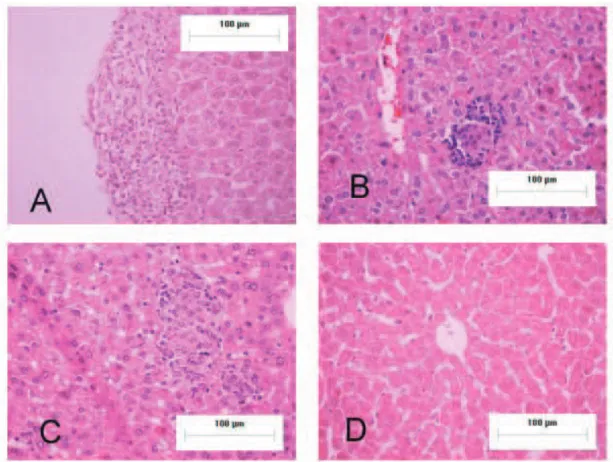
ield. The granulomas were also classiied according to the following criteria: Ex= exudative; corresponding to a
mixed and non-organized concentration of leukocytes with the appearance of an exudate but which differs form a pure exudate by the presence of some adjacent macrophages
(data not shown), M=mixed; a geometrically well shaped
cluster of epithelioid cells and lymphocytes with random
localization of these species (Figure 1A), O=organized; a
geometrically well shaped cluster of epithelioid cells and lymphocytes where the lymphocytes form a rim (Figure
1B), Mφ = Macrophagic granuloma; a well shaped cluster
that contains predominantly macrophages (including
epithelioid cells) (Figure 1C), and F = Fibrotic granuloma with some collagenous ibers (data not shown).
Tests of anti-mycobacterial action of the essential oil were performed in vitro employing the interferometric method (Jardim et al., 2003), where bacterial activity is monitored by measuring the change in refractive index of the nutrient solution caused by bacterial metabolism. Five experiments were performed with (2.5±0.1) mL of Bacto Middlebrook 7H9 culture media enriched with oleic acid albumin dextrose catalase (OADC) in the interferometer.
All ive experiments used 4 x 10⁶ CFU of BCG. The
experiments BCGa) BCGb) contained no essential oil and in the remaining three experiments, essential oil was mixed with the samples in the following concentrations: SCa (2.4±0.2) x10-3 μL oil/mL solution, and experiments
SCb and SCc (0.24±0.02) μL oil/mL solution. In order
to compare our results with a conventional antibiotic, interferometric measurements were also performed with samples containing rifampicin (Lot 780773, SIGMA) with concentrations zero (control), and 4 and 250 ng/mL (positive control). This experiment used smaller sample holders with a smaller stock of oxygen. The changes in refractive index were monitored continuously. At the beginning of the experiments, the same specimen of BCG was also seeded in 7H10 agar culture medium enriched with OADC in order to have a conventional control of bacterial viability. Furthermore, at the end of each experiment the content of the sample holder was seeded to observe whether there existed any contamination. All experiments were found to be free of contamination. At the beginning of experiment SCb conventional experiments were also conducted to evaluate bacterial growth in the presence of the essential oil: four test tubes with liquid culture medium and BCG were prepared with 4x10⁶ CFU of BCG each. In
two of the test tubes, essential oil was added to achieve a
inal concentration of (0.24±0.02) μL/mL. The four tubes
were incubated at 37 oC and monitored regularly by visual
inspection. Tests with solid culture medium 7H10 enriched with OADC in Petri dishes of 5 cm diameter were also performed. Seven dishes were inoculated with 4x10⁶ CFU
of BCG. All dishes had a central disc of sterile paper with 10 mm diameter. Five paper discs were impregnated with
1.2 μL S. cumini oil and the dishes were held at 37 oC.
Results
Enhancement of essential oil yield
Upon extraction of the essential oil from fresh or dried leaves of S. cumini, we obtained very low yields (less
than 10 μL/kg). After freezing the fresh leaves prior to
extraction, it was possible to increase the yield to 0.5 mL/ kg. We suspect that the freeze-thaw cycle ruptures vesicles or other plant structures which contain the steam volatile components and that this method may also be useful for other plants with low essential oil yields.
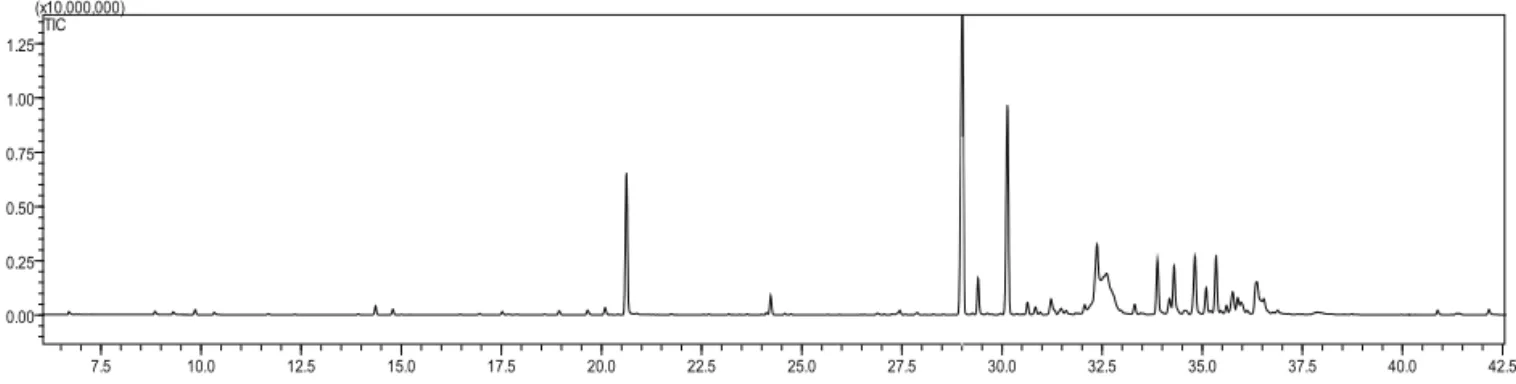
Analysis of principal chemical constituents
We have identiied and conirmed, with a high
degree of certainty, the principal constituents of the essential oil of S. cumini used for this study. The majority of the constituents are sesquiterpenes of the carane family,
namely α-caryophyllene and β-caryophyllene, its alcohol
and epoxide as well as terpineol (Figure 2). Table 2 summarizes the constituents we were able to identify, their retention times (Rt), relative % composition based on peak area, and the % correlation to the NIST 9.0 library.
Table 2. Principal components of the essential oil of fresh
Syzygium cumini leaves, their retention times on GC-MS chromatography and relative percent composition.
RT Area % Substance
14.360 1.33 trans-β-ocimene
20.649 9.08 α-terpineol
24.226 1.99 bornyl acetate
29.068 25.24 α-caryophyllene
29.412 2.00 carveol
30.173 16.00 β-caryophyllene
30.643 1.19 α-muurolene
32.377 4.90 iso-caryophyllene
33.892 3.90 caryophyllenyl alcohol
34.306 3.83 caryophyllene oxide
34.830 4.82 β-humulene
36.362 5.23 epiglobulol
Histopathologic analysis
In all four test groups the hepatic parenchyma showed interstitial edema with perivascular granulomas composed of T-cells and epithelioid cells. However, a normal pattern of hepatic parenchyma was also observed in different regions (Figure 1D). The polymorphonuclear cell content was limited to very small numbers of cells (typically 1 to 7 cells) although roughly 30% of the
granulomas did have these cells. No statistically signiicant
difference in the apoptotic process among the four groups
was found (see Table 3). In the group C28, numerous macrophagic granulomas were found. As described in the
previous section, the granulomas were classiied in order
to judge their patterns. Figures 1A, B, C show examples
of the categories M, O, and Mφ. No granulomas with
predominant polymorphonuclear leukocyte populations were observed, however, the presence of such cells was also detected.
Table 3. Comparison of mean values of percentage of granulomatous area, number of apoptosis per area of granuloma
and type ratio Mφ/M for the control and treated groups
corresponding to 21 and 28 days after infection.
Test group %GA α [μm-2] Mφ/M
C21 1.3±0.3 (1.9±0.4) x 10-3 0.33±0.07 T21 1.0±0.2 (1.9±0.5) x 10-3 0.6±0.1
C28 5.6±1.9* (1.3±0.4) x 10-3** 1.4±0.4*** T28 1.3±0.4* (2.7±0.5) x 10-3** 0.4±0.1*** *p-value 0.026; **p-value 0.082; ***p-value 0.009.
Figure 3 shows the relative frequencies of the types of granulomas. As can be seen, the relative frequency of the organized type is practically the same in all groups. On the other hand, the categories M and Mφ show an interesting behavior. Therefore, it would seem to be
appropriate to deine the quotient of relative frequencies
Mφ/M (number of Macrophagic granulomas divided by
the number of mixed granulomas) as a relevant parameter.
In this fashion, the morphology of the granulomas can be characterized using a single parameter. Table 3 shows the mean values of the following characteristic quantities:
%GA=percentage of granulomatous area in a ield; α=number of apoptosis per granulomatous area, and
Mφ/M.
The groups sacrificed 21 days after infection do not show any difference between treated and control animals. However, the control experiment shows a significant growth of granulomatous area in the time interval from 21 to 28 days, where, for the treated animals, %AG remained approximately constant during that time interval. The comparison of the groups C28 and T28 shows significant differences in the granulomatous area %AG and the type ratio Mφ/M. This indicates that essential oil treatment was able to dramatically inhibit the growth of granulomatous areas which occurs during the fourth week post infection in non-treated animals.
Analysis of antimycobacterial action
Figures 4A and B show the negative time derivative of the refractive index of the nutrient solution of
the ive interferometric experiments. This quantity, -dn/dt,
can be correlated to the state of bacterial activity. As can be
seen from Figure 4A, the experiments with 0.24 μL/mL of
S. cumini oil have no, or a very low bacterial activity and no bacterial growth is visible. The curves without S. cumini
oil and the one with the low concentration (0.0024 μL/mL)
show a pronounced bacterial activity and bacterial growth. The interferometric assays show that a concentration of
0.24 μL/mL of essential oil has an antibiotic action similar to 0.25 μg/mL of rifampicin (Figure 4B). The conventional
experiment in liquid culture medium showed abundant colonies after seventeen days in the test tubes without S. cumini oil and no visible colonies in the tubes with 0.24
μL/mL of S. cumini oil. After 21 days, the control Petri
dishes without oil were densely occupied with colonies visible to the unaided eye and the dishes with oil had a halo that occupied almost the entire dish (Figure 5).
0,0
0,1
0,2
0,3
0,4
0,5
0,6
F
M
φ
O
M
Ex
C21 T21 C28 T28
relative frequency
Figure 3. Mean relative frequencies of granuloma types for the four test groups: star: C21; cross: T21; full circle: C28 and full triangle: T28. The type pattern of the groups C21, T21 and T28
are approximately the same and the proile of the group C28 is signiicantly different, revealing an enhancement of Macrophagic
granulomas (see the quotient Mφ/M in Table 3). Granuloma
type classiication: Ex= exudative; M=mixed; O=organized;
Mφ= Macrophagic; F = Fibrotic. The graph serves to deine and illustrate the relevant parameter Mφ/M, which reveals a difference
in the inlammatory development with 95% conidence interval
according to a 2-tailed Mann-Whitney test (compare Table 3).
Discussion
The results obtained in the present work demonstrate conclusively that the essential oil of
Syzygium cumini (L.) Skeels, Myrtaceae, has an antimicrobial action capable of stopping the proliferation of Mycobacterium bovisin vitro and that the development
of granulomatous inlammation caused by BCG in the hepatic parenchyma of mice is modiied after 21 days
of infection. The dramatic increase of granulomatous area that occurs in the time interval (21 days, 28 days) in non-treated animals is not observed in the treated group. The increase of granulomatous area in the control group during the fourth week after infection can be explained assuming that an exponential growth of bacteria occurs and bacteria are subsequently released. Thereafter, resident activated macrophages and/or recruited monocytes from the bloodstream phagocytize these bacilli, which results in the formation of predominantly macrophagic granulomas (Harrison et al., 2005). This explains the observed increase of the parameter Mφ/M. The release of bacteria from macrophages may be due to lysis of macrophages caused by internal multiplication of the bacteria (Harrison et al., 2005) or by apoptosis of macrophages (Raja, 2004). Although the apoptosis of infected macrophages is a mechanism of host defense, this
mechanism is not suficiently eficient to guarantee that
all bacteria contained in the dying macrophage are killed (Condos et al., 1997; Tan et al., 1997). The net effect of apoptosis is an inhibition of bacterial growth (Oddo et al., 1998; Keane et al., 2000) and the main contribution to the growth of granulomatous areas during the fourth week after infection is mostly likely due to lysis.
The principal constituents identiied in the
essential oil of S. cumini are caryophyllene isomers,
α-terpineol, and oxygenated derivitaves of caryophyllene
(alcohol and epoxide). These are ubiquitous in the plant kingdom and their biological activity has been studied
extensively. The α-caryophyllene and the β-caryophyllene are known for their anti-inlammatory potential (Fernandes et al., 2007; Gertsch et al., 2008). The anti-inlammatory
Figure 2. A typical gas chromatogram of the essential oil from the fresh leaves of Syzygium cumini.
7.5 10.0 12.5 15.0 17.5 20.0 22.5 25.0 27.5 30.0 32.5 35.0 37.5 40.0 42.5
0.00 0.25 0.50 0.75 1.00 1.25
as a speciic CB2 receptor agonist. Our results indicate that the activation of immune cells by β-caryophyllene may have an anti-inlammatory effect, which is in
accordance with observations in carrageenan-induced
inlammation (Gertsch et al., 2008). The exact mechanism
of action in the present model system is not completely understood, but may be mediated by CB2 receptors or by the cytotoxic effect of one or more of its components. The interferometric experiments demonstrated the antimycobacterial action of the S. cumini essential oil
in vitro. Therefore, one might surmise that the presence of the oil inhibits the bacterial growth in macrophages and the liberation of bacilli through lysis is stopped. The
effects of α-caryophyllene were shown to be comparable
with dexamethasone (Fernandes et al. 2007). The possible
mechanisms of action of β-caryophyllene have been
studied already. β-caryophyllene is recognized as a CB2
receptor agonist (Gertsch et al., 2008; Bento et al., 2011), while its oxide has proven to be cytotoxic (Neung et al., 2011).
According to Galiègue et al. (1995), CB2 receptors are present in the principal immune defense cells such as polymorphonuclear neutrophil cells, T-lymphocytes and macrophages that are involved in
chronic inlammation investigated in the present work. Moreover, Gertsch et al. (2008) described β-caryophyllene
Figure 4. Negative time derivative of refractive index of the liquid culture medium as a function of time with BCG containing
samples. A. Full squares: BCGa’ and BCGb’=BCG+culture medium (no essential oil); open squares: SCa’=BCG+culture medium+essential oil with concentration 0.0024 μL/mL; crosses: SCb’=BCG+culture medium+essential oil with concentration 0.24 μL/mL; open triangles: SCc’=BCG+culture medium+essential oil with concentration 0.24 μL/mL. The curves corresponing to the concentration 0.24 μL/mL show no or very low bacterial activity and no bacterial exponential growth. B. full squares: BCG’=BCG+culture medium (no rifampicin); open squares: RIFa’=BCG+culture medium+rifampicin with concentration 0.004 μg/mL; open triangles: RIFb’=BCG+culture medium+rifampicin with concentration 0.25 μg/mL. The sudden decrease in the
curves BCG’ and RIFa’ is due to oxygen depletion.
0 5 10 15 20 25 30 35 40
0,0 2,0x10⁻⁵ 4,0x10⁻⁵ 6,0x10⁻⁵
BCGa' BCGb' SCa' SCb' SCc'
-dn/dt [d
-1
]
t
[d]
0 5 10 15 20 25 300,0
2,0x10⁻⁵
4,0x10⁻⁵
6,0x10⁻⁵
BCG' RIFa' RIFb'
-dn/dt
[d
-1
]
t
[d]
Figure 5. Petri dishes with 7H10+OADC (enrichment) seeded with 4 x 10⁶ CFU of BCG with central discs of sterile paper, three of which were wetted with S. cumini essential oil. The picture shows the state 21 days after seeding without and with Syzygium cumini
essential oil. The two dishes to the left show abundant colonies whereas the dishes with essential oil show a halo the covers almost
average number of apoptosis per area of granuloma (α) in
the group T28 is about two times larger than in the group C28. The statistic analysis does not permit the conclusion
that the oil treatment increases the α-value. Nevertheless,
it does show that the presence of the essential oil did not decrease apoptosis. In this manner, the apoptotic host defense mechanism is maintained in the presence of the oil.
Conclusions
The assays permit us to conclude that the essential oil of S. cumini show relevant anti-inlammatory
activity in vivo and antimycobacterial action in vitro. The analyses of the essential oil composition revealed major quantities of caryophyllenes and its isomers and oxygenated derivatives. As a possible explanation, based on the literature, we suggest that the high percentage of
β-caryophyllene identiied in the essential oil is likely to be responsible for the anti-inlammatory activity. The
antimycobacterial action observed in vitro may be related to the cytotoxic effect present in the caryophyllene oxide also detected in the oil. The mechanism of action present
in anti-inlammatory effect of α-caryophyllene has to
be better investigated. A method to enhance the yield of essential oil of S. cumini by freezing leaves prior to hydro-distillation was discovered. Thus, the study of the essential oil of S. cumini showed that this plant deserves further investigation of its biological activities.
Acknowledgements
The Authors would like to thank Dr. Lyderson Facio Viccini (Department of Biology, UFJF) for help with microscopy, Dr. José Antônio da Silva Reis (Statistics Department UFJF) for important help with statistical analysis and the CBR and Botany Department of UFJF for experimental facilities. Financial support from FAPEMIG, CNPq and logistical support from FADEPE is gratefully acknowledged in realizing this multi-disciplinary collaboration. The results of this work are part of the master degree of RRPM.
Authors contributions
RRPM colleted leaves of S. cumini, extracted
the essential oil, infected, treated and sacriiced the mice,
processed histological slides and performed histopathologic analyses. RRPM also performed antimicrobial sensibility tests including the interferometric ones, participated in statistical analysis and manuscript edition. DFJ developed and applied the interferometric measurements. ARS participated in the animal experiments. ES and RLF collected plant samples for exsiccate preparation and performed toxicity tests of the essential oil. AGC and
RMG performed the chromatographic analysis of the essential oil. RMG suggested mechanisms of action of the oil and revised the manuscript. JPRFM participated in the development of the interferometric method. BL designed and developed the interferometric method advising DFJ.
He also participated in the classiication of granulomas,
statistical analysis, performed interferometric antimicrobial sensibility tests and participated in manuscript edition. FMA designed the project to study the effect of the essential
oil in the granulomatous chronic inlammation advising
RRPM to perform the animal assays. All the authors have
read the inal manuscript and approved the submission.
References
Aarestrup FM, Gonçalves-da-Costa SC, Sarno EN 1995. The effect of thalidomide on BCG-induced granulomas in mice. Braz J Med Biol Res 28: 1069-1076.
Aarestrup FM, Sampaio EP, de Moraes MO, Albuquerque EC, Castro AP, Sarno EN 2000. Experimental Mycobacterium leprae infection in BALB/c mice: effect of BCG administration on TNF-alpha production and granuloma development. Int J Lepr Other Mycobact Dis 68: 156-166.
Achrekar S, Kaklij GS, Pote MS, Kelkar SM 1991. Hypoglycemic activity of Eugenia jambolana and Ficus bengalensis:
mechanism of action. In Vivo 5: 143-147.
Ayyanar M, Subash-Babu P 2012. Syzygium cumini (L.) Skeels: A review of its phytochemical constituents and traditional uses. Asian Pac J Trop Biomed 2: 240-246.
Bento AF, Marcon R, Dutra RC, Claudino RF, Cola M, Leite DF, Calixto JB 2011. Beta-caryophyllene inhibits dextran sulfate sodium-induced colitis in mice through CB2 receptor activation. Am J Pathol 178: 1153-1166.
Brasseur T 1989. Anti-inlammatory properties of lavonoids. J Pharm Belg 44: 235-241.
Chirvan-Nia P, Ratsimamanga AR 1972. Regression of cataract and hyperglycemia in diabetic sand rats (Psammomys obesus) having received an extract of Eugenia jambolana
(Lamarck). CR Acad Sci Hebd Seances Acad Sci D 274: 254-257.
Condos R, Rom WN, Liu Y, Schluger NW 1997 Local immune responses correlate with presentation and outcome in tuberculosis. Am J Respir Crit Care Med 157: 729-735. Jardim DF, Batista Santiago Neto R, Machado RRP, Aarestrup
FM, de Mendonça JPRF, Lesche B 2003.Observing bacterial activity interferometrically. Eur Biophys J 32: 159-162.
Fernandes ES, Passos GF, Medeiros R, da Cunha FM, Ferreira J, Campos MM, Pianowski LF, Calixto JB 2007.
Anti-inlammatory effects of compounds alpha-humulene and
(-)-trans-caryophyllene isolated from the essential oil of
Carayon P, Bouaboula M, Shire DLE, Fur G, Casellas P 1995. Expression of central and peripheral cannabinoid receptors in human immune tissues and leukocyte subpopulations. Eur J Biochem 232: 54-61.
Gertsch J, Leonti M, Raduner S, Racz I, Chen JZ, Xie XQ, Altmann KH, Karsak M, Zimmer A 2008. Beta-caryophyllene is a dietary cannabinoid. Proc Natl Acad Sci USA 105: 9099-9104.
Harrison Braunwald E, Fauci AS, Kasper DL, Hauser SL, London DL, Jameson JL 2005. Harrison’s Principles of Internal Medicine. 16th ed. New York: McGraw-Hill’s.
Keane J, Remold HG, Kornfeld H 2000. Virulent Mycobacterium tuberculosis strains evade apoptosis of infected alveolar macrophages. J Immunol 164: 2016-2020.
Ling-Ling Y, Chih-Ying L, Kun-Ying Y 2000. Induction of apoptosis by hidrolyzable tannins from Eugenia jambos
L. on human leukemia cells. Cancer Lett 157: 65-75. Mahmoud II, Marzouk MAS, Moharram FA, El-Gindi MR,
Hassan AMK 2001. Acylated lavonol glycosides from
Eugenia jambolana leaves. Phytochemistry 58: 1239-1244.
Martin S, Ocete MA, Galvez J, Jimenez J, Zazuelo A 1993.
Anti-inlammatory activity of the essential oil of Bupleurum fruticescens. Planta Med 59: 533-536.
McLaughlin JL, Rogers LL, Andrson JE 1998.The use of biological assays to evaluate botanicals. Drug Inf J 32: 513-525.
Menezes-de-Lima Jr O, Wernek-Barroso E, Cordeiro RSB, Henriques MGMO 1997. Effects of inhibitors of
inlammatory mediators and cytokines on eosinophil
and neutrophil accumulation induced by Mycobacterium bovis bacilus Calmete-Guérin in mouse pleurisy. J Leukoc Biol 62: 778-785.
Ministério da Saúde, 2004. Conselho Nacional de Saúde. Comissão Nacional de Ética em Pesquisa. Manual Operacional para Comitês de Ética em Pesquisa. Brasília: Ministério da Saúde, p. 124.
Modi DC, Patel JK, Shah BN, Nayak BS 2010. Antiinlammatory
activity of seeds of Syzygium cumini Linn. J Pharm Educ Res 1: 68-70.
Muruganandan S, Srinivasan K, Chandra S, Tandan SK, Lal
J, Raviprakash V 2001. Antiinlammatory activity of
Syzygium cumini bark. Fitoterapia 72: 369-377. Muruganandan S, Pant S, Srinivasan K, Chandra S, Tandan SK,
Lal J, Prakash RV 2002. Inhibitory role of Syzygium cumini on autacoid-induced inlammation in rats. Indian J Physiol Pharmacol 46: 482-486.
NC3Rs 2012. National Center for the Replacement Reinement
and Reduction of Animals in Research. http://www.nc3rs. org.uk, accessed May 2012.
Neung JJ, Ashik M, Jeong YM, Ki-Chang J, Dong-Sun L, Kwang SA, Somi KC 2011. Cytotoxic Activity of
β-caryophyllene oxide isolated from jeju guava (Psidium cattleianum Sabine) Leaf. Rec Nat Prod 5: 242-246 Ocete MA, Risco S, Zarzuelo A, Jimenez J 1989.
Pharmacological activity and effects of the essential oil of Bupleurum gibraltaricum: antiinflammatory activity and effects on isolated uteri. J Ethnopharmacol 25: 305-313.
Oddo M, Renno T, Attinger A, Bakker T, MacDonald HR, Meylan PRA 1998. Fas ligand-induced apoptosis of infected human macrophages reduces the viability of intracellular Mycobacterium tuberculosis. J Immunol 160: 5448-5454.
Pandey M, Khan A 2002. Hypoglycemic effect of defatted seeds and water soluble fiber from the seeds of
Syzygium cumini (Linn.) skeels in alloxan diabetic rats. Indian J Exp Biol 40: 1178-1182.
Raja A 2004. Immunology of tuberculosis. Indian J Med Res 120: 213-232.
Scharma SB, Nasir A, Prabhu KM, Murthy OS, Dev G 2003. Hypoglycemic and hypolipidemic effect of ethanolic extract of seeds of Eugenia jambolana in alloxan-induced diabetic rabbits. J Ethnopharmacol 85: 201-206. Siani AC, Sampaio ALF, Sousa, MC, Henriques, MGMO, Ramos
MFS 2000. Óleos essenciais. Biotecnologia Ciência e Desenvolvimento Ano III: 38-43.
Slowing K, Söllhuber M, Carretero E, Vilar A 1994. Flavonoid glicosides from Eugenia jambos. Phytochemistry 37: 225-258.
Stanely Mainzen Prince P, Kamalakkannan N, Menon VP 2003.
Syzygium cumini seed extracts reduce tissue damage in diabetic rat brain. J Ethnopharmacol 84: 205-209. Tan JS, Canaday DH, Boom WH, Balaji KN, Schwander SK,
Rich EA 1997. Human alveolar T lymphocyte responses to Mycobacterium tuberculosis antigens: role for CD4 and CD8 cytotoxic T cells and relative resistance of alveolar macrophages to lysis. J Immunol 159: 290-297. WHO 2011. The sixteenth global report on tuberculosis. World
Health Organization http://www.who.int/tb/publications/ global_report/2011/gtbr11_executive_summary.pdf, accessed May 2012.
*Correspondence
Rachel R. P. Machado
Faculdade de Ciências Médicas e da Saúde de Juiz de Fora, Suprema
Alameda Salvaterra, nº 200, Bairro Salvaterra, 36.033-003 Juiz de Fora-MG, Brazil