What influences Hb Fetal production in adulthood?
Laboratory of Hemoglobin and Genetics of Hematologic Diseases, Universidade Estadual Paulista – UNESP/IBILCE, São José do Rio Preto, SP, Brazil
Gisele Cristine de Souza Carrocini
Paula Juliana Antoniazzo Zamaro
Claudia Regina Bonini-Domingos
Human hemoglobin genes are located in α and β globin gene clusters in chromosomes 16 and 11, respectively. Different types of hemoglobin are synthesized according to the stage of development with fetal hemoglobin (α2γ2) (Hb F) being the main hemoglobin in the fetal period. After birth, there is a reduction (to about 1%) in Hb F levels and adult hemoglobin, Hb A (α2β2), increases to more
than 96% of total hemoglobin. However, some genetic conditions whether linked to the β-globin gene cluster or not are associated with high Hb F levels in adults. Among those linked to β-globin are hereditary persistence of fetal hemoglobin, delta-beta thalassemia (δß-Thalassemia) and the XmnI polymorphism (-158 C > T). Other polymorphisms not related to ß-globin gene cluster are known to influence the γ-globin gene expression in adulthood. The most relevant polymorphisms that increase concentrations of Hb F are the HMIP locus on chromosome 6, the BCL11A locus on chromosome 2, the Xp22.2 region of the X chromosome and the 8q region on chromosome 8. Findings from our research group studying genetic factors involved in γ-globin gene regulation in adults without anemia in the northwestern region of São Paulo State showed that high Hb F levels are influenced by the presence of hereditary persistence of fetal hemoglobin mutations and the XmnI polymorphism, suggesting that both genetic alterations characterize the molecular basis of the evaluated population.
Keywords: Fetal hemoglobin; Polymorphism, genetic; gamma-Globins
Organization of globin genes
Human globin genes are found in two gene clusters designated α (alpha) and β (beta) globin. The α-globin gene cluster is located on chromosome 16 with the resulting polypeptide chain totaling 141 amino acids. It consists of three inactive genes (ψζ, ψα and ψα2), three functional genes (ζ, α1 and α2) and two genes with unidentified functions in vivo (µ and
θ1).(1) The β-globin gene cluster is located on chromosome 11 with the synthesized products
having 146 amino acids. This cluster consists of a pseudogene (ψβ) and five functional genes expressed at different development stages in the same order as they are arranged in the DNA segment (5' - εγGγA δ β - 3').(2,3)
During the ontogeny of globin chains, red blood cells express different genes belonging to the α and β-globin cluster; changes in their expression occur in two distinct stages of development: 1) from the embryonic to fetal period in early pregnancy which involves changes in the expressions of alpha-type globin (from ζ to α) and beta-type globin (from
ε to γ) and 2) approximately six months after birth with just one change in the beta-type globin (from β to γ).(4)
Fetal Hemoglobin and its benefits for individuals with abnormal
hemoglobins
Fetal Hemoglobin (Hb F), formed by two α and two γ-globin chains (α2 γ2), is produced at high levels in the fetal period due to a high expression of γ-globin genes.(5)
The γ-globin gene originates from a 5 kilobase (kb) tandem repeat. These genes differ from one another by only one amino acid [glycine (γG) or alanine (γA)] at position 136 of the
polypeptide chain. At birth, γG chains are more abundant while γA chains predominate in
adulthood.(6)
In adults, the β-globin gene is predominant; approximately 98% of all hemoglobin is comprised of adult hemoglobin, Hb A (α2β2). Thus, γ-globin genes are poorly expressed; less than 1% of adult hemoglobin is made up of Hb F.(7) Hb F levels can be evaluated by
counting the number of F cells, that is, adult erythrocytes that contain measurable amounts of this hemoglobin. The Hb F and F cell levels vary considerably in healthy adults but usually there is a good correlation between the two.(8,9)
Conflict-of-interest disclosure: The authors declare no competing financial interest
Submitted: 2/3/2011 Accepted: 3/22/2011
Corresponding author:
Claudia Regina Bonini-Domingos Laboratório de Hemoglobinas e Genética das Doenças Hematológicas – LHGDH -Ibilce - Unesp
Rua Cristóvão Colombo, 2265 – Jd. Nazareth 15054-000 – São José do Rio Preto, SP, Brazil
Phone: 55 17 3221-2392 [email protected]
www.rbhh.org or www.scielo.br/rbhh
When γ-globin genes are highly expressed, higher Hb F levels in red blood cells may compensate defective β-globin products and significantly reduce the symptoms of hemoglobinopathies such as sickle cell anemia.(7) The Hb F
concentration and its distribution in red blood cells are major genetic modulators of disease and high levels of this hemoglobin dilute the amount of Hb S thereby inhibiting or delaying the polymerization process, the result of which is fewer harmful events.(10) In beta thalassemia, an induced
increase in gamma chain production has a beneficial effect on the clinical status of homozygotes, not only by reducing the imbalance in the α-type/non-α-type chains, but also by increasing the synthesis of total hemoglobin.(11) Thus, an
increased γ-globin gene expression has clinical relevance in the treatment of diseases related to the β-globin gene.(7) Some
genetic conditions are known to influence γ-globin gene expression during adulthood, including hereditary persistence of fetal hemoglobin (HPFH) and delta-beta thalassemia (δβ-thalassemia).(12,13)
Hereditary persistence of fetal hemoglobin
HPFH is usually a benign condition characterized by a lack of changes in the synthesis of β-globin resulting in a substantial increase in gamma chains and consequently high Hb F levels.(14) This condition can occur due to single point
mutations in the γ-globin gene promoter or β-globin gene cluster deletions, which remove the δ and β-globin genes.(15)According to the literature, Hb F levels in deletional
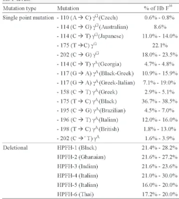
HPFH are, in most cases, higher compared to single point mutations. HPFH mutations are associated to different ethnic groups. Individuals that present HPFH mutations have normal erythrocytes.(16) Studies by our research group on mutations
related to increases in Hb F levels in adults without anemia in the northwestern region of São Paulo State show that 26.7% of cases were heterozygous for deletional HPFH (HPFH1 -Black; and HPFH-2 - Ghanaian); HPFH-2 is the most common mutation. These mutations were found in individuals who had Hb F levels above 15% of the total hemoglobin (unpublished data). In relation to erythrocyte morphology, the presence of mild alterations was observed thus different to published findings.
Table 1 shows different Hb F concentrations for the point mutation and deletional mutations in HPFH and the associated ethnic groups.
Delta-Beta thalassemia [(
δβ
δβ
δβ
δβ
δβ
)
0-thalassemia]
[γG (γAδβ)0-thalassemia].(1,16-19) Heterozygous individuals forWhat influences Hb Fetal production in adulthood?
mutation in three individuals with Hb F levels of 9.6%, 9.8% and 10.7% (unpublished data); these values are within the range expected for this mutation.(16)
Genetic polymorphisms involved in Hb F
regulation
Family studies show that high Hb F levels tend to be inherited but the pattern of inheritance is often unclear and this trait does not seem to be correlated with the β-globin gene cluster.(20,21) The persistence of Hb F and F cells in
populations suggests that variability is not from the inheritance of just one gene locus, but from a combination of several genes on different chromosomes. Thus, in genetic terms, high Hb F levels in adulthood is regarded as a quantitative trait, in which the action of multiple genes combined with an environmental component may explain the absence of a clear pattern of Mendelian inheritance.(22)
In some situations, it is not clear if genetic variability in the β-globin gene cluster plays a decisive role in Hb F variations. This variation is attributed to co-inherited polymorphisms in regulatory regions that affect the β-globin gene cluster or regions close to it. In this scenario, studies have been performed to identify candidate genes and also to analyze possible associations that might explain high Hb F levels in adulthood. It is believed that high Hb F levels are inherited as quantitative traits that are dependent on many gene loci.(23)
Genome-wide association studies (GWAS) and family studies have shown that regions outside the β-globin gene cluster, including 2q16, 6q23, 8q and Xp22.2, are implicated in the regulation of Hb F levels.(24) Approximately 45% of
variations in Hb F levels are associated with the presence of three main quantitative trait loci including the XmnI polymorphism on chromosome 11 (11p15), the HMIP locus on chromosome 6 (6q23) and SNPs (single nucleotide polymorphisms) for the BCL11A gene on chromosome 2 (2q16).(9,25)
Xmn
I polymorphism (-158 C > T) –
Chromosome 11
The XmnI polymorphism is known to influence the γG
gene expression, predisposing carriers to increased Hb F concentrations in particular when they are under conditions of erythropoietic stress.(26) In patients with sickle cell disease
and beta thalassemia, the presence of this polymorphism is associated with higher Hb F levels.(27) With this polymorphic
site, there is an increase in the proportion of γG chains
resulting in a γG:γA ratio similar to that seen at birth (70:30).(28,29)
This polymorphism is common in many populations with frequencies as high as 32% to 35% being reported.(27)
Studies by Peri et al.(30) and Garner et al.(22) found similar
frequencies for different populations of healthy Europeans. Our research group, on evaluating this polymorphic site in adults without anemia from the northwestern region of São Paulo, observed a frequency of 33.3%, with Hb F levels ranging from 2.0% to 33.3%. The XmnI polymorphic site was more frequent (60%) in individuals with Hb F below 15% of total hemoglobin (unpublished data).
For populations with changes in hemoglobin levels, the frequency of the XmnI polymorphism differs from that reported for the healthy population. Table 3 shows the literature reports regarding Hb F levels, the phenotype expressed and the presence of this polymorphism in different population groups.
8q region – Chromosome 8
6q23 region (
HMIP
locus) – Chromosome 6
Recent studies show that another QTL, HMIP at 6q23 in the HBS1L-MYB intergenic region, influences Hb F levels. The HBS1L gene is involved in the regulation of several cellular processes and is expressed in hematopoietic cells, whereas the MYB gene is involved in oncogenesis and plays a key role in the erythropoiesis process.(25,37) It is believed
that a gene in the 6q region is responsible for encoding a factor involved in the erythroid maturation pathway resulting in increased Hb F levels and F cell production.(38) The results
obtained by Garner et al.(39) show the influence of 6q23 QTL
on the modulation of Hb F levels and F cell production in Indian patients with beta thalassemia. However, it is not clear if the effect of this QTL is direct, that is, acting as a factor that interacts with γ-globin genes, or indirect.
In Caucasian populations, this region is presented in three blocks of linkage disequilibrium, where the second block is more strongly associated with the Hb F variation. The variability in block 2 of the HMIP region appears to play a pleiotropic effect on some hematological parameters in relation to the number of cells and erythrocyte content of healthy European individuals. This characteristic suggests that the HBS1L-MYB intergenic region contains distal regulatory sequences that act on precursor erythroid lineages.(38)
Studies on Indian families identified that the 6q23 locus had a greater association with high Hb F levels in patients with beta thalassemia. These studies also demonstrated that for healthy subjects and individuals heterozygous for beta thalassemia, the homozygous presence of this site is linked to higher Hb F concentrations (from 1.1% to 3% for healthy individuals and 10% to 24% for patients with thalassemia).(38)
In healthy individuals from Northern Europe, the presence of the 6q23 locus contributes to approximately 19% of the variation in Hb F levels.(25) In other populations
this locus is present in healthy Afro-Americans and in patients with sickle cell disease from Britain and Brazil, contributing to about 3% to 7% of the variation in Hb F of these populations.(40)
2q16 region (BCL11A) – Chromosome 2
Another QTL associated with high Hb F levels located in the 2q16 region of chromosome 2 is known as BCL11A. Studies by Menzel et al.(9) found that, besides the XmnI
polymorphism and HMIP locus, a third region, 2q16, was associated with Hb F concentrations accounting for
with sickle cell anemia in Afro-Americans and Brazilians, who had Hb F variations strongly associated to the presence of the BCL11A QTL.
From these findings, some researchers suggest that the
BCL11A gene product, a zinc-finger transcription factor, is the key mediator in the process of silencing the γ-globin gene expression and in switching to the β-globin gene.(42) Studies
using K562 cells showed that the BCL11A QTL binds to a central region of the γ-globin gene promoter to form a repressor thus acting as a silencing agent of the expression of this gene.(43) Studies by Xu et al.(44) showed that the
silencing of γ-globin genes is the result of an interaction that occurs between the BCL11A transcription factor and the β-globin gene cluster interacting with chromatin to form a chromosomal loop in combination with the SOX6 transcription factor in γ-globin gene promoter regions. These findings show that the BCL11A transcription factor may be a regulator in the development and genetic control of γ-globin gene silencing in adults.
Xp22.2 region – Chromosome X
Dover et al.,(45) on analyzing healthy subjects and sickle
cell disease patients of African origin, showed that the percentage of F cells present in women was significantly higher than in men. It was thus suggested that regions located in the X chromosome could be responsible for this difference. These authors found that Xp22.2 was associated with F cell production and suggested that 70% of the variation in Hb F levels in sickle cell anemia patients is associated with variations in the percentages of F cells, which are partially controlled by the Xp22.2 locus on chromosome X.
Conclusions
Much evidence shows that co-inheritance of genetic determinants related to Hb F levels may play an important role in modulating the phenotype of patients with sickle cell anemia and beta thalassemia.(36) Thus, the identification
of genes and genetic variants that contribute to the variability of Hb F production and the molecular mechanisms involved in this variation may help to discover new therapeutic strategies and develop pharmacological agents that increase the levels of this hemoglobin.(25) Our findings
the expression of the γ-globin gene, showing that high Hb F levels in adulthood may result from a range of genetic factors which may explain the observed variations in Hb F of healthy subjects and patients with hemoglobinopathies.(46,47)
References
1. Weatherall DJ, Clegg JB. Inherited haemoglobin disorders: an increasing global health problem. Bull World Health Organ. 2001; 79(8):704-12.
2. Grosveld F, Dillon N, Higgs D. The regulation of human globin gene expression. Baillieres Clin Hematol. 1993;6(1):31-55. 3. Dillon N, Trimborn T, Strouboulis J, Fraser P, Grosveld F. The
effect of distance on long-range chromatin interactions. Mol Cell. 1997;1(1):131-9.
4. Pace BS, Zein S. Understanding mechanisms of γ-globin gene regulation to develop strategies for pharmacological fetal hemoglobin induction. Dev Dyn. 2006;235(7):1727-37. 5. Stamatoyannopoulos G. Control of globin gene expression during
development and erythroid differentiation. Exp Hematol. 2005; 33(3):259-71.
6. Zago MA, Falcão RP, Pasquini R. Hematologia: fundamentos e prática. Ed rev atual. São Paulo: Editora Ateneu; 2004. 245 p.
7. Xu XS, Hong X, Wang G. Induction of endogenous γ-globin gene expression with decoy oligonucleotide targeting Oct-1 transcription factor consensus sequence. J Hematol Oncol. 2009;2(15):1-11. 8. Miyoshi K, Kaneto Y, Kawai H, Ohchi H, Niki S, Hasegawa K, et
al. X-linked dominant controlo f F-cells in normal adult life: characterization of the Swiss type as hereditary persistence of fetal hemoglobin regulated dominantly by gene(s) on X chromosome. Blood. 1988;72(6):1854-60.
9. Menzel S, Garner C, Gut I, Matsuda F, Yamaguchi M, Heath S, et al. A QTL influencing F cell production maps to a gene encoding a zinc-finger protein on chromosome 2p15. Nat Genet. 2007;39 (10):1197-9.
10. Steinberg MH. Genetic etiologies for phenotypic diversity in sickle cell anemia. Scient World J. 2009;9(1):46-67.
11. Galanello R, Cao A. Relationship between genotype and phenotype. Ann N Y Sci. 1998;850:325-33.
12. Craig JE, Barnetson RA, Prior J, Raven JL, Thein SL. Rapid detection of deletions causing δβ thalassemia and hereditary persistence of fetal hemoglobin by enzymatic amplification. Blood. 1994;83(6):1673-82.
13. Panyasai S, Fucharoen S, Surapot S, Fucharoen G, Sanchaisuriya K. Molecular basis and hematologic characterization of δβ-thalassemia and hereditary persistence of fetal hemoglobin in Thailand. Haematologica. 2004;89(7):777-81.
14. Nussbaum RL, McInnes RR, Willard HF. Genética médica. 7th ed. Rio de Janeiro: Guanabara koogan 2007.
15. Olave IA, Doneanu C, Fang X, Stamatoyannopoulos G, Li Q. Purification and identification of proteins that bind to the hereditary persistence of fetal hemoglobin-198 mutation in the γ-globin gene promoter. J Biol Chem. 2007;282(2): 8 5 3 - 6 2 .
16. Huisman HJ, et al. HbVar: A database of human hemoglobin variants and Thalassemias. Summaries of mutation categories. Pennsylvania University USA and McMaster University in Canada, 1996. [Internet]. [cited 2010 May 20]. Available at: http://globin.cse. psu.edu/>
17. Collins FS, Stoeckert CJJr, Serjeant CR, Forget BG, Weissman Sm. Gamma beta+ hereditary persistence of fetal hemoglobin: cosmid cloning and identification of a specific mutation 5' to the Agamma gene. Proc Natl Acad Sci USA. 1984;81(15):4894-8.
18. Craig JE, Rochette J, Sampietro M, Wilkie Ao, Barnetson R, Hatton CS, et al. Genetic heterogeneity in heterocellular hereditary persistence of fetal hemoglobin. Blood.1997;90(1):428-34. 19. Forget BG. Molecular basis of hereditary persistence of fetal
hemoglobin. Ann N Y Acad Sci. 1998;850:38-44.
20. Martinez G, Novelletto A, Di Rienzo A, Felicetti L, Colombo B. A case of hereditary persistence of foetal haemoglobin caused by a gene not linked to the β-globin cluster. Hum Genet. 1989;82(4): 335-7.
21. Thein SL, Weatherall DJ. A non-deletion hereditary persistence of foetal hemoglobin (HPFH) determinant not linked to the beta-globin gene complex. In: Hemobeta-globin Switching; New York: Alan R Liss; 1989.
22. Garner C, Tatu T, Reittie JE, Littlewood T, Darley J, Cervino S, et al. Genetic influences on F cells and other hematologic variables: a twin heritability study. Blood. 2000;95(1):342-6.
23. Thein SL, Menzel S. Discovering the genetics underlying foetal haemoglobin production in adults. Br J Haematol. 2009;145(4): 455-67.
24. Nguyen TKT, Joly P, Bardel C, Moulsma M, Bonello-Palot N, Francina A. The XmnI Gγ polymorphism influences hemoglobin F
synthesis contrary to BCL11A and HBS1L-MYB SNPs in a cohort of 57 β-thalassemia intermedia patients. Blood Cells Mol Dis. 2010;45(2):124-7.
25. Thein SL, Menzel S, Lathrop M, Garner C. Control of fetal hemoglobin: new insights emerging from genomics and clinical implications. Hum Mol Genet. 2009;18(R2):216-23.
26. Grosso M, Amendolara M, Rescigno G, Danise P, Todisco N, Izzo P, et al. Delayed decline of γ-globin expression in infant age associated with the presence of Gγ -158 (CÆT) polymorphism.
Int J Lab Hem. 2008;30(3):191-5.
27. Thein SL. Genetic insights into the clinical diversity of β -thalassaemia. Br J Haematol. 2004;124(3):264-74.
28. Sampietro M, Thein SL, Contreras M, Pazmany L. Variation of Hb F and F-cell number with the Gγ XmnI (CÆT) polymorphism
in normal individuals. Blood. 1992;79(3):832-33.
29. Nemati H, Rahimi Z, Bahrami G. The XmnI polymorphic site 5' to the Gγ gene and its correlation to the Gγ:Aγ ratio, age at the first
blood transfusion and clinical features in β-Thalassemia patients from Western Iran. Mol Biol Rep. 2010;37(1):159-64. 30. Peri KG, Gagnon J, Gagnon C, Bard H. Association of 158 (CÆT)
(XmnI) DNA polymorphism in G [gamma]-globin promoter with delayed switchover from fetal to adult hemoglobin synthesis. Pediatr Res.1997;41(2):214-7.
31. Gibney GT, Panhuysen CIM, So JCC, Ma ES, ha SY, Li CK, et al. Variation and heritability of Hb F and F-cells among β -thalassemia heterozygotes in Hong Kong. Am J Hematol. 2008; 83(6):458-64.
32. Karimi M, Yarmohammadi H, Farjadian S, Zeinali S, Moghaddam Z, Capellini MD, et al. Beta thalassemia intermedia from southern Iran: IVSII.1 (G:A) is the prevalent thalassemia intermedia allele. Hemoglobin. 2002;26(2):147-54.
33. Neishabury M, Azarkeiven A, Najmabadi H. Frequency of positive XmnI Ggamma polymorphism and coinheritance of common alpha thalassemia mutations do not show statistically significant difference between thalassemia major and intermedia cases with homozygous IVSII-1 mutations. Blood Cells Mol Dis. 2010;44 (2):95-9.
34. Nadkarni A, Gorakshakar AC, Lu CY, Krishnamoorthy R, Ghosh k, Colah R, et al. Molecular pathogenesis and clinical variability of beta-thalassemia syndromes among Indians. Am J Hematol. 2001;68(2):75-80.
35. El-Hazmi MA. Xmn I polymorphism in the Gamma-globin gene region among Saudis. Hum Hered. 1989;39(1):790-2.
36. Garner CP, Tatu T, Best S, Creary L, Thein SL. Evidence of genetic interaction between the β-blobin complex and chromosome 8q in the expression of fetal hemoglobin. Am J Hum Genet. 2002;70 (3):793-9.
37. Wahlberg K, Jiang J, Rooks H, Jawaid K, Matsuda F, Yamaguchi M, et al. The HBS1L-MYB intergenic interval associated with elevated Hb F levels shows characteristics of a distal regulatory region in erythroid cells. Blood. 2009;114(6):1254-62.
38. Craig JE, Rochette J, Fisher CA, Weatherall DJ, Marc S, Lathrop GM, et al. Dissecting the loci controlling fetal haemoglobin production on chromosomes 11p and 6q by the regressive approach. Nat Genet. 1996;12(1):58-64.
39. Garner C, Mitchell J, Hatzis T, Reittie J, Farrall M, Thein SL. Haplotype mapping of a major quantitative-trait locus for fetal hemoglobin production on chromosome 6q23. Am J Hum Genet. 1998;62(6):1468-74.
40. Lettre G, Sankaran VG, Bezerra MA, Araújo AS, Uda M, Sanna S, et al. DNA polymorphisms at the BCL11A, HBS1L-MYB, and γ -globin loci associate with fetal hemo-globin levels and pain crises in sickle cell disease. Proc Natl Acad Sci U S A. 2008;105(33): 11869-74.
41. Uda M, Galanello R, Sanna S, Lettre G, sankaran VG, Chen W, et al. Genome-wide association study shows BCL11A associated with persistent fetal hemoglobin and amelioration of the
xxx
phenotype of beta-thalassemia. Proc Natl Acad Sci USA. 2008; 105(5):1620-5.
42. Sankaran VG, Menne TF, Xu J, Akie TE, Lettre G, Van Handel B, et al. Human foetal hemoglobin expression is regulated by the developmental stage-specific repressor BCL11A. Science. 2008; 322(5909):1839-42.
43. Chen Z, Luo HY, Steinberg MH, Chui DH. BCL11A represses HBG transcription in K562 cells. Blood Cells Mol Dis. 2009;42 (2):144-9.
44. Xu J, Sankaran VG, Ni M, Menne TF, Puram RV, Kim W, et al. Transcriptional silencing of γ-globin by BCL11A involves long-range interactions and cooperation with SOX6. Genes Dev. 2010; 24(8):783-98.
45. Dover GJ, Smith KD, Chang YC, Purvis S, Mays A, Meyers DA, et al. Fetal Hemoglobin Levels in Sickle Cell Disease and Normal Individuals Are Partially Controlled by an X-Linked Gene Located at Xp22.2. Blood. 1992;80(3):816-24.
46. Bianchi N, Zuccato C, Lampronti I, Borgatti M, Gambari R. Expression of miR-210 during erythroid differentiation and induction of γ-globin gene expression. BMB Rep. 2009;42(8): 493-9.