Cytogenetic studies in six species of
Scinax
(Anura, Hylidae)
clade
Scinax ruber
from northern and northeastern Brazil
Lídia Nogueira
1, Juliani Bruna Zanoni
2, Mirco Solé
3, Paulo Roberto Antunes de Mello Affonso
2,
Sérgio Siqueira
2and Iracilda Sampaio
11
Instituto de Estudos Costeiros, Universidade Federal do Pará, Bragança, PA, Brazil.
2
Departamento de Ciências Biológicas, Universidade Estadual do Sudoeste da Bahia, Jequié, BA, Brazil.
3
Departamento de Ciências Biológicas, Universidade Estadual de Santa Cruz, Ilhéus, BA, Brazil.
Abstract
Scinax species are still underrepresented in cytogenetic studies, mainly with respect to populations from northeast-ern and northnortheast-ern Brazil. In this study, we provide new chromosomal information on Scinax boesemani, S. camposseabrai, S. garbei, S. pachycrus, S. trilineatus and S. x-signatus, all belonging to clade S. ruber. They were collected at two locations in the Caatinga biome (northeastern Brazil) and at one in the Amazon (northern Brazil) biomes. Chromosomes were analyzed by conventional staining, C-banding, Ag-NOR staining, and fluorochrome staining. All species shared a modal diploid value of 2n = 24 and fundamental arm number (FN) of 48. Moreover, both chromosomal size and morphology were similar to other species in this Scinax clade. C-banding revealed centromeric heterochromatin in all species, along with terminal species-specific C-bands in some species. Active nu-cleolar organizer regions (Ag-NORs) were identified at 11q in most species, except forS. boesemani and S. garbei (Ag-NORs at interstitial region of 8q). Differing from most anurans, GC-rich regions were not restricted to NORs, but also coincident with some centromeric and terminal C-bands. These data contribute to the cytotaxonomy ofScinax by providing chromosomal markers and demonstrating the occurrence of microstructural rearrangements and inver-sions on chromosomal evolution ofScinax.
Keywords: amphibians, chromosomes, fluorochromes, heterochromatin, rDNA.
Received: September 26, 2014; Accepted: January 22, 2015.
Introduction
The genus Scinax (Anura: Hylidae) comprises 113 species widespread from southern Mexico to Argentina, Uruguay, Trinidad and Tobago and Santa Lucia islands (Frost, 2014). Based on molecular markers, morphology, osteology, myology, reproductive biology and chromo-somes, this genus was divided into two clades: S. catharinae (which includes two species groups - S. catharinaeandS.perpusillus) andS.ruber(species groups S.rostratus,S.uruguayusand the remaining species) (Fai-vovich, 2002; Faivovichet al., 2005). The cladeS.ruber comprises nearly 65 species (Frost, 2014) widespread over open ares of tropical and subtropical regions (Faivovich, 2002).
So far, karyotypes are known for only 39 species of Scinax and only few studies included banding methods,
such as Ag-NOR staining, C-banding, BrdU, fluorochrome staining, and in situ hybridization (Pombal et al., 1995; Kasaharaet al., 2003; Nunes and Fagundes, 2008; Cardozo et al., 2011). These cytogenetic studies showed that all spe-cies in this genus share a modal diploid value of 2n = 24.
In spite of numerical conservativeness in diploid number, the two clades can be differentiated by cytogenetic analyses. In the cladeS. catharinae, the first and second chromosomal pairs are submetacentric and NORs are usu-ally located on the sixth pair, while, in the cladeS.ruber, the first and second pairs are metacentric and NORs have been frequentle been detected on pair 11 (Cardozoet al., 2011).
SinceScinaxspecies and populations are still under-represented in terms of cytogenetic data, and species-specific or population differences might be overlooked, we herein provide the first chromosomal information about samples of the following species within the cladeS.ruber: S.boesemani,S.camposseabrai,S.garbei,S.pachycrus,S. trilineatus and S. x-signatus, collected in northern and northeastern Brazil.
Sende correspondence to Lídia Nogueira. Instituto de Estudos Costeiros, Universidade Federal do Pará, Alameda Leandro Ri-beiro s/n, Aldeia, 68600-000 Bragança, PA, Brazil. E-mail: lidia.nogueira@yahoo.com.br.
Materials and Methods
Twenty one individuals of six species ofScinaxfrom the cladeS.rubercollected in distinct localities in northern (Bragança, PA) and northeastern (Maracás and Jequié, BA) Brazil were cytogenetically analyzed (Table 1). The speci-mens were deposited in the Herpetological Collection at Universidade Estadual de Santa Cruz (UESC). Metaphasic chromosomes were obtained from cells of intestinal epithe-lium as described by Schmid (1978). Slides were stained with 10% Giemsa solution in phosphate buffer 0.1 M (pH 6.8) for about 10 min, washed in distilled water and air dried. The best metaphases were selected and photo-graphed for karyotyping and chromosomal measurements. Active nucleolar organizer regions (Ag-NORs) were lo-cated by silver nitrate staining (Howell and Black, 1980), and heterochromatin was detected by C-banding according to Sumner (1972), modified by Siqueira et al. (2008). Fluorochrome staining using chromomycin A3(CMA3) and
4’,6-diamidino-2-phenylindole (DAPI) was performed to detect GC- and AT-rich regions, respectively (Schmid, 1980). All images were captured by epifluorescence mi-croscopy (Olympus BX-51 equipped with digital image software Image Pro-Plus version 6.2). The chromosomes were classified based on centromere position as: m (meta-centric), sm (submetacentric) and st (subtelo(meta-centric), ac-cording to Green and Sessions (1991).
Results
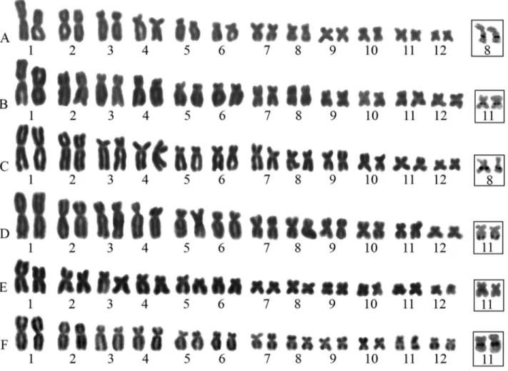
All analyzed species presented a modal diploid num-ber of 2n = 24 and FN = 48. The chromosomal morphology was similar in nearly all species, comprising eight meta-centric pairs (1, 2, 7, 8, 9, 10, 11, and 12) and four sub-metacentric ones (3, 4, 5, and 6). However, pair 7 inS. boesemaniwas submetacentric and pairs 11 and 12 inS. x-signatuswere submetacentric (Figure 1 and Table 2).
Silver nitrate staining showed that NORs were usu-ally located on the long arms of pair 11 (11q), either at an interstitial position (S. campossabrai,S. trilineatusandS. x-signatus) or in the terminal region (S.pachycrus).NORs were also detected at interstitial positions on 8q in S. boesemani and S. garbei. It is worthy of note that the
Ag-NORs were coincident with secondary constrictions observed in Giemsa-stained karyotypes ofS.boesemani,S. campossabraiandS.x- signatus(Figure 1).
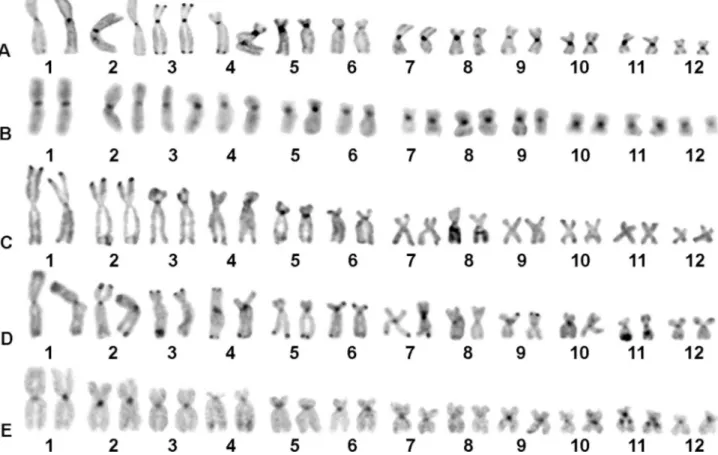
Heterochromatin was visualized at centromeric re-gions of most chromosomes in all species (Figure 2). In ad-dition, some species-specific C-bands represented hetero-chromatin segments at telomeric regions of pairs 3 and 4 in S. boesemani(Figure 2A).S. campossabraiwas character-ized by the presence of exclusively centromeric C-bands in all chromosomes (Figure 2B).S. garbeiandS. pachycrus presented terminal C-bands on arms of pairs 1 and 2 (Figu-re 2C) and pairs 1, 2, 3, 4, 5, 6, 7, and 9 (Figu(Figu-re 2D), (Figu- respec-tively.S. x-signatusalso presented heterochromatin at ter-minal regions of pair 4 (Figure 2E). Heterochromatic blocks interspersed with NORs were observed inS. garbei, S. pachycrusandS. x-signatus(Figure 2C,D,E).
Fluorochrome staining was successful in S. boesemani,S. pachycrusandS. x-signatus. In these species, GC-rich segments (CMA3+ and DAPI-) were coincident
with NORs (Figure 3). In addition, all centromeric regions of S. boesemani were positively stained, following the C-banding pattern (Figure 3A), while fluorescent signals were observed in several centromeres and terminal regions in 3q and 5 of S. pachycrus (Figure 3B) and in most centromeric regions ofS.x-signatus(Figure 3C).
Interestingly, no chromosomal differences could be found among samples ofS. x-signatus, in spite of a high de-gree of isolation by distance of collection sites from distinct biomes (Amazon in Bragança-PA and caatinga in Jequié, BA).
Discussion
We herein provide the first karyotypic data aboutS. boesemani,S. camposseabrai,S. garbei,S. pachycrus,S. trilineatusandS. x-signatus. The present results confirm the modal diploid values of 24 chromosomes and FN = 48 proposed for Scinax and most species in Hylinae (Duellman, 2001; Faivovich, 2002; Kasaharaet al., 2003; Cardozoet al., 2011).
In general, the karyotype formulae are similar among species of theS.ruberclade. Nonetheless, we noted
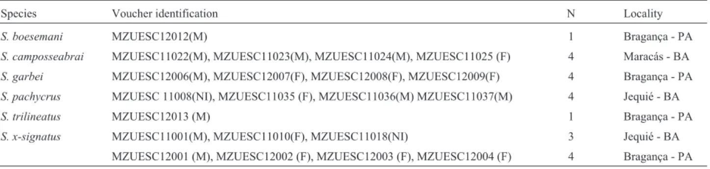
varia-Table 1- Analyzed species, number and sex of specimens (N) and collection site.
Species Voucher identification N Locality
S. boesemani MZUESC12012(M) 1 Bragança - PA
S. camposseabrai MZUESC11022(M), MZUESC11023(M), MZUESC11024(M), MZUESC11025 (F) 4 Maracás - BA
S. garbei MZUESC12006(M), MZUESC12007(F), MZUESC12008(F), MZUESC12009(F) 4 Bragança - PA
S. pachycrus MZUESC 11008(NI), MZUESC11035 (F), MZUESC11036(M) MZUESC11037(M) 4 Jequié - BA
S. trilineatus MZUESC12013 (M) 1 Bragança - PA
S. x-signatus MZUESC11001(M), MZUESC11010(F), MZUESC11018(NI) 3 Jequié - BA
tion in morphology has been reported for pairs 7, 11 and 12 from submetacentric to metacentric (Cardozoet al., 2011), as well as in pair 7 ofS. boesemani(submetacentric) and pairs 11 and 12 (submetacentric) inS. x-signatus(Figure 1 and Table 1). These results suggest that microrearrange-ments such as deletion/duplication of chromosomal seg-ments or inversions may have taken place independently in the abovementioned species.
The results also should prove to be useful for cyto-taxonomy and systematics ofScinaxspecies. Brusquettiet al.(2014) revisedS.fuscomarginatusand related species, suggesting that S. trilineatus is a synonym for S. fuscomarginatus, since the genetic differences in 16S and COI sequences could be associated with geographic dis-tance among populations. Moreover, these authors found no signification differences in vocalization and morpho-metric patterns between the two putative species. On the other hand, we observed chromosomal differences between S. trilineatus(8 metacentric and 4 submetacentric pairs) (Table 2), and S. fuscomarginatus (6 metacentric and 6 submetacentric pairs) (Cardozo et al., 2011). Thus, the
cytogenetic data support the separation of both cryptic spe-cies based on structural rearrangements, such as inversions. The nucleolar organizer regions are also considered efficient markers for the identification of new species (Bruschi et al., 2012) or cryptic forms (Siqueira et al., 2008), as well as to establish infragenus subdivisions (Raber and Carvalho, 2004; Cardozoet al., 2011). In this sense, the Ag-NOR located at 11q is regarded as an ances-tral condition inScinax(Cardozoet al., 2011). In the pres-ent study, two species of the Amazon region (S. boesemani andS. garbei) showed GC-rich NORs on 8q, as confirmed by Ag-NOR, C-banding and fluorochrome staining, sug-gesting a derived feature. An additional nucleolar region in pair 8 was also reported by Cardozoet al.(2011) in another species ofS.ruberclade,Scinax hayii. The two species did not come out as closely related in the phylogenetic analysis carried out by Wienset al.(2010) and, hence, the changes in Ag-NOR location should be regarded as independent events leading to convergent distribution pattern of NORs.
A single NOR-bearing chromosome was visualized inScinax trilineatus, and this may be related to the small size of ribosomal cistrons in one homologous or an inactive
site, since silver nitrate staining can only reveal previously active NORs (Schmid, 1978; Kasahara, 2009). This Ag-NOR pattern is similar to that reported inScinax alterand Scinax hiemalis(Cardozoet al., 2011).
Heterochromatin at centromeric region, as detected in this study, has been commonly reported in species of theS. ruberclade, such asS. acuminatus,S. alter,S. curicica,S. duartei,S. eurydice,S. fuscovarius,S. granulatus,S. hayii, S. nasicus, S. perereca, S. similis and S. squalirostris (Kasaharaet al., 2003; Cardozoet al., 2011). Another
com-mon feature observed in the analyzed species, except forS. camposseabrai, was the presence of heterochromatin inter-spersed with NORs, as described inS. argyreornatus,S. curicica, S. eurydice, S. hiemalis, S. similis and S. squalirostris(Cardozoet al., 2011).
A distinguishable C-banding pattern was observed in S. boesemani,S. garbei,S. pachycrusandS. x-signatus, as these species presented additional heterochromatic seg-ments at terminal regions of some chromosomes. Even though terminal C-bands have been reported in other hylids
Figure 2- C-banded karyotypes ofScinaxspecies: (A)S. boesemani, (B)S. camposseabrai, (C)S. garbei, (D)S. pachycrusand (E)S. x-signatus.
(Businet al., 2006; Kasaharaet al., 2003; Gruber et al., 2012), this is the first description in Scinax. Therefore, C-banding patterns can be potentially used to identify spe-cies in this genus, and more studies based on this technique should be performed withinScinax.
Fluorochrome staining in amphibians usually identify GC-rich regions associated with NORs and more occasion-ally C-bands (Ananiaset al., 2007; Camposet al., 2008). Indeed, NORs and several centromeric regions in the spe-cies anayzed herein were positively stained by CMA3. In
the case ofS.pachycrus, C-bands at telomeric regions of pairs 3 and 5 were also GC-rich, indicating a homogeneous base composition of heterochromatin. However, there are only few studies on CMA3/DAPI staining inScinaxto
pro-vide a reliable scenario of heterochromatin composition.
In conclusion, the present work increased the number of karyotyped species inScinaxby providing the first chro-mosomal data in S. boesemani, S. camposseabrai, S. garbei,S. pachycrus,S. trilineatusandS. x-signatus. Com-parisons with previous reports suggest that chromosomal evolution inScinax(S.ruberclade) may have been mainly driven by microstructural rearrangements and inversions associated with stable karyotype fomulae, particularly among species within a same clade (e.g. S.camposseabrai andS.pachycrus). As indicated by Cardozoet al.(2011), the chromosomal inversions in the cladeS.ruber are re-stricted to pairs 7, 11 and 12. Therefore, banding techniques should prove essential to provide cytotaxonomic markers,
as observed in relation to NOR location of the Amazon spe-cies and heterochromatin distribution.
Acknowledgments
We thank Hoogmoed M. for identifying the biologi-cal material from the Amazon site. Financial support to this work was provided by Fundação de Amparo à Pesquisa do Estado da Bahia (FAPESB: PET 035/2010).
References
Ananias FAL, Bombeiro CDB, Silva APZ, Haddad CFB and Kasahara S (2007) Cytogenetics of Eupemphix nattereri Steindachner, 1863 (Anura, Leiuperidae) and karyotypic similarity with species of related genera: Taxonomic impli-cations. Acta Zool Sin 53:285-293.
Bruschi DP, Busin CV, Siqueira S and Recco-Pimentel SM (2012) Cytogenetic analysis of two species in the Phyllomedusa hypochondrialis group (Anura, Hylidae). Hereditas 40:34-40.
Brusquetti F, Jansen M, Barrio-Amorós C, Segalla M and Had-dad, CFB (2014) Taxonomic review of Scinax fuscomarginatus(Lutz, 1925) and related species (Anura, Hylidae). Zool J Linnean Soc Society 171:783-821. Busin CS, Lima AP, Prado CPA, Strüssmann C, Siqueira S and
Recco-Pimentel MS (2006) Chromosomal differentiation of populations ofLysapsus limellus limellus,L. l. bolivianus, and of Lysapsus caraya (Hylinae, Hylidae). Micron 37:355-362.
Campos JRC, Ananias F, Haddad CFB and Kasahara S (2008) Karyotypic similarity amongBarycholos ternetziand five Table 2- Chromosomal measurements of analyzed species (RL - relative length; CI = centromeric index and CT = chromosomal type) according to Green and Sessions (1991).
Species Chromosomal pairs
1 2 3 4 5 6 7 8 9 10 11 12
S. boesemani RL 12.34 10.71 10.05 8.90 7.51 6.21 5.82 5.63 5.62 4.65 4.21 3.84
CI 0.49 0.41 0.29 0.31 0.25 0.32 0.30 0.42 0.47 0.45 0.47 0.46
CT M M SM SM SM SM SM M M M M M
S. camposseabrai RL 14.42 11.58 10.51 9.67 7.45 7.25 6.61 6.30 5.14 4.71 4.52 4.08
CI 0.46 0.38 0.28 0.31 0.25 0.28 0.41 0.41 0.44 0.46 0.45 0.49
CT M M SM SM SM SM M M M M M M
S. garbei RL 16.17 13.57 11.51 10.55 8.96 8.82 8.81 7.02 6.85 5.56 5.47 3.61
CI 0.46 0.41 0.29 0.33 0.25 0.3 0.38 0.46 0.39 0.48 0.46 0.47
CT M M SM SM SM SM M M M M M M
S. pachycrus RL 11.33 9.48 8.85 8.04 6.66 6.61 5.11 4.80 3.88 3.78 3.55 2.60
CI 0.46 0.39 0.25 0.26 0.26 0.28 0.42 0.45 0.45 0.47 0.4 0.4
CT M M SM SM SM SM M M M M M M
S. trilineatus RL 9.76 8.36 7.18 6.76 5.68 5.29 4.24 4.02 3.63 3.43 3.41 3.02
CI 0.47 0.43 0.31 0.3 0.32 0.28 0.38 0.45 0.44 0.47 0.46 0.49
CT M M SM SM SM SM M M M M M M
S. x-signatus RL 11.77 9.26 8.50 8.41 6.76 6.45 5.45 4.80 4.11 4.06 3.87 3.00
CI 0.48 0.41 0.27 0.35 0.27 0.27 0.41 0.38 0.47 0.42 0.28 0.3
species of the genusEleutherodactylus from southeastern Brazil (Anura, Brachycephalidae). Micron 39:151-159.
Cardozo DE, Leme DM, Bortoleto JF, Catroli GF, Baldo DF, Faivovich J, Kolenc F, Silva APZ, Borteiro C, Haddad CFB et al.(2011) Karyotypic data on 28 species ofScinax (Am-phibia, Anura, Hylidae): Diversity and informative varia-tion. Copeia 2:251-263.
Duellman WE (2001) The Hylid Frogs of Middle America. Soci-ety for the Study of Amphibians and Reptiles, Ithaca, 1180 pp.
Faivovich J (2002) A cladistic analysis of Scinax (Anura, Hylidae). Cladistics 18:367-393.
Faivovich J, Haddad CFB, Garcia PC, Frost DR, Campbell J and Wheeler WC (2005) Systematic review of the frog family Hylidae, with special reference to Hylinae: Phylogenetic analysis and taxonomic revision. Bull Am Mus Nat Hist 294:6-75.
Green DM and Sessions SK (1991) Nomenclature for chromo-somes. In: Green DM and Sessions SK (eds) Amphibian Cytogenetics and Evolution. Academic Press, San Diego, pp. 431-432.
Gruber SL, Zina J, Narimatsu H, Haddad CFB and Kasahara S (2012) Comparative karyotype analysis and chromosome evolution in the genusAplastodiscus. BMC Genet 13:e28.
Howell WM and Black DA (1980) Controlled silver-staining of nucleolus organizer regions with a protective colloidal de-veloper. Experientia 8:1014-1015.
Kasahara S (2009) Introdução à Pesquisa em Citogenética de Vertebrados. Sociedade Brasileira de Genética, Ribeirão Preto, 160 pp.
Kasahara S, Silva AP, Gruber SL and Haddad CFB (2003) Com-parative cytogenetic analysis on four tree frog species (Anura, Hylidae, Hylinae) from Brazil. Cytogenet Genome Res 103:155-162.
Nunes RRA and Fagundes V (2008) Cariótipos de oito espécies de anfíbios das subfamílias Hylinae e Phyllomedusinae (Anura, Hylidae) do Espírito Santo, Brasil. Bol Mus Biol Mello Leitão 23:21-36.
Pombal JJP, Haddad CFB and Kasahara S (1995) A new species ofScinax(Anura, Hylidae) from southeastern Brazil, with comments on the genus. J Herpetol 29:1-6.
Raber SC and Carvalho KA (2004) Chromosomal characteriza-tion ofHyla bischoffiandHyla guentheri(Anura, Hylidae). Phyllomedusa 3:43-49.
Schmid M (1978) Chromosome banding in Amphibia I. Constitu-tive heterochromatin and nucleolus organizer regions in BufoandHyla.Chromosoma 66:361-388.
Schmid M (1980) Chromosome banding in Amphibia. IV. Differ-entiation of GC- and AT-rich chromosome regions in Anura. Chromosoma 77:83-103.
Siqueira S, Aguiar O, Strussmann C, Del-Grande ML and Recco-Pimentel MS (2008) Chromosomal analysis of three Brazil-ian “Eleutherodactyline” frogs (Anura, Terrarana), with suggestion of a new species. Zootaxa 1860:51-59.
Sumner AT (1972) A simple technique for demonstrating centro-meric heterochromatin. Exp Cell Res 75:304-306.
Wiens JJC, Kuczynski CA, Hua X and Moen DS (2010) An ex-panded phylogeny of treefrogs (Hylidae) based on nuclear and mitochondrial sequence data. Mol. Phylogenet Evol 55:871-882.
Internet Resources
Frost DR (2014) Amphibian Species of the World: An Online Reference. Version 6.0. American Museum of Natural His-tory, New York, USA. http://research.amnh.org/herpetol-ogy/amphibia/index.html. Accessed July 24, 2014.
Associate Editor: Yatiyo Yonenaga-Yassuda