ISOLATION OF MICROSPORUM GYPSEUM IN SOIL SAMPLES FROM DIFFERENT GEOGRAPHICAL REGIONS OF BRAZIL, EVALUATION OF THE EXTRACELLULAR PROTEOLYTIC ENZYMES ACTIVITIES (KERATINASE
AND ELASTASE) AND MOLECULAR SEQUENCING OF SELECTED STRAINS
Mauro Cintra Giudice1,3*, Adriana Araújo Reis-Menezes1, Glauce Mary Gomes Rittner1, Adolfo José Mota2, Walderez Gambale1
1
Instituto de Ciências Biomédicas, Departamento de Microbiologia, Universidade de São Paulo, SP, Brasil; 2Laboratório de
Genética Molecular e Genomas, São José dos Campos, SP, Brasil; 3 Laboratório de Bacteriologia, Laboratório de Investigação
Médica/54, Hospital das Clínicas, Faculdade de Medicina, Instituto de Medicina Tropical, Universidade de São Paulo, São Paulo,
SP, Brasil.
Submitted: January 20, 2011; Approved: June 07, 2012.
ABSTRACT
A survey of Microsporum gypseum was conducted in soil samples in different geographical regions of
Brazil. The isolation of dermatophyte from soil samples was performed by hair baiting technique and the
species were identified by morphology studies. We analyzed 692 soil samples and the recuperating rate was
19.2%. The activities of keratinase and elastase were quantitatively performed in 138 samples. The
sequencing of the ITS region of rDNA was performed in representatives samples. M. gypseum isolates
showed significant quantitative differences in the expression of both keratinase and elastase, but no
significant correlation was observed between these enzymes. The sequencing of the representative samples
revealed the presence of two teleomorphic species of M. gypseum (Arthroderma gypseum and A.
incurvatum). The enzymatic activities may play an important role in the pathogenicity and a probable
adaptation of this fungus to the animal parasitism. Using the phenotypical and molecular analysis, the
Microsporum identification and their teleomorphic states will provide a useful and reliable identification
system.
Key words: Microsporum gypseum; Brazil; Elastase; Keratinase; Sequencing.
INTRODUCTION
Microsporum gypseum is a geophilic dermatophyte, and in
this group, is the main representative that affects humans and
other animals (4, 21). The human infections are rare which may
suggest a natural resistance to this infection or limited
mechanisms of virulence of the fungus (12), furthermore, some
outbreaks have been reported in different parts of the world
(21). Atypical clinical manifestations, refractory to either topic
or systemic treatments have already been described in literature
in patients HIV positive, epidemiologically related to the
source of geophilic infection (10, 11, 24).
*Corresponding Author. Mailing address: Bacteriology Laboratory – LIM 54/ HCFMUSP and Institute of Tropical Medicine, University of São Paulo, São Paulo, SP, Brazil, Av. Dr. Enéas de Carvalho Aguiar, 470 São Paulo, SP. CEP 05403000, Brazil.; Tel.: +55-11-3061-7030 Fax: +55-11-30617043.; E-mail:
Giudice, M.C. et al. M. gypseum evaluation of the extracellular proteolytic enzymes
The isolation of M. gypseum and other geophilic
dermatophytes from soil can be obtained through the
hair-baiting technique (Vambreuseghem method) (19). Several
reports describe the M. gypseum on soils with distinct
characteristics in different parts of the world (9, 10, 16, 25, 31,
41, 44).
The dermatophytes are capable of producing enzymes as a
mechanism to degrade various substrates. Several enzymes
have been described and eventually correlated with
dermatophytes virulence (28, 37), like the keratinase (2, 14, 20,
26, 39, 42, 45), elastase (2, 18, 33, 37, 42), DNase (22, 42) and
collagenase (17, 23, 30, 32). The keratinase seems to have a
direct relation to the pathogenicity (13, 41). The elastase has an
important role in the pathogenicity of other fungi such as
Aspergillus fumigatus (8, 20, 39) and also, probably, to the
dermatophytes (14, 17, 30).
The objectives of this study were evaluate the frequency
of M. gypseum in Brazil’s soil, demonstrate quantitatively the
activities of both keratinase and elastase from these isolates,
and identify representative samples through the sequence to
examine the distribution of the teleomorphic states.
MATERIAL AND METHODS
Collection of soil samples and fungal identification
Samples of soil from different states of Brazil were
collected in the cities’public places (parks, squares, bus-stops
etc.) and at peripheral regions with a spatula and transferred to
a plastic bag. The fungi were isolated from the soil with the
Vambreuseghem technique (19) .Macroscopic and microscopic
characterization, through giant colony and microculture
techniques, respectively were performed (19).
Determination of the strain enzymatic profile
The quantitative evaluation for both enzymes was made in
line with the procedures previously described, with some
modifications (42,43).The M. gypseum samples were cultivated
in Sabouraud dextrose broth (Oxoid) at 25ºC for 15 days. The
conidia were harvested in sterile saline, and using a Newbauer
Chamber, the conidial suspension was adjusted to 1.0 x 10⁶
conidia/ml.
Keratinase: One milliliter of suspension was inoculated
in 50 ml of basal medium (6 g MgSO4, 1.0 ml Protovit vitamin
mixture (Roche, São Paulo, Brazil), 0,111 g CaCl2, in 1000 ml
distilled water) with 3 g keratin powder from hooves and
horns (ICN, Montreal, Canada) and incubated at 25º for 28
days in the dark (36,43). One milliliter of 1500 g centrifuged
culture supernatant was then mixed with 2 ml of buffer 200
mM Tris-HCI, CaCl2 100 mM, pH 8.0 plus 10 mg of Keratin
Azure (Sigma, St. Louis, MO, USA) plus 2.0 mL of and
afterwards incubated at 37 ºC for 24 h. Substrate degradation
was measured in a spectrophotometer at 595 nm using
uninoculated substrate buffer as a negative control. A
keratinase unit (UK) was defined as an increase of 0.01 in the
absorbance, when compared to the control.
Elastase: One milliliter of adjusted suspension was
inoculated in 50 mL of the same basal medium with 3.0 g of
elastin (Sigma St. Louis, MO, USA) and incubated at 25ºC for
28 days in the dark (43). Two milliliters of 1500 g centrifuged
culture supernatant was then mixed with 2.0 mL of phosphate
buffer (10 mM pH, 7.0) with 20 mg of Elastin Congo-Red
(Sigma St. Louis, MO, USA) was incubated at 37ºC for 2
hours. The elastin degradation was evaluated by
spectrophotometer in 495 nm wavelength. A unit of elastase
(UE) was defined as the 0.1 increase in the absorption reading,
when compared to the control.
DNA extraction
Extraction protocol was modified from Bir et al., 1995 (5),
with some modifications. M. gypseum samples were incubated
in Sabouraud broth medium for 14 days at 25ºC and the
mycelia was powdered with liquid nitrogen and 3.0 mL of 10
mM phosphate buffer pH 6.0. Volumes of 20 µL chitinase
(5U/mL) (Sigma) were added and it was incubated for 90
minutes at 25ºC temperature, shaking it many times. Equal
volume of lysis buffer (50 mM Tris HCl, 50 mM EDTA, 3%
SDS, 1% β-mercaptoethanol, pH 7.2) was added and the
suspension was incubated at 55ºC for one hour, shaking it
many times. The products were then subjected to heat
Extraction with phenol:chloroform:isoamylalcohol (25:24:1)
was performed and the DNA was precipitated with isopropyl
alcohol at -20ºC for about 18 hours. DNA was washed with
70% ethanol and it was dried at environment temperature,
rehydrated in 25 µL of TE buffer (10 mM Tris-HCl, 1 mM
EDTA, pH 8.0) with RNase (10 µg/mL) and it was incubated
in a dry bath for 30 minutes at 37ºC. The product was again
drawn up with isoamilic phenol-chloroform-alcohol and the
DNA was dissolved in 50 µL of sterile ultrapure water. The
DNA concentration was determined spectrophotometrically.
Quality of the DNA was evaluated by agarose 0.8 % in TAE
buffer (1X) gel electrophoresis and stained with ethidium
bromide. The visualization for confirmation of the extraction
was done in the chamber of the GEL DOC 1000 equipment
under ultraviolet rays (Bio-Rad).
PCR amplification
The amplification of the conserved regions of rDNA
(ITS-PCR Internal Transcribed Spacers - Primer Chain Reaction)
was performed with a pair of primes ITS1 (5’
TCCGTAGGTGAACCTGCGG 3’), and ITS 4 (5’
TCCTCCGCTTATTGATATGC 3’) (6).
The mixture for a PCR was prepared in total volume of 25
µL containing the reaction buffer (10 mM Tris-HCl in pH 8.0,
50 mM of KCl, 1.5 mM of MgCl2 10 mM of each DNTP
(Fermentas, USA), 0.125 µM of each primer, 2 U of Taq
Polimerase and 50 ng of DNA). The amplification was
performed in thermocycler (PTC – 100, MJ Research, INC.) on
the following conditions: 94ºC, 5 min (94ºC 1 min, 56ºC 30 s,
72ºC 1 min.) per 25 cycles and final extension at 72º 5 min.
Sequencing
Five specimens (three from Rondônia and two from São
Paulo states) were arbitrarily selected to the sequencing. These
isolates were chosen because they represent respectively the
most sampled state (RO) and state where we have had the
highest recuperating rate (SP) of Microsporum gypseum . After
amplification, the PCR products were purified using QIAquick
Spin Columns (Qiagen Corp.., Chatsworth, Calif.), and were
reconstituted with 30 µL of distilled water. The sequencing
was carried out in platforms for dna sequencing PDTIS, which
we used Big Dye reagent (Applied Biosystem, Foster City CA,
USA) and automated sequencer Applied Biosystems ABI
Prism 3730 (Applied Biosystem, Foster CA, USA).. The
sequences were submitted to a search on the BLASTn by using
the databank of the National Center of Biotechnology (NCBI;
http://www.ncbi.nlm.nih.gov/BLAST).
RESULTS
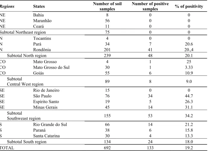
The soil samples were collected from all Brazilian
regions. The largest Microsporum gypseum positivity
percentual was obtained from the samples originated in the
Southeast (SE), followed by the North (N), Southern (S) and
Center-West (CW). In Northeast region, in the states evaluated,
there was no isolation of M. gypseum (Table 1, Fig. 1). The
number of soil samples collected was differed in each region,
as well as the positivity percentage. In total, 692 soil samples
were analyzed and the recuperating rate was 19.2%. In
samples from some states it was not possible to obtain growth
of M. gypseum (Bahia, Tocantins, Maranhão, Ceará and Rio de
Janeiro). The state of São Paulo demonstrated larger fungus
recuperation from the soil samples, proportionally, with a
recuperating rate of 44.7%. The state of Rondônia contributed
with the largest number of soil samples, but the recuperating
rate was only 20.4% (Table 1, Fig. 1).
The evaluation of both the elastinolytic and keratinolytic
activities was performed in Microsporum gypseum (n=121)
isolated from the soil from different states. The means and the
standard deviations of the two enzymatic activities (UK and
UE) were 6.90 ± 8.48 to UK and 1.60 ± 2.20 to UE. The
median were 5.0 UK and 0.92 UE
The results of absolute and relative frequencies from both
enzymatic activities for 121 samples of M. gypseum, are
showed on Fig. 2 (a and b).
The Spearman’s coefficient of correlation from two
enzymatic activities (UK and UE) was very weak. In soils
Giudice, M.C. et al. M. gypseum evaluation of the extracellular proteolytic enzymes
negative, which suggests the absence of correlation among the
enzymatic activities.
The sequencing of the representative samples revealed the
presence of two teleomorphic species of M. gypseum
(Arthroderma gypseum and A. incurvatum) and A. incurvatum
was identified in only one of the isolated.
Table 1. Distribution of the number of analyzed samples of soil, absolute and relative frequencies of recuperation by
administrative region and by States of the Federation of Brazil.
Regions States Number of soil
samples
Number of positive
samples % of positivity
NE Bahia 8 0 0
NE Maranhão 56 0 0
NE Ceará 11 0 0
Subtotal Northeast region 75 0 0
N Tocantins 4 0 0
N Pará 34 7 20.6
N Rondônia 201 41 20.,4
Subtotal North region 239 48 20.1
CO Mato Grosso 4 1 25
CO Mato Grosso do Sul 30 1 3.33
CO Goiás 55 6 10.9
Subtotal
Central West region 89 8 9.0
SE Rio de Janeiro 15 0 0
SE São Paulo 76 34 44.7
SE Espírito Santo 19 5 26.3
SE Minas Gerais 45 14 31.1
Subtotal
Southweast region 155 53 34.2
S Rio Grande do Sul 66 14 21.2
S Paraná 38 6 15.8
S Santa Catarina 30 4 13.3
Subtotal South region 134 24 18.0
TOTAL 692 133 19.2
Figure 2. Percentual distribution of elastinolytic activity (UE - unit of elastase) (a) and keratinolytic activity (UK - unit of
keratinase) (b), in Microsporum gypseum strains (n=121) isolated from soils of different geographical regions of Brazil.
DISCUSSION
The isolation of M. gypseum can be obtained from human
clinic sources, as well as from veterinarian or from the soil.
The recuperating rate of this dermatophyte in clinic cases is
very limited (21). However, reports of outbreaks, extensive
cases and refractory therapeutics are already presented by the
literature (10, 21). In this research, 692 samples of soil of
different Brazilian regions were evaluated and the recuperating
rate was 19.2%. Microsporum gypseum was isolated in almost
all sampled regions, except for the Northeast. The great
majority of the samples were collected both in urban
environments and public parks places (parks and squares), at
random and in different times of the year. The absence of
homogeneity in the sampled areas and in the collecting period
makes it difficult to compare the analyzed areas.
In the Northeast, a very extensive geographic area, which
comprises different type of soil and population distribution
variable, a more thorough study, must be done studying soil
Giudice, M.C. et al. M. gypseum evaluation of the extracellular proteolytic enzymes
local environmental variables.
Some studies demonstrate that the recuperation of this
fungus can change with the sampled environment. When one
evaluates either public areas of both parks or public squares or
areas of confinement of animals (breeding, slaughtering and
zoos) the isolating rates may be very high, differently from
those found in closed environments (domestic or hospital) (4,
9, 16, 21, 25, 31, 41, 44).
Both the synthesis and the enzymes secretion are
important metabolic activities in filamentous fungi and their
production is an indispensable condition to the fungi’s
development (1). Proteolytic enzymes, such as the keratinases,
collagenases and elastases are important in various processes,
some of that are implicated in the pathogenesis of fungal
diseases, thereby causing damages to the host’s tissue (2, 14,
34, 42).
The dermatophytes can produce various proteolytic
enzymes and their lythic activities on different substrates, have
already been described (3, 7, 8, 15, 17, 27, 29, 33, 36, 40, 42).
The keratinase, among other enzymes, was considered as a
virulence factor, which correlated with clinic forms of
dermatophytosis (13).
In M. gypseum, T. mentagrophytes, T. verrucosum,
Muhsin et al. (27) demonstrated the presence of elastase.
However, the elastinolytic activity was not observed in strains
of M. canis.
The data obtained from this work refer to the first report
in the literature on hydrolytic enzymes activities in samples of
Microsporum gypseum isolated from Brazilians soil samples,
This research was made to evaluate the degree of
correlation as well as the description of both keratinase and
elastase activities. The results suggested that the expressions of
these enzymes, which are produced by M. gypseum, occur in
samples isolated from environmental fonts.
The mean and median values obtained were higher in the
keratinolytic activity than in the elastinolytic one.
In strains isolated from soil originated in 16 Brazil’s
different geographic regions, the larger intensity of
keratinolytic activity occurred in one samples (sample 19, with
77.7 UK) and the same occurred to the elastinolytic activity
(sample 26, with 12.85 UE), both recuperated in São Paulo
state, but from different places. The correlation grade,
however, was both low and negative between the two
enzymatic activities evaluated.
More detailed studies with the samples that showed high
grade of enzymatic activities and with other samples with low
enzymatic activities must be performed, including both
experimental infection and evaluation of the tissue
compromise, with the intention of evaluating the host’s
compromising degree with the fungus and their metabolites.
In a previous study, which was performed to
quantitatively evaluate both the keratinolytic and elastinolytic
activities in M. canis, strains isolated from animals with and
without clinic lesion, the results showed statistically significant
differences, suggesting that the elastase like the keratinase, can
influence the tissue reactions in the dermatophytosis (6).
There was no correlation among the strains with the
production of each enzyme, suggesting that there is not only a
proteinase with keratinolytic, elastinolytic activities, but
specific enzymes to each substrate. Brouta et al. (7)
demonstrated that two described proteases can exert activity on
the keratin, the elastin and collagen by suggesting that these
two proteases should be responsible for the observed
keratinase, elastase and collagenase activities.
The differentiation among six species of the
Microsporum, based on the characterization of ITS of rDNA
region, was evaluated, demonstrating to be sufficient to
separate them (35, 38) and, despite the fact that the
Microsporum gypseum is endemic in different parts of the
world, few studies of molecular analysis have been performed
in this specie.
Molecular methods based on the rDNA amplification and
associated with the enzymatic restriction have been used both
to differentiate species and to biotype microorganisms (14, 35).
The use of different restriction enzymes has afforded results
which, when associated with phenotypic characteristics, can
help the identification of isolated ones (7).
species has often been used. Sharma et al. (35), when using the
RFLP sequencing of the ITS region of rDNA plus phenotypic
described a new taxon, Microsporum appendiculatum which is
highly related to M. gypseum.
In our study, when evaluated the sequencing, a high
identity was noted, showing that the ITS region is highly
preserved in this specie. The sequencing revealed the presence
of two teleomorphic species of M. gypseum (Arthroderma
gypseum and A. incurvatum). Despite the fact that only few
samples have been sequenced, A. incurvatum was identified in
one of the isolated. The obtaining of the teleomorphic stage in
vitro and the characterization of the species are very time
consuming and, in the great majority, the results depend upon a
very accurate morphological evaluation. Therefore the
molecular methods of sequencing are promising.
In spite of the fact that the asexual reproduction among
the dermatophytes is much more frequent than the sexual
reproduction, constituting their principal way propagation, the
molecular methods utilized here are still limited to characterize
differences in these isolated and, possibly, the expression of the
enzymatic activities must be related to genotypes not yet
characterized by the techniques used in this study.
Geophilic dermatophytes are very common in Brazil soils
and the relationship between the characterization based on
morphological, biochemical and molecular aspects seems to be
very important in demonstrate the real role in its pathogenicity.
However, this fungal and its biological relationship requires to
be better clarified once these fungi are found as human and
veterinarian pathogens causing distinct clinic manifestations
that are sometimes refractory to the available treatments at
present.
ACKNOWLEDGEMENTS
We are grateful to the financial Foundation of Research
Support of São Paulo – Program for Centers of
(FAPESP-PRONEX), National Council for Scientific and Technological
(CNPq) and the group of Biomedical Center of University of
São Paulo in Monte Negro (RO), Brazil.
REFERENCES
1. Alvarez, D.P.; Luque, A.G.; Bracalenti, B.C. (1986). La influencia de factores ecológicos in el aislamento de dermatofitos queratinolíticos y geofílicos. Rev. Latinoam. Microbiol. 28, 351-354.
2. Apodaca, G; Mckerrow J.H. (1989). Purification and characterization of a 27,000 – Mr extracelullar proteinase from Trichophyton rubrum.
Infect. Immun. 57(10), 3072-3080.
3. Aubaid, A.H.; Muhsin, T.M. (1998). Partial purification and kinetic studies of exocellular proteinase from Trichophyton mentagrophytes var.
erinacei. Mycoses. 41, 163168.
4. Bársena-Asensio, .M.C.; Cabo, J.F.G. (1996). Ecología de los dermatofitos. Rev. Iberoam. Micol. 13(2), 47-54.
5. Bir, N.; Paliwal, A.; Muralidhar, K.; Reddy, P.; Usha-Sarma, P. (1995) A rapid method for the isolation of genomic DNA from Aspergillus
fumigatus. Prep. Biochem. 25(4), 171-181.
6. Blanz, P.; Buzia, W.; Ginter, G.; Gräser, Y. (2000). Molekularbiologische methoden und ihre konsequenzen für taxonomie und diagnostik dei dermatophyten. Mycoses. 43(suppl. 1), 11-16. 7. Brouta, F.; Descamps, F.; Feti, T., et al. (2001). Purification and
characterization of a 43.5 kDa keratinolytic metalloprotease from
Microsporum canis. Med. Mycol. 39, 269-275.
8. Chattaway, F.W.; Ellls, D.A.; Barlow, E.J. A.( 1963). Peptidases of Dermatophytes. The J. Invest. Dermatol. 41(1), 31-37.
9. Deshmukh, S.K. (2004). Isolation of dermatophytes and other keratinophilic fungi from the vicinity of salt pan soils of Mumbai, India.
Mycopathologia.157:265–267.
10. Fernandes, N.C.; Lamy, F.; Akiti, T.; Da Barreiros, M.G.C. (1998).
Microsporum gypseum infection in AIDS patient: a case report. An.
Bras. Dermatol. 73, 39-41.
11. Giudice, M.C.; Szeszs, M.W,; Scarpini, R.L. et al.(1997). Clinical and epidemiological study in an AIDS patient case with Microsporum
gypseum infection. Rev. Iberoam. Micol. 14(4), 184-187.
12. Gordon, M.A.; Perrin, N.; Little, G.N. (1967). Differences in pathogencit between M. gypseum and M. fulvum. Sabouraudia .5,366-390.
13. Grappel, S.F.; Blank, F. Role of keratinase in dermatophytosis. (1972).
Dermatologica. 145, 245-255.
14. Gupta, R.; Ramnani, P. (2006) Microbial keratinases and their prospective applications: an overview. Appl. Microbiol. Biotechnol. 70, 21-33.
15. Hamaguchi, T.; Morishita, N.; Usui, R.; Takiuchi, I. (2000). Characterization of an extracellular keratinase from Microsporum canis.
Japanese J. Med. Mycol. 41, 257-262.
16. Hedayat, M.T.; Mohseni-Bandpi, A.; Moradi, S. ( 2004). A survey on the pathogenic fungi in soil samples of potted plants from Sari hospitals, Iran. J. Hosp. Infect. 58 (1), 59-62.
Giudice, M.C. et al. M. gypseum evaluation of the extracellular proteolytic enzymes
Expression of PZ-peptidases by cultures of several pathogenic fungi. Purification and characterization of a collagenase from Trichophyton
schoenleinii. J.Med. Vet. Mycol. 34, 83-90.
18. Kothary, M.H.; Chase, T.; Macmillan, J.D. (1984) Correlation of elastase production by strain of Aspergillus fumigatus with ability to cause pulmonary invasive aspergillosis in mice. Infect. Immun.43(1), 320-325.
19. Lacaz, C.S.; Porto, E.; Martins, J.E.C.; Heins-Vaccari, E.M.;, Melo, N.T. (2002) Tratado de Micologia Médica. 9th ed. Sarvier, São Paulo, 2002. 20. Lee, K.H.; Park, K.K.; Park, S.H.; Lee, J.B. (1987). Isolation,
purification and characterization of keratinolytic proteinase from
Microsporum canis. Yonsei Med. J. 28(2), 131-138.
21. Londero, A.T.; Ramos, C.D. (1989). Agentes de dermatofitoses humanas no interior do Estado do Rio Grande do Sul, no período de 1960-1987. An. Bras. Dermatol. 64(3), 161-164.
22. Lópes-Martinez, R.; Manzano-Gayosso, P.; Mier, T.; Mendez-Tovar, L.J.; Hernández-Hernández, F. (1994). Exoenzimas de dermatofitos aislados de tiñas agudas y crônicas. Rev. Latinoam. Microbiol. 36, 17-20.
23. Lupan, D.M.; Nziramasanga, P. (1986). Collagenolytic Activity of
Coccidioides immitis. Infect. Immun. 51(1), 360-361.
24. Luque, A.G.; Biasoli, M.S.; Ortino, M.A.; Lupo, S.H.; Bussy, R.F. (2001). Atypical tinea corporis caused by Microsporum gypseum in a subject with acquired immune deficiency syndrome. J. Eur. Acad.
Dermatol. Venereol.15, 374-375.
25. Mercantini, R.; Marsella, R.; Cervellati, M.C. (1989).Keratinophylic fungi isolated from Antartic soil. Mycophatologia.106, 47-52.
26. Mignon, B.R.; Nikkles, A.F.; Piérard, G.E.; Losson, B.J. (1998). The in vitro and in vivo production of a 31.5 kDa keratinolitic subtilase from
Microsporum canis and the clinical status in naturally infected cats.
Dermatology.196, 438-441.
27. Muhsin, T.M.; Aubaid, A.H.; Al-Duboon, A.H. (1997). Extracellular enzyme activities of dermatophytes and yeast isolates on solid media.
Mycoses. 40, 465-469.
28. Muhsin, T.M.; Salih, T.H. (2001). Exocellular enzyme activity of dermatophytes and other fungi isolated from ruminants in Southern Iraq.
Mycopathologia.150(2), 49-52.
29. Okafor, J.; Ngwogu, A. (2000). Keratinolytic activity of five human isolates of dermatophytes. J. Commun. Dis. 32(4), 300-305.
30. Okeke, C.N.; Müller, J. (1991). Production of extracellelar collagenolytic proteinases by Histoplasma capsulatum var. duboisii and
Histoplasma capsulatum var. capsulatum in the yeast phase. Mycoses.
34, 453-460.
31. Periasamy, A.; Hilda, A.; Gopinath, S.C.B. (2004). Keratinophilic fungi
of poultry farm and feather dumping soil in Tamil Nadu, India.
Mycopathologia.158, 303–309.
32. Rippon, J.W.; Varadi, D.P. (1968). The elastases of pathogenic fungi and actinomycetes. The J. Invest. Dermatol. 50(1), 54-58.
33. Rippon, J.W. (1967). Elastase production by ringworm fungi.
Science.157, 947.
34. Scott, J.A,.; Untereiner, W.A. (2004). Determination of keratin degradation by fungi using keratin azure. Med. Mycol. 42, 239-246. 35. Sharma, R.; Rajak, R.C.; Pandey, A.K.; Gräser, Y. (2006)Internal
Transcribed Spacer (ITS) of rDNA of appendaged and non-appendaged strains of Microsporum gypseum reveals Microsporum appendiculatum
as its synonym. Antonie van Leeuwenhoek. 89(1), 197-202.
36. Siesenop, U.; Bohm, K. H. (1995). Comparative studies on keratinase production of Trichophyton mentagrophytes strains of animal origin.
Mycoses.38, 205-209.
37. Simpanya, M.F.; Baxter, M. (1996). Multiple proteinases from two
Microsporum species. J. Med. Vet. Mycol. 34, 31-36.
38. Summerbel, R.C.; Moore, M.K.; Starink-Willemse, M.; Van Iperen, A. (2007) ITS barcodes for Trichophyton tonsurans and T. Equinum. Med.
Mycol. 45, 193-200.
39. Takiuchi, I.; Higuchi, D.; Sei, Y.; Koga, M. (1982). Isolation of an extracellular proteinase (keratinase) from Microsporum canis.
Sabouraudia. 20, 281-288.
40. Takiuchi, I.; Youshihiro, S.; Hisae, T.; Makoto, N. (1984). Partial characterization of the extracelular keratinase from Microsporum canis.
Sabouraudia. 22(3), 219 - 224.
41. Ulfig, K.; Plaza, G.; Sztyler, A.; Bronder, J.; Terakowiski, M.; Guarro, J. (2000) General assessment of the influence of a municipal landfill site and environmental factors on the occurrence of keratinolytic fungi in soil. Rocz. Panstw. Zakl. Hig. 51(2), 167-81.
42. Viani, F.C.; Dos Santos, J.I.; Paula, C.R.; Larsson, C.E.; Gambale, W. (2001) Production of extracellular enzymes by Microsporum canis and their role in its virulence. Med. Mycol. 39 (5), 463-468.
43. Viani, F.C.; Viani, P.R.C.; Rivera, I.N.G.; Silva, E.G.; Paula, C.R.; Gambale, W. (2007). Extracellular proteolytic activity and molecular analysis of Microsporum canis strains isolated from symptomatic and asymptomatic cats. Rev. Iberoam. Micol. 24(1), 19-23.
44. Vidyasagar, G.M.; Narayan, H.; Shivkumar, D. (2005) Keratinophilic fungi isolated from hospital dust and soils of public places at Gulbarga, India. Mycopathologia.159, 13–21.
45. Yu, R.J.; Harmon, S.R, (1968). Isolation and purification of an extracellular keratinase of Trichophyton mentagrophytes. J. Bacteriol.
96(4), 1435-1436.