online | memorias.ioc.fiocruz.br
Reduced susceptibility to vancomycin and biofilm formation in
methicillin-resistant Staphylococcus epidermidis
isolated from blood cultures
Luiza Pinheiro/+, Carla Ivo Brito†, Valéria Cataneli Pereira, Adilson de Oliveira, Carlos Henrique Camargo, Maria de Lourdes Ribeiro de Souza da Cunha
Laboratório de Microbiologia, Departamento de Microbiologia e Imunologia, Instituto de Biociências de Botucatu, Universidade Estadual Paulista, Botucatu, SP, Brasil
This study aimed to correlate the presence of ica genes, biofilm formation and antimicrobial resistance in 107 strains of Staphylococcus epidermidis isolated from blood cultures. The isolates were analysed to determine their methicillin resistance, staphylococcal cassette chromosome mec (SCCmec) type, ica genes and biofilm formation and the vancomycin minimum inhibitory concentration (MIC) was measured for isolates and subpopulations grow-ing on vancomycin screen agar. The mecA gene was detected in 81.3% of the S. epidermidis isolated and 48.2% carried SCCmec type III. The complete icaADBC operon was observed in 38.3% of the isolates; of these, 58.5% produced a biofilm. Furthermore, 47.7% of the isolates grew on vancomycin screen agar, with an increase in the
MIC in 75.9% of the isolates. Determination of the MIC of subpopulations revealed that 64.7% had an MIC ≥ 4 μg
mL-1, including 15.7% with an MIC of 8 μg mL-1 and 2% with an MIC of 16 μg mL-1. The presence of the icaADBC operon, biofilm production and reduced susceptibility to vancomycin were associated with methicillin resistance. This study reveals a high level of methicillin resistance, biofilm formation and reduced susceptibility to vancomycin in subpopulations of S. epidermidis. These findings may explain the selection of multidrug-resistant isolates in hos-pital settings and the consequent failure of antimicrobial treatment.
Key words: Staphylococcus epidermidis - icaADBC - mecA - SCCmec - vancomycin - MIC
Staphylococcus epidermidis, a member of the coag-ulase-negative staphylococci (CoNS) group, is the main bacterium found on human skin and causative agent of medical device-associated infections. The contamination of prostheses and intravenous devices with this patho-gen is related to the production of a virulence factor, which represents an important pathogenic mechanism of implant infections. This substance, known as slime or biofilm, permits microorganisms to adhere to different materials (Costerton et al. 1995).
Biofilm formation is a process that involves micro-bial adhesion to and colonisation of a surface, cell pro-liferation and accumulation in multilayers, maturation and, finally, biofilm detachment and the release of cells (Houston et al. 2011). In addition to non-specific interac-tions such as electrostatic and hydrophobic interacinterac-tions, specific adhesins, including two staphylococcal surface proteins (SSP-1 and SSP-2), are involved in the initial attachment to the polymer surface. When a certain ma-terial is implanted into an individual, the components of body fluids, such as serum proteins and platelets, start
doi: 10.1590/0074-0276140120
Financial support: FAPESP (2010/18720-1), CNPq † In memoriam
+ Corresponding author: [email protected] Received 6 April 2014
Accepted 10 September 2014
to cover the catheter or implant, modifying its surface properties and facilitating bacterial adhesion (Patti et al. 1994, von Eiff et al. 2002). S. epidermidis and Staphy-lococcus aureus express dozens of proteins on their surfaces. These proteins, called microbial surface com-ponents recognising adhesive matrix molecules, specifi-cally bind to extracellular matrix proteins of the host, such as fibrinogen, collagen, fibronectin and vitronectin (Patti et al. 1994, von Eiff et al. 2002).
One of the main steps of biofilm formation is the production of polysaccharide intercellular adhesin (PIA), which is responsible for intercellular adhesion and the accumulation of cells in multilayers. The pro-duction of PIA is mediated by the products of the chro-mosomal intercellular adhesion genes (ica), which are organised into an operon that contains icaADBC genes, responsible for biosynthesis, and the icaR gene, which exerts a regulatory function (Otto 2008). PIA is synthe-sised from UDP-N-acetylglucosamine by N-acetylglu-cosamine transferase, an enzyme encoded by icaA and
In addition to PIA, adhesive proteins such as accu-mulation-associated protein and biofilm-associated pro-tein (Bhp in S. epidermidis and Bap in S. aureus) may contribute to biofilm formation (Rupp et al. 2001, Von Eiff et al. 2002), even in the absence of the icaADCB
operon. However, catheter-related and other nosocomial infections caused by biofilm-producing S. epidermidis
are related to the presence of the ica operon, which is the main factor responsible for biofilm formation in this species (Cafiso et al. 2004).
The bacteria present inside a biofilm are protected against the action of the host immune system and an-timicrobial drugs, thus permitting their survival (Mah & O’Toole 2001, Donlan & Costerton 2002). Biofilm-associated bacteria are usually less susceptible to antibi-otics than planktonic bacteria; this can be explained by different mechanisms, such as the binding of antibiotics to biofilm components, reduced penetration of the anti-biotic, slower growth of the microorganisms in the bio-film, high bacterial density and altered gene expression in the bacteria present in the biofilm (Stewart & Coster-ton 2001, Singh et al. 2010).
Oxacillin, one of the antibiotics most commonly used in Brazil for the treatment of staphylococcal infections, is no longer effective because of the high prevalence of resistant strains. Resistance to methicillin is generally conferred by the mecA gene, which produces a peni-cillin-binding protein (PBP2a or PBP 2’) with reduced affinity for beta-lactam antibiotics when compared to other PBPs (Chambers et al. 1985).
The mecA gene is carried by a mobile genetic element, staphylococcal cassette chromosome mec (SCCmec). Eleven different types of SCCmec have been identified in S. aureus (I-XI) (sccmec.org); however, types VI, VII, IX-XI have not yet been described in CoNS (Zong et al. 2011, Vitali et al. 2014). SCCmec elements are more di-verse in methicillin-resistant-CoNS (MR-CoNS) and new variants of the ccr genes continue to be identified, which cannot be typed with the currently available schemes. Thus, classification schemes of SCCmec in MR-CoNS are needed (Zong et al. 2011). SCCmec types III-V are prevalent in MR-CoNS (Zong et al. 2011). Type IV is the smallest, which reduces the cost of transfer between strains and selectively favours this type (Ito et al. 2001).
The high prevalence of methicillin resistance has led to the use of toxic antibiotics, such as the glycopeptide vancomycin for the treatment of Gram-positive infec-tions. However, intermediate resistance to this drug was described in Staphylococcus haemolyticus in the 1980s (Schwalbe et al. 1987) and two strains (1 each of
S. epidermidis and S. haemolyticus) with intermediate vancomycin resistance were found in Brazil in 1996 (Del’Alamo et al. 1999). This is a matter of concern be-cause few other options are currently available for the treatment of staphylococcal infections.
In view of the importance of biofilm formation and antimicrobial resistance in infections caused by S. epidermidis, the objective of the present study was to characterise S. epidermidis strains isolated from blood cultures of patients hospitalised at a Brazilian teaching hospital with regard to their methicillin resistance, SC-Cmec type, reduced susceptibility to vancomycin, pres-ence of the ica genes and biofilm formation.
SUBJECTS, MATERIALS AND METHODS
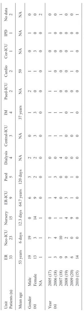
A total of 107 S. epidermidis strains isolated from blood cultures of patients hospitalised at a teaching hos-pital at Botucatu Medical School (FMB), São Paulo State University (UNESP), state of São Paulo, Brazil, between 2005-2010 were studied. The University Hospital of FMB provides tertiary care and possesses 385 beds, including 52 in intensive care units (ICUs) (30 adult, 15 neonatal and 7 paediatric ICU beds). The isolates are stored in the Cul-ture Collection of the Department of Microbiology and Immunology, Botucatu Institute of Biosciences, UNESP, and were obtained from adult and paediatric patients of both genders hospitalised in different units of the hospi-tal. Table I describes the characteristics of the patients and the year of S. epidermidis isolation. The criteria pro-posed by the Centers for Disease Control and Prevention/ National Healthcare Safety Network (CDC/NHSN 2014) were used to determine the inclusion of blood cultures.
Species of the genus Staphylococcus were isolated and identified as described by Baker (1984) and Kone-man et al. (1997). The simplified method proposed by Cunha et al. (2004) was used for strain identification. DNA was extracted from isolates identified as S. epider-midis using the Illustra® kit (GE Healthcare).
Amplifica-tion of the internal transcribed spacer region-polymerase chain reaction (PCR), as described by Barry et al. (1991) and Couto et al. (2001), was used to confirm that the isolates belonged to the species S. epidermidis.
The disk diffusion test employing oxacillin (1 μg) and cefoxitin (30 μg) disks was used for the phenotypic
detection of methicillin resistance according to the rec-ommendations of the Clinical and Laboratory Standards Institute (CLSI 2007, 2012). For genotypic analysis, PCR was used to detect the mecA gene (Murakami et al. 1991). International reference strains were included in all reac-tions as positive (S.aureus ATCC 33591) and negative (S. aureus ATCC 25923) controls, according to the CLSI (2012). The strains that were positive for the mecA gene were subjected to SCCmec typing by multiplex PCR, ac-cording to Machado et al. (2007).
The presence of the icaA, icaD, icaB and icaC genes was determined by PCR (Arciola et al. 2001, 2005) and the results were validated using international reference strains as positive (S. epidermidis ATCC 35983) and negative (S. epidermidis ATCC 12228) controls.
Biofilm production by the isolates that were posi-tive for icaADBC was detected by the polystyrene plate method described by Christensen et al. (1985) and modi-fied by Oliveira and Cunha (2010). All isolates were screened for reduced susceptibility to vancomycin by growth on agar containing 4 and 6 μg mL-1 of the
antibi-otic, as described by Hiramatsu (2001) and CLSI (2012), but using an inoculum size of 2.0 McFarland standards. International reference strains (Enterococcus faecalis
ATCC 29212 and ATCC 51299) were used as negative and positive controls, respectively. The presence of the
vanA and vanB genes was determined by PCR (Clark et al. 1993) and the results were validated using inter-national reference strains as positive (E. faecalis ATCC 51299) and negative (E. faecalis ATCC 29212) controls.
Vancomycin minimum inhibitory concentrations (MICs) were determined by a standardised broth mi-crodilution method according to CLSI recommendations (CLSI 2012) and using panels prepared in-house. The following vancomycin concentrations were prepared with cation-adjusted Mueller-Hinton broth: 16, 8, 4, 2,
1, 0.5, 0.25 and 0.125 μg mL-1. After inoculation, each
well contained approximately 5 x 10⁵ colony-forming
unit mL-1. The MIC was defined as the concentration
that completely inhibited bacterial growth after 24 h of incubation at 35ºC. However, for comparison, the plates were also analysed after 48 h. The following interna-tional reference strains were used to validate the results:
E. faecalis ATCC 51299, E. faecalis ATCC 29212 and S. aureus ATCC 29213.
The results were compared by the chi-squared test, adopting a level of significance of < 0.05 (Curi 1997).
RESULTS
Seventy-nine (73.8%) of the 107 S. epidermidis iso-lates studied were resistant to methicillin based on the disk diffusion method using cefoxitin or oxacillin disks; 87 (81.3%) of the isolates were positive for the mecA
gene. The sensitivity and specificity of the two pheno-typic methods were 87.3% and 85%, respectively.
The mecA gene-positive isolates were subjected to SCCmec typing. Twenty-one (24.1%) of the 87 isolates were classified as SCCmec type I, one (1.1%) as type II, 42 (48.2%) as type III and 18 (20.7%) as type IV; five (5.7%) could not be typed by this technique. A decrease in strains carrying SCCmec type III was observed over the period from 2005-2007 (56.8%) and from 2008-2010 (39.5%), whereas the prevalence of type IV increased al-most threefold (from 11.3 to 30.3%).
Vancomycin susceptibility testing showed that 51 (47.7%) isolates were able to grow on agar plates
contain-ing 4 μg mL-1 of the antibiotic, whereas three (2.8%)
iso-lates were able to grow on agar containing 6 μg mL-1. The
colonies were confirmed to be CoNS by Gram staining as well as catalase and coagulase tests to rule out the pos-sibility of contamination. DNA was extracted from the colonies to determine the presence of the vanA and vanB
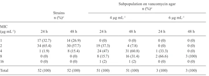
genes and all of them were negative for these genes. The vancomycin MICs obtained for subpopulations that grew on vancomycin screen agar and for the original
populations are shown in Table II. The MIC was ≥ 4 μg
mL-1 in 64.7% of the isolates that grew on agar plates
con-taining 4 μg mL-1 vancomycin, with an MIC of 8 μg mL-1
in 15.7% of these isolates and an MIC of 16 μg mL-1 in
one isolate (2%). MICs of 8 and 4 μg mL-1 were observed
in two (66.6%) and one of the three strains that grew on
agar plates with 6 μg mL-1 vancomycin, respectively. A
comparison of MICs between the original isolates and the subpopulations that grew on vancomycin screen agar showed an increase in MIC in 75.9% of the isolates after 24 h of incubation. A two, four or eight-fold increase was observed in 96.3% of the strains after 48 h.
Regarding the presence of ica genes, at least one ica
gene was detected in 96 (89.7%) of the isolates. Forty-one (38.3%) S. epidermidis isolates carried the complete ica
(43.9%) concomitantly carried the icaA and icaD genes and 11 (10.9%) were negative for all genes of the operon. All icaA gene-positive isolates were positive for icaD
(Table III). Twenty-four (58.5%) of the isolates carrying the complete ica operon produced a biofilm; of these, 15 (36.6%) were classified as weakly adherent and nine (22%) as strongly adherent.
Eight (33.3%) of the isolates that produced a biofilm and carried the icaADBC operon exhibited vancomycin
MICs ≥ 8 μg mL-1, whereas this level of resistance was
observed in only nine (8.4%) of all isolates studied. The only isolate that exhibited a vancomycin MIC equal to
16 μg mL-1 produced a strongly adherent biofilm.
Comparisons of the isolates that exhibited an in-crease in the vancomycin MIC after 24 h of incubation (n = 39; 36.4%) with those that did not grow (n = 55; 51.4%) and those that did grow, but maintained the same MIC (n = 13; 12.1%), revealed that the increase in van-comycin MIC was positively associated with methicillin resistance using oxacillin or cefoxitin disks (p = 0.02) and the presence of SCCmec type III (p = 0.0003).
An association was observed between the presence of the mecA gene and the presence of the icaA+icaD genes (p = 0.001): most isolates carrying the mecA gene were also positive for the icaA+icaD+icaC genes (p = 0.002) and also for the complete icaADBC operon (p = 0.004). In
ad-TABLE III
Frequency of the mecA and ica genes, biofilm formation, reduced susceptibility to vancomycin, increased vancomycin minimum inhibitory concentrations (MICs) on screen agar and associations found
Isolates (n)
icaa
(96)
icaA+icaD (47)
icaA+icaD+icaC (43)
icaADBC (41)
icaADBC/ biofilm production
(24)
Reduced susceptibility to vancomycin
(52)
Subpopulation with increased vancomycin MIC
(39)
MRSE Total (87) 79 45b 41b 39b 24b 44 33
I (21) 17 6 6 6 3 7 3
II (1) 1 1 1 1 1 1 1
III (42) 39 24c 20 19 14 28 24c
IV (18) 17 11 11 10 6 8 5
NT (5) 5 3 3 3 0 0 0
MSSE (20) 17 2 2 2 0 8 6
MRSE: methicillin-resistant Staphylococcus epidermidis based on the presence of the mecA gene; MSSE: methicillin-sensitive S. epidermidis; NT: no typed; a: at least one ica gene; b: significantly associated with mecA gene positivity(p < 0.05); c: sig-nificantly associated with the presence of staphylococcal cassette chromosome mec type III(p < 0.05).
TABLE II
Vancomycin minimum inhibitory concentrations (MICs) obtained after 24 h and 48 h of incubation of Staphylococcus epidermidis strains and subpopulations that grew on vancomycin screen agar
Strains n (%)a
Subpopulation on vancomycin agar n (%)a
4 μg mL-1 6 μg mL-1
MIC
(μg mL-1) 24 h 48 h 24 h 48 h 24 h 48 h
1 17 (32.7) 14 (26.9) 0 (0) 0 (0) 0 (0) 0 (0)
2 34 (65.4) 30 (57.7) 19 (37.3) 4 (7.8) 0 (0) 0 (0)
4 1 (1.9) 8 (15.4) 24 (47) 31 (60.8) 1 (33.3) 0 (0)
8 0 (0) 0 (0) 8 (15.7) 16 (31.4) 2 (66.6) 3 (100)
16 0 (0) 0 (0) 1 (2) 1 (2) 0 (0) 0 (0)
Total 52 (100) 52 (100) 51 (100) 51 (100) 3 (100) 3 (100)
dition, the presence of icaA and icaD was associated with SCCmec type III (p = 0.030) (Table III). Furthermore, the presence of the complete ica operon and biofilm produc-tion were significantly associated with the presence of the
mecA gene (100%; p = 0.006) and with growth on vanco-mycin screen agar (66.7%; p = 0.05) when compared to isolates that did not carry the complete operon or that did carry the operon, but did not produce a biofilm.
DISCUSSION
Formerly considered to be an innocuous commensal bacterium of human skin, S. epidermidis is now recog-nised as an important opportunistic pathogen. This mi-croorganism is one of the most common causative agents of medical device-related infections (Sievert et al. 2013). We isolated 107 S. epidermidis strains from blood cultures of patients seen at a Brazilian teaching hospital and ana- lysed antimicrobial resistance patterns, the presence of ica
operon genes and biofilm formation. The results showed a high prevalence of methicillin resistance as detected by both phenotypic and genotypic methods (73.8% and 81.3%, respectively). This finding was expected because the prevalence of methicillin resistance is high (70-80%) among CoNS in Latin America (Diekema et al. 2001).
The prevalence of SCCmec types III (48.2%) and IV (20.7%) was high, which is a finding that was also re-ported by Wisplinghoff et al. (2003). SCCmec type IV is smaller and the metabolic cost of its transfer is thus lower; as a consequence, its prevalence is expected to increase over time. Indeed, in the present study, the pro-portion of SCCmec type IV increased three-fold over a period of six years. Conversely, the prevalence of type III decreased during the same period, although its preva-lence continues to be high. This high proportion of
SCC-mec type III might be related to the selection of this type in the hospital environment due to the multidrug resist-ance of these strains. These facts should be considered in infection control policies.
SCCmec type II was detected in only one isolate. A similar frequency was reported by Machado et al. (2007), suggesting that MR S. epidermidis (MRSE) carrying this SCCmec type is not prevalent in Brazil. SCCmec type I, a hospital-associated type, was frequent (24.1%); this finding stands in contrast to previous studies in which type I was found to be rare (Ibrahem et al. 2009, Barbier et al. 2010, Zong et al. 2011), but is similar to the find-ings reported in other Brazilian studies (Machado et al. 2007, Pereira & Cunha 2013, Ternes et al. 2013). These findings might be related to the local epidemiology of MRSE, indicating a higher frequency of SCCmec type I in Brazilian hospitals. These data indicate the exist-ence of a vast reservoir of SCCmec among S. epider-midis strains (Wisplinghoff et al. 2003). Although these similarities prevail, the distribution of SCCmec types depends on several factors, including geographic loca-tion and the use of antimicrobial agents.
The incidence of the ica genes was high in the present study, with 90.6% of the isolates being positive for at least one ica gene and 38.3% being positive for all genes. Biofilm production was detected by the pheno-typic method in 58.5% of the isolates carrying the
com-plete ica operon. Similarly, Oliveira and Cunha (2010) observed that 56.6% of strongly adherent CoNS carried the icaA+icaC+icaD genes. The regulation of the ica
operon is complex and the expression of the ica genes is variable: it can be activated or deactivated according to in vivo conditions. Some events, such as the addition of the insertion sequence IS256, appear to be associated with this phenomenon (Ziebuhr et al. 1999).
In the present study, all isolates that were positive for the icaA gene were also positive for icaD, in agreement with Arciola et al. (2001) and Gad et al. (2009). Cafiso et al. (2004) showed that the icaD gene was always ex-pressed in S. epidermidis, but that phenotypic biofilm production only occurred when icaA was expressed si-multaneously. This relationship might be explained by the selective pressure exerted by biofilm production be-cause, when expressed, the icaD gene alone does not in-duce transferase activity and icaA induces little activity; in contrast, the combined expression of icaA and icaD
produces large amounts of PIA (Gerke et al. 1998). Al-though the icaA and icaD genes overlap and are co-tran-scribed, the fact that the isolates were more frequently positive for icaD than icaA, icaB and icaC in the present study and in previous studies (Zhou et al. 2013) may be related to the loss or mutation of one of these genes.
The mecA gene was found to be associated with the presence of the icaADBC operon and biofilm produc-tion (p < 0.05). In addiproduc-tion, the concomitant presence of
icaA+icaD was associated with SCCmec type III. Zhou et al. (2013) also detected icaD more frequently than icaA
and observed a significant association between icaD and the mecA gene in S. epidermidis. Furthermore, other studies have demonstrated that methicillin resistance is correlated with biofilm production and the presence of
ica genes (Alcaráz et al. 2003, Abassi et al. 2008, Koksal et al. 2009). These associations might be a consequence of the intimate contact between biofilm bacteria, facili-tating the horizontal transfer of genetic material such as antimicrobial resistance genes (Klingenberg et al. 2005), which would explain the horizontal transfer of mecA to
ica gene-positive strains (Kozitskaya et al. 2004). The present results suggest the presence of the icaADBC
operon in conjunction with biofilm production and the presence of the mecA gene to be an important marker of nosocomial strains with the potential to cause infec-tion. These markers are therefore useful for evaluating the clinical significance of CoNS.
The indiscriminate use of antibiotics has led to the selection of bacterial populations that contain cells with distinct levels of susceptibility to antimicrobial agents, including resistant cells. Intermediate and complete re-sistance to vancomycin in S. epidermidis has been re-ported (Palazzoet al. 2005) and biofilm formation is known to significantly reduce the effects of this antibi-otic on bacteria (Raad et al. 2007). In the present study, 47.7% and 2.8% of the S. epidermidis isolates grew colo-nies on screen agar plates containing 4 and 6 μg mL-1
vancomycin, respectively. One-third of the isolates that were biofilm producers also exhibited vancomycin MICs
≥ 8 μg mL-1 after growing on vancomycin screen agar,
The term vancomycin heteroresistance refers to strains that contain a subpopulation of cells with differ-ent levels of susceptibility to this antibiotic, including resistant cells and cells with intermediate susceptibility (Raad et al. 2007). Our analysis of colonies grown on vancomycin screen agar showed an increase in MICs
in 75.9% of the isolates, with an increase from 1 to ≥
4 μg mL-1 in 64.7% of the isolates. MIC increases were
also observed in 100% of the colonies that grew on agar with 6 μg mL-1 vancomycin. Using isolates that grew on
screen agar with 4 μg mL-1 vancomycin, Ma et al. (2011)
reported slightly lower rates: 64.7% of strains showed
increased MICs and 35.3% showed MICs ≥ 4 μg mL-1.
MICs ≥ 4 μg mL-1 were observed in 85.7% of the
subpop-ulations that grew on screen agar with 6 μg mL-1
vanco-mycin. In our study, increased MICs were detected in 96.3% of the isolates after 48 h of incubation. This find-ing confirms the existence of heteroresistant isolates.
The existence of vancomycin heteroresistance in nosocomial S. epidermidis is an alarming finding. Furthermore, the observation of subpopulations with MICs > 4 μg mL-1 (64.7%), as well as MICs of 8 μg mL-1
(15.7%) and 16 μg mL-1 (2%), classifying the isolates as
intermediate resistant, indicates that infections caused by these microorganisms will be difficult to treat. In-deed, reports have shown higher mortality among pa-tients infected with heteroresistant bacteria and strains with higher MICs compared to those infected with sus-ceptible strains (Wong et al. 1999). However, therapeutic alternatives for infections caused by staphylococci with reduced vancomycin resistance have been approved by the US Food and Drug Administration and include li-nezolide, daptomycin, tigecycline and quinupristin/ dalfopristin (Micek 2007). Two next-generation cepha-losporins, ceftobiprole and ceftaroline fosamil, are cur-rently available for the treatment of patients carrying MR and vancomycin-intermediate staphylococci (Sara-volatz et al. 2011, Lovering et al. 2012).
In the present study, reduced susceptibility to van-comycin was significantly associated with methicillin resistance detected by phenotypic methods (p < 0.05). Other studies (Wong et al. 1999, Loomba et al. 2010) have shown that the isolation of MR staphylococci is significantly more common among patients with bacter-aemia due to vancomycin-heteroresistant staphylococci. Although the mechanism underlying the association be-tween methicillin resistance and reduced vancomycin susceptibility remains unclear, it might be related to the treatment of infections caused by MR bacteria with van-comycin, thereby selecting heterogeneous populations and strains with low susceptibility.
The proportion of strains with reduced susceptibility to vancomycin was higher among the isolates harbour-ing SCCmec type III. Differing results have been report-ed by Moise et al. (2007), who found that higher vanco-mycin MICs were significantly associated with SCCmec
type II in S. aureus, whereas lower MICs were related to SCCmec type IV. The association between reduced van-comycin susceptibility and SCCmec type III observed in the present study may be due to the high prevalence of SCCmec type III in the hospital environment and the frequent use of vancomycin.
The presence of subpopulations with higher vanco-mycin MICs and intermediate resistance demonstrates the heterogeneity of S. epidermidis strains in terms of susceptibility to this antibiotic. Because the main genes responsible for vancomycin resistance (vanA and vanB) were not detected, this reduced susceptibility may be re-lated to a thickening of the bacterial cell wall that prevents uptake of the antibiotic by the microorganism (Billot-Klein et al. 1996). However, further studies are needed to elucidate how these events occur in staphylococci.
The present study demonstrated an association be-tween the presence of the icaADBC operon, biofilm for-mation and antimicrobial resistance in S. epidermidis
strains isolated from blood cultures. Furthermore, the detection of S. epidermidis subpopulations with inter-mediate vancomycin resistance associated with methi-cillin resistance highlights the role of this species as an important multidrug-resistant microorganism and the consequent need to implement measures for the control of antibiotic use.
REFERENCES
Abassi MS, Bouchami O, Touati A, Achour W, Hassen AB 2008. Clonality and occurrence of genes encoding antibiotic resistance and biofilm in methicillin-resistant Staphylococcus epidermidis strains isolated from catheters and bacteremia in neutropenic pa-tients. Curr Microbiol 57: 442-448.
Alcaráz LE, Satorres SE, Lucero RM, Puig de Centorbi ON 2003. Species identification, slime production and oxacillin suscepti-bility in coagulase-negative staphylococci isolated from nosoco-mial specimens. Braz J Microbiol 34: 45-51.
Arciola CR, Baldassarri L, Montanaro L 2001. Presence of icaA and icaD genes and slime production of staphylococcalstrains from catheter-associated infections. J Clin Microbiol 39: 2151-2156.
Arciola CR, Gamberini S, Campoccia D, Visai L, Speziale P, Baldas-sari L, Montanaro L 2005. A multiplex PCR method for the de-tection of all five individual genes of ica locus in Staphylococcus epidermidis. A survey on 400 clinical isolates from prosthesis-associated infections. J Biomed Mater Res A75: 408-413.
Baker JS 1984. Comparison of various methods for differentiation of staphylococci and micrococci. J Clin Microbiol 9: 875-879.
Barbier F, Lebeaux D, Hernandez D, Delannoy AS, Caro V, Fran-çois P, Schrenzel J, Ruppé E, Gaillard K, Wolff M, Brisse S, An-dremont A, Ruimy R 2010. High prevalence of the arginine cata-bolic mobile element in carriage isolates of methicillin-resistant Staphylococcus epidermidis. J Antimicrob Chemother 66: 29-36.
Barry T, Colleran G, Glennon M, Dunican LK, Gannon F 1991. The 16S/23S ribosomal spacer region as a target for DNA probes to identify eubacteria. PCR Methods Appl 1: 51-56.
Billot-Klein D, Gutmann L, Bryant D, Bell D, Van Heijenoort J, Gre-wal J, Shlaes DM 1996. Peptidoglycan synthesis and structure in Staphylococcus haemolyticus expressing increasing levels of re-sistance to glycopeptides antibiotics. J Bacteriol 178:4696-4703.
Cafiso V, Bertuccio T, Santagati M, Campanile F, Amicosante G, Perilli MG, Selan L, Artini M, Nicoletti G, Stefanii S2004. Presence of the ica operon in clinical isolates of Staphylococcus epidermidis and its role in biofilm production. Clin Microbiol Infect 10: 1081-1088.
Chambers HF, Hartman BJ, Tomasz A 1985. Increased amounts of a nov-el penicillin-binding protein in a strain of methicillin-resistant Sta-phylococcus aureus exposed to nafcillin. J Clin Invest 76: 325-331.
Christensen GD, Simpson WA, Yonger JJ, Baddor LM, Barrett FF, Melton DM, Beachey EH 1985. Adherence of coagulase-negative staphylococci to plastic tissue culture plates: a quantitative model for the adherence of staphylococci to medical devices. J Clin Microbiol 22: 996-1006.
Clark NC, Cooksey RC, Hill BC, Swenson JM, Tenover FC 1993. Characterization of glycopeptide-resistant enterococci from US hospitals. Antimicrob Agents Chemother 37:2311-2317.
CLSI - Clinical and Laboratory Standards Institute 2007. Perfor-mance standards for antimicrobial susceptibility testing. Ap-proved Standard Document M100-S17. Available from: micro-biolab-bg.com/CLSI.pdf.
CLSI - Clinical and Laboratory Standards Institute 2012. Performance standards for antimicrobial susceptibility testing. Approved Stan-dard Document M100-S22. Available from: antimicrobianos. com.ar/ATB/wp-content/uploads/2012/11/M100S22E.pdf.
Costerton JW, Lewandowski Z, Cadwell DE, Korber DR, Lappin-Scott HM 1995. Microbial biofilms. Ann Rev Microbiol 49: 711-745.
Couto I, Pereira S, Miragaia M, Sanches IS, Lencastre H 2001. Identi-fication of clinical staphylococcal isolates from humans by inter-nal transcribed spacer PCR. J Clin Microbiol 39: 3099-3103.
Cunha MLRS, Sinzato YK, Silveira LVA 2004. Comparison of meth-ods for identification of coagulase-negative staphylococci. Mem Inst Oswaldo Cruz 99: 855-860.
Curi PR 1997. Metodologia e análise da pesquisa em ciências bi-ológicas,Tipomic, Botucatu, 263 pp.
Del’Alamo L, Cerada RF, Tosin I, Miranda EA, Sader HS 1999. Anti-microbial susceptibility of coagulase-negative staphylococci and characterization of isolates with reduced susceptibility to glyco-peptides. Diagn Microbiol Infect Dis 34: 185-191.
Diekema DJ, Pfaller MA, Schmitz FJ, Smayevsky J, Bell J, Jones RN, Beach M, SENTRY Participants Group 2001. Survey of infec-tions due to Staphylococcus species: frequency of occurrence and antimicrobial susceptibility of isolates collected in the United States, Canada, Latin America, Europe and the Western Pacific region for the SENTRY Antimicrobial Surveillance Program, 1997-1999. Clin Infect Dis 32: 114-132.
Donlan RM, Costerton JW 2002. Biofilms: survival mechanisms of clinically relevant microorganisms. Clin Microbiol Rev 15: 167-193.
Gad GF, Feky MA, Rehewy MS, Hassan MA, Abolella H, El-Baky RM 2009. Detection of icaA, icaD genes and biofilm pro-duction by Staphylococcus aureus and Staphylococcus epider-midis isolated from urinary tract catheterized patients. J Infect Dev Ctries 3: 342-351.
Gerke C, Kraft A, Sussmuth R, Schweitzer O, Götz F 1998. Character-ization of the N- acetylglucosaminyltransferase activity involved in the biosynthesis of the Staphylococcus epidermidis polysac-charide intercellular adhesion. J Biol Chem 273: 1856-1893.
Hiramatsu K 2001. Vancomycin-resistant Staphylococcus aureus: a new model of antibiotic resistance. Lancet Infect Dis 1: 147-155.
Houston P, Rowe SE, Pozzi C, Waters EM, O’Gara JP 2011. Essential role for the major autolysin in the fibronectin-binding protein-mediated Staphylococcus aureus biofilm phenotype. Infect Im-mun 79: 1153-1165.
Ibrahem S, Salmenlinna S, Virolainen A, Kerttula AM, Lyytikäinen O, Jägerroos H, Broas M, Vuopio-Varkila J 2009. Carriage of methicillin-resistant staphylococci and their SCCmec types in a long-term-care facility. J Clin Microbiol 47: 32-37.
Ito T, Katayama Y, Asada K, Mori N, Tsutsumimoto K, Tiensasitorn C, Hiramatsu K 2001. Structural comparison of three types of staphylococcal cassette chromosome mec integrated in the chro-mosome in methicillin-resistant Staphylococcus aureus. Antimi-crob Agents Chemother 45: 1323-1336.
Klingenberg C, Aarag E, Ronnestad A, Sollid JE, Abrahamsen TG, Kjeldsen G, Flaegstad T 2005. Coagulase-negative staphylococ-cal sepsis in neonates: association between antibiotic resistance, biofilm formation and the host inflammatory response. Pediatr Infect Dis 24: 817-822.
Koksal F, Yasar H, Samasti M 2009. Antibiotic resistance patterns of coagulase-negative staphylococcus strains isolated from blood cul-tures of septicemic patients in Turkey. Microbiol Res 164: 404-410.
Koneman EW, Allen SD, Janda WM, Schreckenberger PC, Winn Jr WC 1997. Color atlas and textbook of diagnostic microbiology, 5th ed., Lippincott Williams & Wilkins, Philadelphia, 1395 pp.
Kozitskaya S, Cho SH, Dietrich K, Marre R, Naber K, Ziebuhr W 2004. The bacterial insertion sequence element IS256 occurs preferentially in nosocomial Staphylococcus epidermidis isolates: association with biofilm formation and resistance to aminoglyco-sides. Infect Immun 72: 1210-1215.
Loomba PS, Taneja J, Mishra B 2010. Methicillin and vancomycin resist-ant S. aureus in hospitalized patients. J Glob Infect Dis 2: 275-283.
Lovering AL, Gretes MC, Safadi SS, Danel F, Castro L, Page MGP, Natalie CJ, Strynadka NCJ 2012. Structural insights into the anti-methicillin-resistant Staphylococcus aureus (MRSA) activity of ceftobiprole. J Biol Chem 287: 32096-32102.
Ma XX, Wang EH, Liu Y, Luo EJ 2011. Antibiotic susceptibility of coagulase-negative staphylococci (CoNS): emergence of teico-planin non-susceptible CoNS strains with inducible resistance to vancomycin. J Med Microbiol 60:1661-1668.
Machado ABMP, Reiter KC, Paiva RM, Barth AL 2007. Distribution of staphylococcal cassette chromosome mec (SCCmec) types I, II, III and IV in coagulase-negative staphylococci from patients attending a tertiary hospital in southern Brazil. J Med Microb 56: 1328-1333.
Mack D, Riedewald J, Rohde H, Magnus T, Feucht HH, Elsner HA, Laufs R, Rupp ME 1999. Essential functional role of the polysac-charide intercellular adhesin of Staphylococcusepidermidis in hemagglutination. Infect Immun 67: 1004-1008.
Mah TF, O’Toole GA2001. Mechanisms of biofilm resistance to anti-microbial agents. Trends Microbiol 9: 34-39.
McCann MT, Gilmore BF, Gorman SP 2008. Staphylococcus
epider-midis device-related infections: pathogenesis and clinical man-agement. J Pharm Pharmacol 60: 1551-1571.
Micek ST 2007. Alternatives to vancomycin for the treatment of me-thicillin-resistant Staphylococcus aureus infections. Clin Infect Dis 45 (Suppl. 3):S184-S190.
Moise PA, Sakoulas G, Forrest A, Schenag JJ 2007. Vancomycin in vitro bactericidal activity and its relationship to efficacy in clear-ance of methicillin-resistant Staphylococcus aureus bacteremia. Antimicrob Agents Chemother 57: 2582-2586.
Murakami K, Minamide K, Wada K, Nakamura E, Teraoka H, Watanabe S 1991. Identification of methicillin-resistant strains of staphylococ-ci by polymerase chain reaction. J Clin Microbiol 29: 2240-2244.
Oliveira A, Cunha MLRS 2010. Comparison of methods for the detec-tion of biofilm producdetec-tion in coagulase-negative staphylococci: BMC Res Notes3: 260.
Palazzo ICV, Araujo MLC, Darini ALC 2005. First report of van-comycin-resistant staphylococci isolated from healthy carriers in Brazil. J Clin Microbiol 43: 179-185.
Patti JM, Allen BL, McGavin MJ, Hook M 1994. MSCRAMM-me-diated adherence of microorganisms to host tissues. Ann Rev Mi-crobiol 48: 585-617.
Pereira VC, Cunha MLRS 2013. Coagulase-negative staphylococci strains resistant to oxacillin isolated from neonatal blood cul-tures. Mem Inst Oswaldo Cruz 108: 939-942.
Raad I, Hanna H, Jiang Y, Dvorak T, Reitzel R, Chaiban G, Sher-ertz R, Hachem R 2007. Comparative activities of daptomycin, linezolid and tigecycline against catheter-related methicillin re-sistant Staphylococcus bacteremic isolates embedded in biofilm. Antimicrob Agents Chemother 51:1656-1660.
Rupp ME, Fey PD, Heilmann C, Gotz F 2001. Characterization of the importance of Staphylococcus epidermidis autolysin and poly-saccharide intercellular adhesin in the pathogenesis of intravas-cular catheter associated infection in a rat model. J Infect Dis 183: 1038-1042.
Saravolatz LD, Stein GE, Johnson LB 2011. Ceftaroline: a novel ce-phalosporin with activity against methicillin-resistant Staphylo-coccus aureus. Clin Infect Dis 52: 1156-1163.
Schwalbe RS, Stapleton JT, Gilligan PH 1987. Emergence of vanco-mycin resistance in coagulase negative staphylococci. N Engl J Med 316: 927-931.
Sievert DM, Ricks P, Edwards JR, Schneider A, Patel J, Srinivasan A, Kallen A, Limbago B, Fridkin S, for the National Healthcare Safety Network (NHSN) Team and Participating NHSN Fa-cilities 2013. Antimicrobial-resistant pathogens associated with healthcare-associated infections: summary of data reported to the National Healthcare Safety Network at the Centers for Dis-ease Control and Prevention, 2009-2010. Infect Control Hosp Epidemiol34: 1-14.
Singh R, Ray P, Das A, Sharma M 2010. Penetration of antibiotics through Staphylococcus aureus and Staphylococcus epidermidis biofilms. J Antimicrob Chemother 65: 1955-1958.
Stewart PS, Costerton JW 2001. Antibiotic resistance of bacteria in biofilms. Lancet 358: 135-138.
Ternes YM, Lamaro-Cardoso J, André MCP, Pessoa VP, Vieira MAS, Minamisava R, Andrade AL, Kipnis A 2013.Molecular epidemiology of coagulase-negative Staphylococcus carriage in neonates admitted to an intensive care unit in Brazil. BMC Infect Dis 13: 572.
Vitali LC, Petrelli D, Lamikanra A, Prenna M, Akinkunmi EO 2014. Diversity of antibiotic resistance genes and staphylococcal cas-sette chromosome mec elements in faecal isolates of coagulase-negative staphylococci from Nigeria. BMC Microbiology14: 106.
Von Eiff C, Peters G, Heilmann C 2002. Pathogenesis of infections due to coagulase-negative staphylococci. Lancet Infect Dis 2: 677-685.
Wisplinghoff H, Rosato AE, Enright MC, Noto M, Craig W, Archer GL 2003. Related clones containing SCCmec type IV predomi-nate among clinically significant Staphylococcus epidermidis isolates. Antimicrob Agents Chemother 47: 3574-3579.
Wong SSY, Ho PL, Woo PCY, Yuen KY 1999. Bacteremia caused by staphylococci with inducible vancomycin heteroresistance. Clin Infect Dis 29: 760-767.
Zhou S, Chao X, Fei M, Dai Y, Liu B 2013. Analysis of S. epidermidis icaA and icaA genes by polymerase chain reaction and slime pro-duction: a case control study. BMC Infect Dis13: 242.
Ziebuhr W, Krimmer V, Rachid S, Lössner I, Götz F, Hacker J 1999. A novel mechanism of phase variation of virulence in Staphylococ-cus epidermidis: evidence for control of the polysaccharide inter-cellular adhesin synthesis by alternating insertion and excision of the insertion sequence element IS256. Mol Microbiol 32: 345-356.