WORK PERFORMED AT:
FOREST BIOTECHNOLOGY LABORATORY
Instituto de Biologia Experimental e Tecnológica Instituto de Tecnologia Química e Biológica Universidade Nova de Lisboa
Av. da República 2780-157 Oeiras Portugal
SHORT-TERM STAYS AT:
Xylem Maturation and Wood Properties Research Group Umeå Plant Science Centre (UPSC), Umeå, Sweden Hormone Signaling and Plant Plasticity Research Group
Instituto de Biología Molecular y Celular de Plantas (IBMCP-UPV),
Valencia, Spain
SUPERVISOR:
Dr. Célia Maria Romba Rodrigues Miguel
Aos meus pais e ao Pedro,
The most exciting phrase to hear in science, the one that heralds
new discoveries, is not 'Eureka!' (I found it!)
but 'That's funny ...'
Isaac Asimov
ACKNOWLEDGEMENTS
I would like to express my deepest gratitude to Célia Miguel, for being the supervisor and advisor any student can ambition to have; for the scientific guidance, for the good advice but also for all the confidence and for giving me some freedom to
experiment. I’m truly thankful for all you’ve taught me and for making it possible for me to learn here and abroad. Thanks for being generously extra-available during the writing period, for the debates and always getting back to so many e-mails! Thank you for believing in me and for fighting through adversity when needed. Thank you for your kind friendship throughout these years.
I would like also to thank Hannele Tuominen, for all her kindness in receiving me at her lab at UPSC. For teaching me so much on xylem development. For the countless hours of debate and e-mails, it has been a great pleasure that I can´t wait to repeat.
To Miguel A. Blázquez, for so kindly receiving me at his lab at IBMCP, for giving good advice and always finding some time and solution to my requests.
To Andreia Matos, for always being willing to help and for taking care of the in vitro plants like no one can. For being so dedicated, if it wasn’t for your help this would have taken me much more time to achieve.
To Jakob Prestele and Benjamin Bollhöner for all the help during and after my stay at UPSC, for guiding me in the lab and also for making me feel welcome and always be willing to teach, help and debate on the results. To Karin Ljung, for kind suggestions and auxin measurements. To Veronica Bourquin, for the hints on trees histology, to Kjell for his advice on observing xylem cells at the microscope, and to Sacha and Maribel, for being such nice company during my stay. Thanks to everyone at UPSC who made me feel very welcome.
polyamine measurements. To José Luis Rambla for performing the samples injections and teaching me how you got that beautiful tiny peak. Thanks to everyone at MAB´s lab for welcoming me at IBMCP.
To Brian Jones and Max Cheng for providing the hybrid aspen T89 and Nisqually-1 clones used throughout this work. To Luís Goulão, for kindly advising on in situ hybridization. To Jörg Becker, for kindly allowing attendance and participation in your microarray workshop and valuable advice on microarray analysis.
To Alan Phillips for kindly reviewing the summary of this thesis.
To Marta Simões for being my lab-mate all these years, for the laughs, the companionship, the complicity in our silliness, for your friendship, for all the
scientific discussions we had and for being such a great “bench any-problem
solver”... I miss you!
To Liliana Marum for being such a nice inspiration, a role model of resilience and
persistence in the “tree-world”, and a very good friend that has grown on me over the years. I will miss you!
To all the current and former members of Forest Biotech lab, Andreia Miguel, Andreia Rodrigues, José de Vega-Bartol, Ilanit, Inês Chaves, Inês Modesto, Raissa, Ana Maria, Marigrazia, Sónia Gonçalves, Susana Tereso, Margarida Rocheta... and many more for the companionship, the help and making the lab a great place to work in!
Alicja... and everyone else, always so kind and helpful.
To Professor Pinto Ricardo, for always caring about my work and for always being available whenever I had some request.
To everyone at DSB, at BCV (especially to Inês and Mara for the chats and many
“Friday´s night weekend wrap-up” at the ITQB balcony), at Prof. Pinto Ricardo’s lab and at LEM, all of which, at some point, provided generous assistance.
To Eugénia, Pilar and all the staff at the washing rooms, without whom this work would have taken forever. To maintenance, administrative and to security staff that helped me with some loose-screw or broken down -80ºC in the middle of the night throughout these years.
To ITQB and IBET and everyone at ITQB and IBET that throughout the years have had the generosity of helping me in some way. A special thanks to Ana Portocarrero and Fátima Madeira for making my life easier in the final steps of delivering the thesis.
À Manela, que no meu imaginário se confunde com o meu ser e que me ensinou o essencial dos 6 aos 10.
À Cris, ao Xavi e à Madalena, e aos amigos espalhados pelo mundo, por saber que apesar das longas ausências, quando nos encontramos afinal somos nós again. Obrigada pela amizade. A todos os amigos que compreenderam a ausência nestes últimos anos.
À minha família, tios e primos, aos meus sogros, Catarina e Zé Manel, e aos cunhados João e Carlinha, pela força, pelo amor e carinho.
por me terem dado sempre a confiança que seria capaz de fazer mais e melhor, por apesar de tudo compreenderem as ausências e as muitas falhas. Por me mostrarem o caminho.
The author of this thesis, Ana Filipa Gonçalves Milhinhos, hereby declares to have had active participation in the following research papers:
Ana Milhinhos and Célia M. Miguel (2013) Hormone interactions in xylem development: a matter of signals. Plant Cell Reports. (doi: 10.1007/s00299-013-1420-7).
Ana Milhinhos participated by performing the experiments, reviewing the literature and writing the paper (Chapter I).
Ana Milhinhos, Jakob Prestele, Benjamin Bollhöner, Andreia Matos, Francisco Vera-Sirera, José L. Rambla, Karin Ljung, Juan Carbonell, Miguel A. Blázquez, Hannele Tuominen and Célia M. Miguel. Thermospermine levels are controlled by an auxin-dependent feedback-loop mechanism in Populus xylem. (revised manuscript submitted)
Ana Milhinhos participated in the experimental design, performing laboratory experiments, analyzing the results and writing the paper (Chapter II).
Ana Milhinhos, Andreia Matos and Célia M. Miguel. Thermospermine-induced transcriptomic responses reveal hormone crosstalk in Populus stems. (submitted)
Ana Milhinhos participated in the experimental design, performing laboratory experiments, analyzing the results and writing the paper (Chapter III).
Ana Milhinhos, Andreia Matos, Francisco Vera-Sirera, Miguel A. Blázquez, Luís Goulão and Célia M. Miguel. Elucidating the regulatory function of PttHB8 on POPACAULIS5 expression. (in preparation)
LIST OF ABBREVIATIONS
ACL5 ACAULIS5
APL ALTERED PHLOEM DEVELOPMENT
ARF AUXIN RESPONSE FACTOR
ARR ARABIDOPSIS RESPONSE REGULATOR
AUX/IAA AUXIN RESISTANT1/INDOLE ACETIC ACID
ATP Adenosine triphosphate
BAP 6-benzylaminopurine
BCIP 5-bromo-4-chloro-3’-indolyphosphate
bp Base pair
BR Brassinosteroid
CaMV 35S Cauliflower mosaic virus 35S promoter
cDNA Complementary DNA
CDS Coding sequence
CK Cytokinin
CNA CORONA/ATHB15
DNA Deoxyribonucleic acid
EDTA Ethylene diamine tetraacetic acid
CG-MS Gas chromatography mass spectrometry
GUS -Glucuronidase
GFP Green fluorescent protein
h Hour
HD-Zip III Homeodomain leucine zipper Class III
IAA Indole-3-acetic acid
IBA Indole butyric acid
KAN KANADI
Mbp Mega base pair
g Microgram
l Microlitre
m Micrometer
M Micromolar
mg Milligram
ml Millilitre
mm Millimetre
m Meter
mRNA Messenger RNA
miRNA MicroRNA
MS Murashige and Skoog medium
nm Nanometer
NBT Nitro blue tetrazolium
NPTII Neomycin phosphotransferase II
PAO Polyamine oxidase
PBS Phosphate-buffered saline
PCR Polymerase chain reaction
PHB PHABULOSA
PHV PHAVOLUTA
Ptt Populus tremulaPopulus tremuloides
Ptr Populus trichocarpa
REV REVOLUTA
RT-qPCR Real-time quantitative reverse transcription PCR
SAM S-adenosyl-methionine
SAM Shoot apical meristem
SD Standard deviation
SDS Sodium dodecyl sulphate
SPDS Spermidine synthase E.C.2.5.1.16
Spd Spermidine
Spm Spermine
SPMS Spermine synthase E.C.2.5.1.22
SSPE Saline-sodium phosphate-EDTA buffer
TDZ Thidiazuron
Tspm Thermospermine
tSPMS Thermospermine synthase E.C.2.5.1.79
SUMMARY
SUMÁRIO
TABLE OF CONTENTS
Acknowledgements ... vii List of abbreviations ... xiii Summary ... xv Sumário ... xix Chapter I.
General introduction ... 1
Chapter II.
Thermospermine homeostasis in Populus xylem... 65 Chapter III.
Thermospermine-induced transcriptomic changes in Populus stems .... 119 Chapter IV.
HD-Zip III regulatory functions in Populus ... 201 Chapter V.
CHAPTER I
GENERAL INTRODUCTION†
†Milhinhos A. and Miguel C. (2013)
Plant vascular development
Early plants were simple and small unicellular or filamentous organisms highly dependent on being submersed in the aquatic environment. It was about 440 million years ago that plants evolved adaptations to land life. One of such remarkable adaptations was the evolution of vascular tissues that allowed vascular plants to colonize vast areas of the earth's terrestrial surface. The appearance of vascular tissues not only made it possible to transport nutrients and water as it granted the plant with tools for increased organismal complexity, allowing growth in height away from the shadow of other plants, for instance. Xylem and phloem thus evolved as a network throughout the plant that connected the plant body and formed the vascular system. While phloem functions to transport and distribute the photoassimilates produced in the shoot, xylem transports water and minerals taken up by the roots and provides structure and girth to the plants. The ability for growth in height was accompanied by lateral growth, which afforded the plant kingdom to evolve the largest organisms on earth. The lateral growth largely originates from the activity of the vascular cambium, a secondary meristem that drives the formation of secondary xylem (commonly named wood). The formation of the wood is modulated by several internal signals that control the vascular cambium activity. However, little is known on the hormonal and genetic background that is behind this secondary growth. Fair to say that in the last decades the bloom of genetic and genomic tools has led to increased understanding of the molecular mechanisms underlying secondary growth, mainly due to this great landmark that was the sequencing of the Populus tree genome (Tuskan et al., 2006).
model plant. This thesis starts by introducing general concepts, with a description of the processes involved in vascular development, with emphasis on xylem specification and differentiation. The organization and establishment of the vascular tissues are also discussed. The current understanding on hormone signaling interactions in xylem development and their role in the regulation of plant meristem activity will be introduced. In addition, a broad view on structure and metabolism of polyamines, as well as their role in plant development are presented, with emphasis on thermospermine role in vascular development. The PhD studies own results will be discussed within this context.
The vascular system
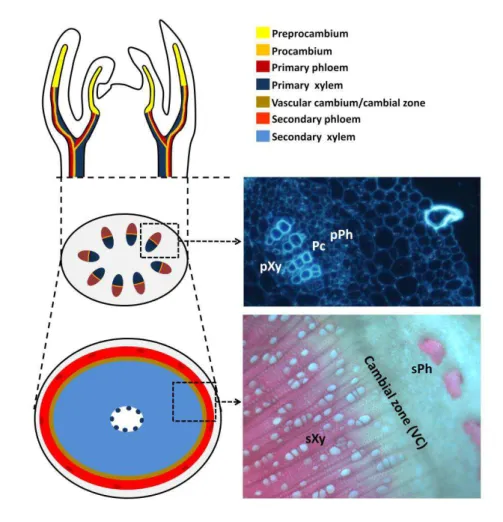
Vascular cells are produced from the apical meristems located at the shoot and root apices. The apical meristem contains the stem cell niche, composed of a few undifferentiated cells, called pre-procambial or provascular cells that give rise to the different cell types present in the plant body, such as procambial and cambial cells, from which xylem and phloem precursor cells develop. Xylem cells differentiate from xylem precursor cells into tracheary elements (tracheary and vessel elements, the later only present in angiosperms), xylem parenchyma cells and xylem fibers. Phloem precursor cells differentiate into sieve elements, companion cells, phloem parenchyma cells and phloem fibers.
Procambium and cambium
(Figure 1; Esau, 1977; Clay and Nelson, 2002). Procambial cells are cytoplasm-dense cells that are organized in continuous strands, the vascular bundles. Coordinated, oriented divisions, parallel to the direction of the vascular bundle axis confer the narrow and elongated shape to procambial cells (Scarpella et al., 2004).
Vascular cambium derives mainly from the procambium within the vascular bundle, but also from the parenchyma between the vascular bundles. Cambial cells divide anticlinally (perpendicularly to the surface of the stem) to produce the derivative initials. These derivative initials then divide periclinally (parallel to the surface of the stem) to produce specialized cells (phloem and xylem) thus promoting growth in the lateral directions (Esau, 1977). These divisions give rise to the secondary meristem, the cylindrical vascular cambium, and mark the start of secondary development (Figure 1; Baucher et al., 2007). The vascular cambium term is used to refer to the cambial initials file of cells, however because it often difficult to distinguish between the initials and their immediate derivatives, the term is usually applied to the group of cambial initials and their immediate derivatives, and instead some authors prefer to name this region as the cambial zone (Raven et al., 2005).
Xylem
possible to elongate in the direction of growth, whereas metaxylem cells have net-like (reticulate) or porous (pitted) wall thickenings and are structurally stronger and cannot be stretched (Chapter II).
transition from primary to secondary growth involves the formation of fascicular cambium, that arises within vascular bundles and interfascicular cambium that arises between vascular bundles (not shown, reviewed in Sanchez et al., 2012). Secondary growth of vascular tissues, shows secondary phloem and xylem that derive from the vascular cambium, a layer of meristematic/cambial cells. The cambial cells at the vascular cambium undergo cell divisions that originate xylem precursor cells (towards the interior of the stem) and phloem precursor cells (towards the exterior of the stem). Xylem precursor cells then differentiate into different xylem cell types (such as vessels, fibers and ray cells). Primary and secondary development is also shown in the corresponding Populus tremula Populus tremuloides stem cross-sections (on the right). Pc, procambium; VC, vascular cambium; pPh and sPh, primary and secondary phloem; pXy and sXy, primary and secondary xylem.
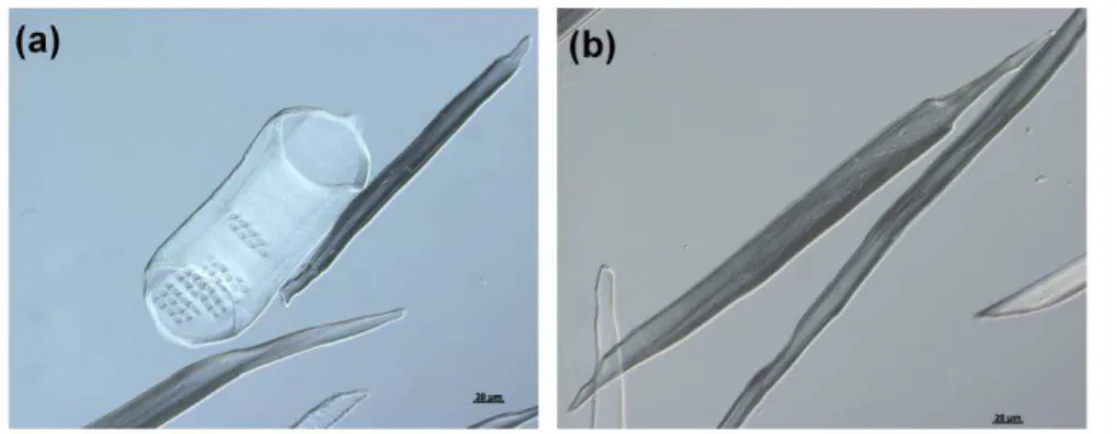
Vascular cambium activity gives rise to secondary xylem (wood). Wood comprises three general types of cells: xylem vessels, xylem fibers and xylem parenchyma (Figure 2). The interconnected xylem vessels consist of lignified secondary cell walled and hollow lumen cells that, joined together at their perforation plates, form the finite conduits. The sap flowing through the lumens in these conduits typically includes nutrient ions, amino acids, hormones, proteins, and traces of carbohydrates (Biles and Abeles, 1991; Davies and Zhang, 1991; Gollan et al., 1992; Satoh et al., 1992; Goodger et al., 2005). Between the adjacent xylem conduits the sap transport occurs via the numerous pits concentrated in the lignified secondary cell walls (Figure 2a). The xylem fibers, which are thick secondary cell walled cells, provide structural support (Figure 2b); and xylem parenchyma cells. The vascular ray cells are mostly parenchymatic cells that transport photosynthesis products and water across the stem between secondary xylem and phloem cells (Esau, 1977).
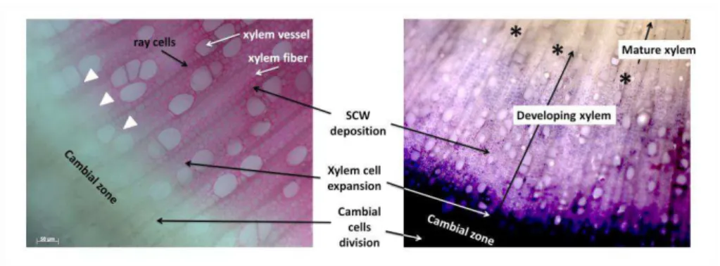
elements die within two days after their specification, fibers stay alive for approximately one month in Populus and ray parenchyma cells stay alive for decades (Bollhöner et al., 2012; Nakaba et al., 2006; 2011). Still, the main events of tracheary elements differentiation include: early differentiation in the cambial zone, cell expansion, secondary cell wall formation, changes in tonoplast permeability and vacuolar rupture, DNA degradation, final autolysis and hydrolysis of the non-lignified primary cell walls (Bollhöner et al., 2012). Maturation of xylem, including lignification of xylem cell wall coincides normally with cell death in time and place (Bollhöner et al., 2012). This is, as lignin accumulation increases, the xylem fiber elements death program is maybe triggered but it is not currently known whether the two processes are coordinated (Figure 3).
Figure 2. Secondary xylem cell types present in Populus wood. (a) secondary xylem vessel and (b) xylem fiber. Examples of primary xylem vessels are illustrated in Chapter II.
Phloem
and mitochondria close to the wall (Raven et al., 2005). It may be primary or secondary in origin as xylem, but contrary to xylem, phloem is living at maturity. Phloem comprises the conducting sieve elements (sieve tubes and sieve cells) and the non-conducting fibers and parenchyma cells. The interconnected sieve elements that are metabolically sustained by adjacent companion cells in source organs, such as leaves, load the photoassimilates and unload to sink organs such as roots and storage tissues (Raven et al., 2005).
Figure 3. Cross section of hybrid-aspen (Populus tremula Populus tremuloides) stem, showing progression of xylem lignification and cell death. (on the left) lignification of xylem as indicated by phloroglucinol staining and (on the right) cell viability test by nitroblue tetrazolium staining observed in developing xylem (methods detailed in Chapter II). The stages of xylem differentiation are indicated. Arrows depict beginning of lignification and asterisks mature xylem.
Populus and Arabidopsis as model systems for vascular development studies
Populus has been the model system of choice. The natural choice of a tree for xylem development studies comes from the utter amount of secondary growth it presents. Populus has become largely accepted in the research
community as the ‘model’ woody plant arguing that such a ‘model’ tree is
needed to complement the genetic resource developed in Arabidopsis. The genus Populus (poplars, cottonwoods and aspens) comprises approximately 30 species of woody plant, all found in the Northern hemisphere and characterized by the fastest growth rates in temperate trees (Taylor, 2002). There are several reasons that elected Populus as a model tree, but the most important is based on the fact that the Populus genome was the first tree genome to be sequenced and made available (Tuskan et al., 2006). The relatively small genome size (450–550 Mbp), the large number of molecular genetic maps and the ease of genetic transformation of Populus are additional reasons for the choice. Populus is also easily propagated vegetatively allowing the mass production of clonal material for experiments. In addition, and contrary to Arabidopsis, Populus trees also have true commercial value for timber, plywood, pulp and paper (Taylor, 2002).
transgenic technologies have also been applied to Populus with success. In vascular development studies, the large size and radial organization of Populus tree stems allows to harvest significant amounts of specialized cells from the cambial zone at different stages of specification. The cambial zone can therefore be divided into separate fractions that correspond to meristematic cambial cells, developing xylem cells, and functional phloem cells. This method is not as easily attained in Arabidopsis stem due to its small scale and the need for specialized technology such as laser microdissection to gather a much reduced amount of cells; while it has been employed successfully in numerous studies using trees (Uggla et al., 1998; Hertzberg et al., 2001; Schrader et al., 2003; Schrader et al., 2004; Nieminen et al., 2008; Chapter II).
Hormone interactions in xylem development: a matter of signals
In the last decades the bloom of genetic and genomic tools has led to increased understanding of the molecular mechanisms underlying the function of the traditional plant hormones in xylem specification and differentiation. Critical functions have been assigned also to novel signalling molecules, such as thermospermine. It is evident that these signals do not function independent of each other but in close interaction in a manner that only now is beginning to be understood.
SHOOTMERISTEMLESS (STM)/ BREVIPEDICELLUS (BP) in Arabidopsis (Groover et al., 2006; Du et al., 2009), as well as in the monocotyledoneous maize and rice plants, suggesting that KNOX genes are conserved mediators of meristematic potential (Scofield and Murray, 2006). Due to such similarities, the following sections review the state of the art integrating data obtained from the study of procambium in the Arabidopsis shoot and root meristem model systems and in inflorescence stems but also data on lateral growth and the emerging knowledge from studies in Arabidopsis, Zinnia elegans xylogenic cultures and Populus trees.
New insights into how signaling molecules direct phloem and xylem differentiation are coming to light. Hormones, synthesised either locally or at a long distance, are important signals in this process, being recognised and integrated into responses during vascular development. Auxin, cytokinin, gibberellins and ethylene were early on identified as regulators of vascular development, but more recently also brassinosteroids, nitric oxide, jasmonic acid and strigolactones have been pointed out involved in the process (Sachs, 1981; Mähönen et al., 2000; Eriksson et al., 2000; Savidge, 1988; Choe et al., 1999a; Gabaldón et al., 2005; Sehr et al., 2010; Agusti et al., 2011a). Other important signals include thermospermine, H2O2 and small peptides
(Vera-Sirera et al., 2010; Ros Barceló et al., 2002; Ito et al., 2006). Even though signaling pathways for some of these compounds are quite well characterized (such as for auxin, cytokinin, ethylene, gibberellin and brassinosteroids) the exact molecular mechanism underlying control of vascular development is not fully understood for any of them.
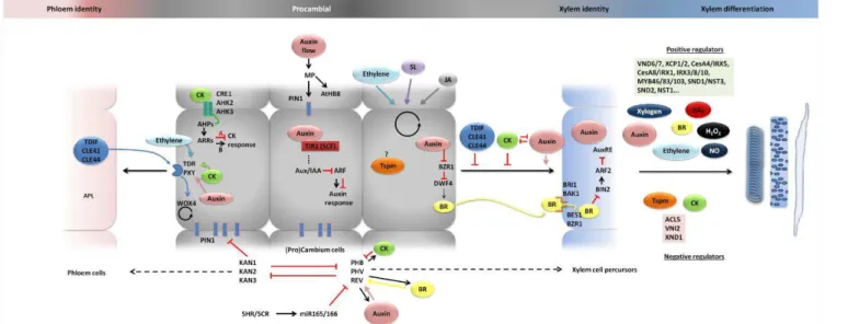
summarizes the hormones action in cambium cells formation, maintenance and in xylem specification and differentiation hereby described.
Auxin role in cambial cell identity: initiate and keeping it cambial
Auxin is a key regulator of almost any aspect of plant development and vascular development is no exception. It has long been known that the initiation of pre-procambial cells depends on auxin signaling and transport. According to the auxin canalisation theory, auxin creates its own transport channels as a result of an initial auxin flux from an auxin source to an auxin sink. Thus, canalization of auxin flow initiates vascular cells, which also promotes further canalization of auxin into these channels. The preferred channels inhibit further canalisation in the surroundings and thus differentiate as narrow vascular strands (Sachs, 1981). Evidence of auxin transport and signaling components functioning in vascular specification, together with pharmacological studies of auxin transport inhibitors, have supported the fundamental role of auxin in the induction of vascular bundles (Galweiler et al., 1998; Mattsson et al., 1999; Sieburth, 1999).
G
en
era
l I
ntr
od
uc
tio
n
15
increase in auxin flow is what determines pre-procambial cell state acquisition. ATHB8 is necessary to stabilize pre-procambial cell specification against auxin transport perturbations (Donner et al., 2009) and probably acts by reducing the sensitivity to auxin, in terms of PIN1 expression, and thereby confining procambium precursor cell state acquisition to narrow regions in the developing leaf (Scarpella et al., 2006; Wenzel et al., 2007; Donner et al., 2009; Ohashi-Ito and Fukuda, 2010). Since procambial cell formation in the athb8 null mutant does not deviate from the pattern observed in the wild- -type, it is also possible that MP acts on several other key components to promote procambium identity (Baima et al., 2001; Prigge et al., 2005). Curiously, while ATHB8 was found to be activated during interfascicular cambium formation, PIN1 and MP were not detected, suggesting the existence of an alternative mechanism of ATHB8 activation, as in the procambium (Agusti et al., 2011b).
CLE peptides: keep it cambial and suppress xylem differentiation
The maintenance of cambial cell identity and activity has been shown to involve another signal: the tracheary element differentiation inhibitory factor (TDIF). TDIF is a CLAVATA3/ENDOSPERM SURROUNDING REGION (CLE)-family peptide produced by the activity of the CLE41 or CLE44 genes and is involved in short-range signaling and cell-to-cell communication (Ito et al., 2006; Fukuda et al., 2007). TDIF also inhibits the transdifferentiation of tracheary elements in Zinnia cell cultures and promotes proliferation of the procambium cells in Arabidopsis hypocotyls and leaves (Hirakawa et al., 2008). The receptor of TDIF is a leucine-rich repeat receptor kinase (LRR-
-RLK) named TRACHEARY ELEMENT DIFFERENTIATION
positioning of xylem and phloem (Fisher and Turner, 2007).
proliferation and the inhibition of xylem differentiation.
The mechanisms behind establishment and maintenance of stem cell populations in the SAM and root apical meristem (RAM) show many similarities (Sarkar et al., 2007). This suggests that the same type of mechanisms that regulate stem cell homeostasis may well exist in the lateral meristem-vascular cambium. In fact, the molecular mechanism involved in the TDIF/CLE41/CLE44-TDR/PXY-WOX4 module closely resembles the signaling module comprising CLAVATA3 (CLV3) peptide and the receptor components CLAVATA1 (CLV1) and CLAVATA2 (CLV2) that operate in the control of stem cell maintenance in the SAM. Despite the differences, surprising parallels can be found in the molecular regulation of the apical and lateral meristems: both pathways comprise a CLE peptide, an LRR-RLK, and a WOX transcriptional regulator. However, the CLV pathway limits the expression of WUS or WOX5, whereas TDIF promotes the expression of the WUS-like gene WOX4 (Hirakawa et al., 2010).
Cytokinin signaling: keep it cambial and suppress xylem differentiation II
Cytokinin (CK) signaling is important in the maintenance and proliferation of cambial cells and in cambial cell specification. CK perception involves a family of CK receptors CYTOKININ RESPONSE1/WOODEN LEG/ARABIDOPSIS HISTIDINE KINASE4 (CRE1/WOL/AHK4) and ARABIDOPSIS HISTIDINE KINASE2 (AHK2) and AHK3 that activate a phosphorilation cascade whereby histidine phosphotransfer proteins (AHPs), in the nucleus, activate type-B ARABIDOPSIS RESPONSE REGULATORS (type-B ARRs), which in turn activate CK responses (Figure 4; reviewed in Bishopp et al., 2006; Kieber and Schaller, 2010). Type-B ARR proteins are also known to activate transcription of type-A ARRs that in turn negatively regulate back CK signaling (Dello Ioio et al., 2008a).
The expression of the key CK catabolic gene CYTOKININ OXIDASE 2 (CKX2), in the Populus cambium, leads to a decrease in CK concentration and results in reduced radial growth (Nieminen et al., 2008). In a similar manner, in Arabidopsis, the disruption of four genes encoding CK biosynthetic isopentenyltransferases results in plants unable to form cambium and with reduced stem and root thickenings (Matsumoto-Kitano et al., 2008). These studies have confirmed that CKs are important cambial regulators. Furthermore, CK receptors are highly expressed in dividing cambial cells. Hejátko et al. (2009) have shown that the histidine kinase CYTOKININ- -INDEPENDENT1 (CKI1), AHK2 and AHK3 are important for vascular development by regulating procambium proliferation and/or the maintenance of its identity in Arabidopsis shoots. Mutations in the CK-induced receptors AHK2 and AHK3 result in defects in vascular tissue formation in the
inflorescence stem that are partially rescued by overexpression of CKI1
Mähönen et al., 2000; 2006). A concrete mechanism was unveiled when Mähönen et al. (2006) found that CK negatively regulates protoxylem specification and that AHP6 (a histidine phosphotransfer protein lacking the histidine residue necessary for phosphorelay) counteracts CK signaling, thus having a positive effect on protoxylem formation. On the other hand, CK signaling negatively regulates the spatial domain of AHP6 expression. The hypothesis is that this balance between proliferation and differentiation of cell lineages directs vascular development in early embryogenesis (Mähönen et al., 2006). Furthermore, auxin was also brought to this equation.
Auxin and cytokinin interplay in protoxylem differentiation
homeostasis (Cui et al., 2011): when SHR is functional it imposes low CK levels to the cell, which promotes xylem differentiation; on the other hand, when SHR is disrupted (shr) or in the presence of exogenous CK, high CK levels suppress xylem cell fate (Cui et al., 2011).
Yet another mechanism of auxin-CK crosstalk is present in the Arabidopsis root meristem. On the one hand, type-B ARRs (ARR1 and ARR12), which are the end points of CK signaling as mentioned above, inhibit the Aux/IAA gene SHORT HYPOCOTYL 2 (SHY2/IAA3). On the other hand, SHY2/IAA3 inhibits PIN gene expression but also feeds-back to repress CK biosynthetic genes. IAA has also been shown to repress back SHY2/IAA expression. These loops that feedback to hormone synthesis (Ruzicka et al., 2009; Dello Ioio et al., 2008b; Jones et al., 2010), the time and space of action, as well as the involvement of other hormones, such as gibberellin and brassinosteroids (Depuydt and Hardtke, 2011), further increase the complexity of this crosstalk.
Jasmonates and strigolactones: new players in keeping it cambial?
of auxin accumulation (Agusti et al., 2011a). More recently, SL signaling was found to trigger PIN1 depletion from xylem parenchyma cells and inhibit bud outgrowth in the stems by counteracting the bud-activating auxin fluxes (Shinohara et al., 2013), suggesting that molecular crosstalk between strigolactones and auxin could be taking place during secondary growth.
HD-Zip III family, KANADIs and hormonal interactions: keeping it cambial and triggering xylem cell fate
2011). The AtHB8 domain of expression coincides with tracheary element precursors and ATHB8/ZeHB10 gain-of-function plants have increased production of tracheary elements in the vascular bundles (Ohashi-Ito et al., 2005; Baima et al., 2001). In Populus, the AtHB8 homolog PttHB8 has increased expression that coincides with the auxin radial concentration gradient in the cambial cells of the stem, thus suggesting a conserved role in promoting xylem specification (Nilsson et al., 2008). The IFL/REV/ZeHB11/ZeHB12 was found expressed in procambium and xylem parenchyma cells, and increased REV expression led to increased production of xylem precursor cells, but not to increased differentiation into tracheary elements (Emery et al., 2003; Ohashi-Ito et al., 2005; Zhong and Ye, 2004). In Populus, the REV ortholog POPREVOLUTA (PRE/PtaHB1) was found localized in cambial cells and has been suggested to have a role in secondary growth, perhaps in the transition between primary to secondary growth, since transgenic poplar for PRE constitutive expression showed reverse polarity of xylem and phloem after abnormal cambial cells were produced from cortical parenchyma cells (Ko et al., 2006; Robischon et al., 2011). These observations mean that REV may function both in cambial maintenance and in xylem specification.
III loss-of-function mutants result in an amphicribal arrangement, wherein phloem surrounds xylem (Emery et al., 2003; McConnell and Barton, 1998; McConnell et al., 2001; Zhong and Ye, 2004). The loss-of-function of all five HD Zip III, observed in the quintuple phb phv rev cna athb8 mutant, and ectopic KAN1 expression result in no vascular development at all. In fact, in the phb phv rev cna athb8 mutant there is suppression of the differentiation of procambial cells into xylem cells and a subsequent increase in procambium cells proliferation (Carlsbecker et al., 2010).
Expression of KAN1, driven by the AtHB15 promoter, has a negative effect on expression and polar localization of PIN1 proteins resulting in the inhibition of procambium formation in early stages of Arabidopsis embryogenesis (Ilegems et al., 2010). It is well known that HD-Zip III genes have an overlapping pattern of expression with the pattern of auxin distribution (Izhaki and Bowman, 2007), which likely indicates that auxin interplays with the HD-Zip III genes. In fact, the expression of ATHB8, REV, PHV and CNA/ATHB15 is known to be induced by auxin, and auxin flux is modulated by HD-Zip III (Baima et al., 1995; Zhou et al., 2007; Izhaki and Bowman, 2007; Yoshida et al., 2009; Ilegems et al., 2010). This suggests that KAN proteins control cambial activity by negatively acting on auxin transport, whereas HD-Zip III promote xylem differentiation by having a role in the canalization of auxin flow (Ilegems et al., 2010).
Mobile signals and HD-Zip IIIs: a matter of signal dosage?
2010). These authors described how crosstalk between the vascular cylinder and the surrounding endodermis is mediated by the cell-to-cell movement of SHR in one direction and miRNAs in the other. SHR, produced in the vascular cylinder, moves into the endodermis to activate SCR and together these transcription factors activate mir165/166. The miRNA 165/166, in turn, migrates from the cells where they are produced, in the endodermis of the Arabidopsis root, into the stele periphery where they act on HD-Zip III PHB levels (Carlsbecker et al., 2010). Thus, this non cell autonomous, dose-dependent, action of miRNA165/166 modulates the PHB gradients in the stele, controlling xylem differentiation: where a high dosage of PHB specifies metaxylem, while a low dosage of PHB specifies protoxylem differentiation (Carlsbecker et al., 2010; Miyashima et al., 2011). Furthermore, SHR is thought to regulate miR165/166 through its effect on CK homeostasis, since high CK levels repress xylem specification in the shr mutant (Cui et al., 2011). Another regulatory model for the interplay between PHB and miRNA165 that also involves CK has been proposed for the Arabidopsis root meristem, wherein PHB induces CK biosynthesis by activating the biosynthetic ISOPENTENYL TRANSFERASE 7 (IPT7) gene, thereby promoting cell differentiation, but CK feedback represses both PHB and mirRNA165, thus negatively regulating both its activator and the activator repressor. This almost non-sense regulatory circuit is proposed to be a mechanism of balancing the division and differentiation of stem cells during root growth (Dello Ioio et al., 2012). Miyashima et al. (2011) recently suggested that miRNA165/166 might act as morphogens, given that they are emitted from a local source, affecting the neighbouring tissues and their HD- -Zips III targets dosage is read by each receiver cell for different cell fates. Morphogen-like action in plants may not follow the exact criteria as defined in animal systems, therefore it has been suggested that a morphogenetic trigger is a factor or signal that induces, through unequal distribution of its
(Benková et al., 2009; Dubrovsky et al., 2008). IAA could, therefore, also be considered a morphogenetic trigger in several contexts, as its local maximum acts like an instructive signal for initiation of organ formation, such as lateral root initiation in Arabidopsis (Benková et al., 2009; Dubrovsky et al., 2008) or the developmental gradient of secondary xylem cell specification found in Pinus and Populus that coincides with the auxin concentration gradient (Uggla et al., 1996; Tuominen et al., 1997). MP (discussed above) is also a genetic switch triggered in response to auxin in a threshold-dependent manner (Lau et al., 2011). MP, which likely regulates SAM (Zhao et al., 2010), links auxin signaling and meristem function. Morphogenetic triggers or morphogen-like action is receiving increased attention, as RNAi-derived small RNAs and auxin embody the required mobile signals (Bhalerao and Bennet, 2003; Benková et al., 2009; Skopelitis et al., 2012). Vascular differentiation may involve yet more substances with morphogen type of action.
Brassinosteroids: are we closer to xylem identity?
biosynthesis, but restored by exogenous application of BR (Ohashi-Ito et al., 2002). Zinnia AtHB15 homolog expression is also induced by BR, which suggests that HD-Zip III genes function in vascular differentiation also in response to BR signaling (Ohashi-Ito and Fukuda, 2003). In addition, BR perception is promoted by HD-Zip III, as increases in HD-Zip III genes expression induce BRL3 and BRI1-associated receptor kinase-1 (BAK1) (Ohashi-Ito et al., 2005).
is still largely unknown.
Xylogen: a mobile signal towards xylem
Xylogen, an extracellular arabinogalactan protein (AGP), is a mobile signal found in procambium and xylem cells that promotes xylem cell differentiation in the vascular tissues (Motose et al., 2004). It was first isolated from Zinnia elegans xylogenic culture medium and found to be secreted from differentiated xylem cells to promote differentiation of uncommitted cells into tracheary elements (Motose et al., 2004). Two Arabidopsis genes, XYLOGEN PROTEIN 1 and 2 (AtXYP1 and AtXYP2), and thirteen xylogen-type genes (XYLP) have been identified. AtXYP2 is proposed to be the best candidate as the Arabidopsis counterpart to the Zinnia xylogen gene, ZeXYP1, responsible for the production of the xylogen peptide. AtXYP1 and AtXYP2 are both expressed in the vascular tissues and the xyp1 xyp2 double mutant, disrupted in xylogen function, displays discontinuous xylem (Motose et al., 2004; Kobayashi et al., 2011). However, the xylogen mode of action in xylem is not yet understood. It is possible that it is a coordinator molecule, secreted from differentiating vascular cells, that induces xylem differentiation in neighbouring uncommitted cells. As for hormonal interactions, it has been shown that the expression of ZeXYP1 is induced by auxin, and that auxin and cytokinin induce the accumulation of xylogen, suggesting that both hormones act synergistically as positive regulators of xylogen, although by unknown mechanisms (Motose et al., 2004).
Ethylene has dual roles: keeping it cambial but also differentiating xylem
(Chang et al., 1993; for detailed reviews on ethylene signaling see Alonso and Stepanova, 2004; Stepanova and Alonso, 2009). Downstream of the ethylene receptors is CONSTITUTIVE TRIPLE RESPONSE 1 (CTR1), a negative regulator of ethylene signaling, and downstream of CTR1 is the positive regulator ETHYLENE-INSENSITIVE2 (EIN2). EIN3, a transcription factor that mediates responses to ethylene, is downstream of EIN2 and is shunt to the proteasomal degradation pathway (Kieber et al., 1993; Alonso et al., 1999). Recent work in transgenic Populus trees with disrupted ethylene perception has demonstrated that ethylene has a stimulatory effect on cambial cell division, at least in trees that are mechanically stimulated (Love et al., 2009). Previously, in the Arabidopsis root meristem, it had already been indirectly shown that the over accumulation of ethylene by the loss of ETHYLENE OVERPRODUCER1 (ETO1) function had a stimulatory effect on cell division at the core of the stem cell niche, the quiescent centre that during normal growth scarcely undergoes cell division (Ortega-Martinez et al., 2007). However, Pesquet and Tuominen (2011) suggested that ethylene has a dual function in vascular development, one stimulating the rate of tracheary elements differentiation and another controlling stem cell pool size during secondary growth in planta.
Gibberellins: late players in xylem differentiation
pathway see for instance Sun, 2011 and Schwechheimer, 2012). The effects of GAs in vascular differentiation suggest that GA is essential for xylem proliferation. Indeed, the overexpression of GIBBERELLIN 20-OXIDASE1 (GA20ox), a GA biosynthetic gene, in Populus results in increased growth and xylem fiber length (Eriksson et al., 2000). GA20ox mRNA and bioactive GAs have also been demonstrated to accumulate in the expansion zone of developing xylem (Israelsson et al., 2005). Moreover, Mauriat et al. (2011) showed that decreased GA precursor levels and reduced bioactive GA levels result in reduced secondary growth. Altogether, these results suggest a role for GAs in xylogenesis.
Crosstalk between GA and auxin pathways has been demonstrated in Populus stems: Björklund et al. (2007) found that GA stimulates auxin transport, and exogenous application of GAs and auxin to decapitated trees had a positive synergistic effect on cambial growth. The application of gibberellin to decapitated Populus trees did not trigger xylogenesis, but instead disrupted the meristematic identity of the cambial cells, again showing that the auxin maxima in cambium cells is indispensable for cambial activity, whereas GA acts later in xylem formation. In this same study, GA treatment induced the expression of PIN1. This coincides with observations made by Willige et al. (2011) demonstrating that Arabidopsis GA biosynthesis and signaling deficient mutants have reduced polar auxin transport. The GA-deficient plants showed a reduced abundance of PIN1, PIN2 and PIN3, but all PIN protein levels recovered to wild-type levels after GA treatment. This suggests that for normal PIN protein accumulation GA promotes the degradation of DELLA proteins via the GID1 pathway (Willige et al., 2011).
differentiation of developing xylem cells differentiation program rather than having a role on cambial activity. Ragni et al. (2011) recently proposed that GA is actually the mobile shoot-derived signal that triggers xylem expansion upon flowering initiation in Arabidopsis. This work however reports that auxin does not appear to be limiting for increased xylogenesis, evidencing discrepancies between Arabidopsis hypocotyls and inflorescence stems and Populus stems.
Xylem differentiation transcriptional network
2010; 2011). So far, no hormonal regulatory mechanism of this transcriptional network has been unveiled.
Several regulatory elements from another NAC family of transcription factors are involved in the terminal stages of xylem development. NAC SECONDARY WALL THICKENING PROMOTING FACTOR1 (NST1), NST2 and SECONDARY WALL-ASSOCIATED NAC-DOMAIN 1 (SND1/NST3) are key regulators of secondary cell wall thickening, particularly in Arabidopsis fibers (Zhong et al., 2006; 2007a; Mitsuda et al., 2007). Examples of characterized transcription factors from Arabidopsis, poplar, pine and eucalyptus suggest that the NAC-mediated transcriptional regulation of secondary wall biosynthesis is a conserved mechanism throughout vascular plants (Zhong et al., 2010a). A number of MYB transcription factors function downstream of SND1 to upregulate the synthesis of secondary cell wall components, such as cellulose and lignin (Zhong et al., 2007b, McCarthy et al., 2009). Hussey et al. (2011) also observed that Arabidopsis SND2 regulates genes involved in secondary cell wall development in Arabidopsis fibers, while overexpression of AtSND2 in Eucalyptus increases fiber cell area.
(Avci et al., 2008).
The transcriptional network in control of secondary cell wall deposition and programmed cell death includes members of the LOB DOMAIN PROTEIN/ASYMMETRIC LEAVES LIKE (LBD/ASL) protein family. For example, the overexpression of LBD30/ASL19 and ASL20 genes induces transdifferentiation of Arabidopsis cells from nonvascular tissues into tracheary element-like cells, similar to those induced upon VND6/7 overexpression (Soyano et al., 2008). Moreover, VND7 transcription factor has been shown to directly target LBD30/ASL19 and LBD15/ASL11 genes (Yamaguchi et al., 2011), whereas the LBD proteins ASL20/LBD18 and ASL19/LBD30 are part of a positive feedback loop that amplifies the expression of VND6 and VND7 (Soyano et al., 2008). These observations reveal an intricate transcriptional regulatory network, but also indicate that most of the regulatory factors involved in secondary cell wall deposition are also implicated in programmed cell death during development.
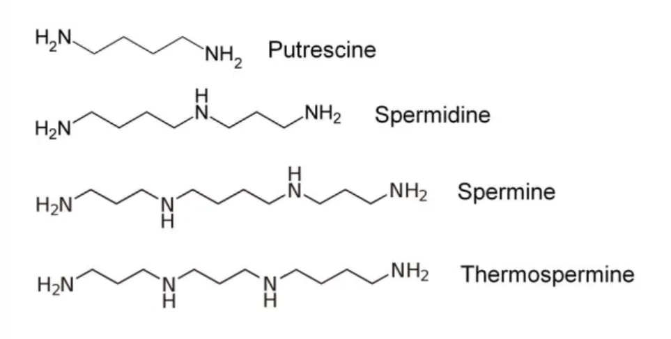
Polyamines: small polycations with major roles
Polyamines are low molecular weight, aliphatic polycations, having two or more primary amine groups and are present in almost all living organisms. Polyamines are involved in a variety of fundamental biological processes, including RNA modification, protein synthesis and the modulation of enzyme activities (Tabor and Tabor, 1999). The most abundant polyamines in flowering plants are the diamine putrescine, the triamine spermidine and the tetramines spermine and thermospermine (Figure 5), each with specific biological functions (reviewed in Takahashi and Kakehi, 2010). The following sections will briefly review the current knowledge on polyamine biosynthesis and catabolism; a special emphasis will be given to the current knowledge on polyamines role in plant vascular development.
Figure 5. Chemical structure of the main polyamines found in plants.
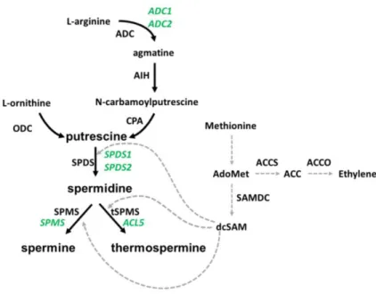
Polyamines biosynthesis and catabolism: a tight control of polyamine homeostasis
decarboxylase (ODC) or from L-arginine in a series of steps catalised by arginine decarboxylase (ADC), agmatine iminohydrolase (AIH) and N- -carbamoylputrescine amidohydrolase (CPA) (Figure 6). The ADC pathway seems specific to bacteria, archaea, and plants, contrary to animal and fungi, in which ODC is the first and rate-limiting enzyme in the synthesis of
Figure 6. Biosynthesis pathway of polyamines. Depicted in green are the genes identified in Arabidopsis thaliana to be involved in polyamine biosynthesis. Depicted in black are the enzymes responsible for the pathway steps. The cross-point to ethylene biosynthesis is indicated, since both pathways share SAM as common substrate. ADC: arginine decarboxylase; AIH: agmatine iminohydrolase; CPA: N-carbamoylputrescine amidohydrolase; ODC: ornithine decarboxylase; SPDS: spermidine synthase; SPMS: spermine synthase; tSPMS: thermospermine synthase; dcSAM: decarboxylated S-adenosylmethionine; SAMDC: SAM decarboxylase; ACCS: 1-amino-cycloproane-1-carboxylic-acid synthase; ACCO: ACC oxidase.
some ODC activity has been detected in this species, suggesting that another gene is responsible for ODC activity (Hanfrey et al., 2001; Tassoni et al., 2003). Putrescine is subsequently converted to the triamine spermidine by spermidine synthase (SPDS), an aminopropyltransferase that transfers an aminopropyl group from the decarboxylated S-adenosylmethionine (dcSAM) donor to the diamine putrescine. Similarly, spermidine is converted to spermine by the activity of spermine synthase (SPMS) or to thermospermine by the activity of thermospermine synthase (tSPMS). The aminopropyl moieties used by SPDS and SPMS are derived from SAM, which also serves as a common substrate for ethylene biosynthesis (Figure 6). In Arabidopsis, several genes encoding the enzymes in polyamines biosynthetic pathway have been described. Two genes have been found to encode for ADC activity (ADC1 and ADC2; Galloway et al., 1998), one that encodes AIH (AIH; Janowitz et al., 2003) and one that encodes CPA (NLP1; Piotrowski et al., 2003). Two genes encode for SPDS (SPDS1 and SPDS2; Hashimoto et al., 1998), one for SPMS (SPMS; Panicot et al., 2002) and one for tSMPS (ACL5; Knott et al., 2007).
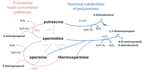
PAOs from Arabidopsis have been shown to oxidise spermine and spermidine in a similar mode as in animals and yeasts (Tavladoraki et al., 2006). Later back-conversion of spermine and thermospermine and its acetylated forms to spermidine, and spermidine to putrescine were suggested to take place in plants (Takahashi et al., 2010; Fincato et al., 2011). These discoveries changed the established idea that plant and animals have distinct catabolic pathways and showed plants also share a polyamine back-conversion pathway. In addition to their free forms, conjugation of polyamines with small molecules, such as amides and hydroxycinnamic acids occurs in plants (Alcázar et al., 2010; and references therein). Thus, it is clear that several mechanisms contribute to polyamine homeostasis, at the same time as polyamines and their catabolic products (e.g. hydrogen peroxide) play important roles during some developmental processes, such as stress responses and plant vascular development.
Polyamines in plant vascular development
Thermospermine, preventing a troubled premature death
Thermospermine is increasingly viewed as a novel plant growth regulator (Kakehi et al., 2010; Yoshimoto et al., 2012; Takano et al., 2012; Chapter II). Although it was first suggested that the defects in acl5 mutant result from deficient auxin transport (Clay and Nelson, 2005) some evidences show that IAA concentrations are actually increased in acl5 seedlings (Vera-Sirera et al., 2010). Similarly, the increased expression of the IAA marker line DR5::GUS in the hypocotyls of acl5 seedlings suggests that thermospermine interacts with auxin in differentiating xylem (Vera-Sirera et al., 2010). The relationship between auxin and thermospermine was elucidated by Yoshimoto et al. (2012), showing that thermospermine is
required to suppress the auxin-inducible xylem differentiation. 2,4- -dichlorophenoxy acid (2,4-D) and other auxin synthetic analogs were shown
thermospermine synthesis ability, closely resembles the acl5 mutant, and displays enlarged vascular tissues and high lignin content (Ge et al., 2006). The BUD2 gene is also induced by auxin (Cui et al., 2010), suggesting that polyamine levels or encoding transcripts might play a role in regulating auxin levels or responses to auxin.
Polyamines, ethylene and NO signals for death
Other polyamine catabolism and biosynthesis products have been shown to influence xylem differentiation. Tisi et al. (2011) demonstrated that after spermidine supply and PAO overexpression, the H2O2 derived from
gradient was inversely related to the degree of xylem differentiation. The authors observed a NO burst associated to the single cell layer of pro- -differentiating thin-walled xylem cells. The scavenging of NO inhibited the tracheary element differentiation but increased cell viability suggesting that NO production is sustained during secondary cell wall synthesis and cell autolysis (Gabaldón et al., 2005). The crosstalk among these signals and signaling pathways will be crucial to understand the trigger mechanism of cell death in xylem differentiation.
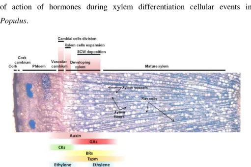
In summary, hormone signaling pathways have been identified in the last 15 years and increasing knowledge on the crosstalks that involve hormone signaling was gained. The developmental context of these cross-talks is also being elucidated. Many mechanisms of cambial state maintenance have been proposed, and new players, like thermospermine, are being brought to an already complex scenario. Figure 8 integrates the place of action of hormones during xylem differentiation cellular events in Populus.
vascular cambium reside the stem cells that are the meristematic cells from which the phloem and xylem precursor cells originate. Since the exact location of the stem cells is not known, this region is also commonly named cambial zone. Xylem development initiates with active cambial cell division, followed by rapid cell expansion and deposition of secondary cell wall and programmed cell death. The xylem cell types in Populus wood (ray parenchymatic living cells, xylem vessel elements and xylem fibers) are indicated. The known or possible function domains of auxin, gibberellin (GAs), cytokinin (CK), brassinosteroids (BRs), thermospermine (Tsmp) and ethylene across the Populus cambial zone are shown. Auxin (Tuominen et al., 1997) and GA (Isrraelsson et al., 2005) labels reflect the concentration of the bioactive hormones and expression pattern of auxin and GAs signaling genes (Moyle et al., 2002; Isrraelsson et al., 2005). CK label depicts the peak of expression of cytokinin signaling genes in the phloem side of the cambial zone (Nieminen et al., 2008). Ethylene label depicts its stimulatory effect on cambial cell division (Love et al., 2009; Pesquet and Tuominen 2011). Tspm label depicts place of action on developing xylem, delaying xylem cell death (Muñiz et al., 2008). BRs are currently not studied in Populus, thus the BR label depicts current knowledge from Arabidopsis and Zinnia showing that BRs are produced in procambial cells, perceived in the xylem precursor cells to induce xylem differentiation (see main text). The cork cell layers cover the surface of the stem, which are produced by the activity of the cork cambium, the other secondary lateral meristem in tree stems.
Research objectives and thesis layout
The present work aims at contributing to the understanding of wood formation, one of the most important and ultimately the defining biological process happening in a tree. Forestry production for wood and bioenergy products is increasingly attractive. Therefore, it is essential to deepen our knowledge of the molecular mechanisms governing wood formation.
signaling pathways in the cell is largely unknown. To help narrow this gap, we proposed to investigate the effects of altering thermospermine metabolism in Populus trees to provide some clues to these fundamental questions. Specifically, this work aims at:
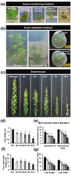
1. Understanding how thermospermine affects xylem formation in woody stems of Populus by altering the thermospermine metabolism through genetic modification of Populus to overproduce thermospermine.
2. Providing a broad view of other hormone crosstalk to thermospermine using a transcriptomic approach to screen for the main genes whose expression is affected by thermospermine metabolic de-regulation, especially those related to hormones and to xylem differentiation.
3. Contributing to the understanding of the regulatory mechanisms of thermospermine homeostasis in the Populus xylem, by studying how the manipulation of the transcript levels of putative upstream regulators affects the dynamics of thermospermine production.
Table I. Methods used in this work.
Method Chapter
Auxin response assays II
Electron microscopy for xylem analysis (II)
Gene identification by database search II, III, IV
Genetic transformation of Arabidopsis using floral dip method IV
Genetic transformation of Populus II, IV
Histological staning for lignin analysis (Phloroglucinol-HCl) II
Histological staining for cell viability (NBT) (II)
Histological general staining (Toluidine blue-O, Hematoxilin-Eosin) I, II, III, IV
Histological GUS staining (II)
IAA quantification by GC-MS (II)
In situ RNA hybridisation IV
Light and fluorescence microscopy I, II, III, IV
Microarray analysis III
Phylogenetic analysis II, IV
Plasmid construction II, IV
Polyamine extraction, purification, derivatization and quantification by GC-MS II, IV
Polymerase chain reaction (PCR) analysis II, IV
Promoter in silico analysis in databases IV
Quantitative real-time PCR analysis II, IV
Sectioning of plastic embedded samples II, III, IV
Site-directed mutagenesis II, IV
Tree growth measurements for growth analysis II, IV
Xylem chemical maceration for cell characterization II
Yeast assays for protein activity assessment (II)
Methods performed by collaborators are indicated in brackets.
Acknowledgements
References
Agusti J, Herold S, Schwarz M, Sanchez P, Ljung K, Dun EA, Brewer PB, Beveridge CA, Sieberer T, Sehr EM, Greb T (2011a) Strigolactone signaling is required for auxin-dependent stimulation of secondary growth in plants. Proc. Natl. Acad. Sci. U.S.A.108, 20242-20247.
Agusti J, Lichtenberger R, Schwarz M, Nehlin L, Greb T (2011b) Characterization of transcriptome remodeling during cambium formation identifies MOL1 and RUL1 as opposing regulators of secondary growth. PLoS Genet. 7, e1001312.
Aichinger E, Kornet N, Friedrich T, Laux T (2012) Plant stem cell niches. Annu. Rev. Plant Biol. 63, 615-636.
Alcázar R, Altabella T, Marco F, Bortolotti C, Reymond M, Koncz C, Carrasco P, Tiburcio AF (2010) Polyamines: molecules with regulatory functions in plant abiotic stress tolerance. Planta, 231, 1237-1249.
Aloni R (1987) Differentiation of vascular tissues. Annu. Rev. Plant Physiol. Plant Mol. Biol. 38, 179-204.
Alonso JM, Hirayama T, Roman G, Nourizadeh S, Ecker JR (1999) EIN2, a bifunctional transducer of ethylene and stress responses in Arabidopsis. Science, 284, 2148-2152.
Alonso JM, Stepanova AN (2004) The ethylene signaling pathway. Science, 306, 1513-1515.
Arabidopsis Genome Initiative (2000) Analysis of the genome sequence of the flowering plant Arabidopsis thaliana. Nature, 408, 796-815.
Avci U, Petzold HE, Ismail IO, Beers EP, Haigler CH (2008) Cysteine proteases XCP1 and XCP2 aid micro-autolysis within the intact central vacuole during xylogenesis in Arabidopsis roots. Plant J. 56, 303-315.
Baba K, Karlberg A, Schmidt J, Schrader J, Hvidsten TR, Bako L, Bhalerao RP (2011) Activity-dormancy transition in the cambial meristem involves stage-specific modulation of auxin response in hybrid aspen. Proc. Natl. Acad. Sci. U.S.A. 108, 3418-3423.
Baima S, Nobili F, Sessa G, Lucchetti S, Ruberti I, Morelli G (1995) The expression of the Athb-8 homeobox gene is restricted to provascular cells in Arabidopsis thaliana. Development, 121, 4171-4182.
Baucher M, El Jaziri M, Vandeputte O (2007) From primary to secondary growth: Origin and development of the vascular system. J. Exp. Bot. 58, 3485-3501.
Benková E, Ivanchenko MG, Friml J, Shishkova S, Dubrovsky JG (2009) A morphogenetic trigger: Is there an emerging concept in plant developmental biology? Trends Plant Sci.14, 189-193.
Berleth T, Jurgens G (1993) The role of the monopteros gene in organizing the basal body region of the Arabidopsis embryo. Development, 118, 575-587.
Bhalerao RP, Bennett MJ (2003) The case for morphogens in plants. Nat. Cell Biol. 5, 939-943.
Biles CL, Abeles FB (1991) Xylem sap proteins. Plant Physiol. 96, 597–601.
Bishopp A, Mähönen AP, Helariutta Y (2006) Signs of change: hormone receptors that regulate plant development. Development, 133, 1857-1869.
Bishopp A, Help H, El-Showk S, Weijers D, Scheres B, Friml J, Benková E, Mähönen AP, Helariutta Y (2011) A mutually inhibitory interaction between auxin and cytokinin specifies vascular pattern in roots. Curr. Biol.21, 917-926.
Björklund S, Antti H, Uddestrand I, Moritz T, Sundberg B (2007) Cross-talk between gibberellin and auxin in development of Populus wood: Gibberellin stimulates polar auxin transport and has a common transcriptome with auxin. Plant J. 52, 499-511.
Bollhöner B, Prestele J, Tuominen H (2012) Xylem cell death: emerging understanding of regulation and function. J. Exp. Bot. 63 1081-1094.
Brand U, Fletcher JC, Hobe M, Meyerowitz EM, Simon R (2000) Dependence of stem cell fate in Arabidopsis on a feedback loop regulated by CLV3 activity. Science, 289, 617-619.
Burrell M, Hanfrey CC, Murray EJ, Stanley-Wall NR, Michael AJ (2010) Evolution and multiplicity of arginine decarboxylases in polyamine biosynthesis and essential role in Bacillus subtilis biofilm formation. J. Biol. Chem. 285, 39224-39238.
Busov V, Yordanov Y, Gou J, Meilan R, Ma C, Regan S, Strauss S (2011). Activation tagging is an effective gene tagging system in Populus. Tree Genet. Genomes 7, 91-101.
Caño-Delgado A, Yin Y, Yu C, Vafeados D, Mora-Garcia S, Cheng JC, Nam KH, Li J, Chory J (2004) BRL1 and BRL3 are novel brassinosteroids receptors that function in vascular differentiation in Arabidopsis. Development, 131, 5341-5351.