The genus
Trichorhina
Budde-Lund in Brazil, with description of seven
new species (Isopoda, Oniscidea, Platyarthridae)
Leila A. Souza1, Joafrâncio P. de Araújo2 & Ivanklin S. Campos-Filho3
1. Laboratório de Carcinicultura, Instituto Superior de Ciências Biomédicas, Universidade Estadual do Ceará,Campus do Itaperi, Av. Paranjana, 1700, 60740-903, Fortaleza, Ceará, Brasil. (leilasouza5000@gmail.com.br)
2. Programa de Pós-Graduação em Ciências Biológicas (Zoologia), Departamento de Sistemática e Ecologia, Centro de Ciências Exatas e da Natureza, Universidade Federal da Paraíba, 58059-900, João Pessoa, Paraíba, Brasil. (joafrancio@gmail.com)
3. Programa de Pós-Graduação em Biologia Animal, Departamento de Zoologia, Instituto de Biociências, Universidade Federal do Rio Grande do Sul, Av. Bento Gonçalves, 9500, prédio 43435, 91501-970, Porto Alegre, Rio Grande do Sul, Brasil. (ivanklin.filho@gmail.com)
Platyarthridae includes seven genera and numerous nominal species. There are 12 species of
Trichorhina Budde-Lund, 1908 recorded for Brazil (SchmalfuSS, 2003; araújo & almerão, 2007).
leiStikow & wägele (1999) included Trichorhina
pearsei (Creaser, 1938) from Mexico as occurring in
Brazil. Perhaps this was a misinterpretation of Souza
-kury (1993), because its eyes could be compared with
those of the species described therein and to comments on its synonymy.
The number of ommatidia forming the eye is used here as a primary diagnostic feature. It appears that there is a general trend in eye reduction (making part of a regressive evolution also evident in the reduction of
body pigmentation) in many species of Trichorhina, not
necessarily related to environment but more generally to an endogeous lifestyle.
There are numerous specimens of anophtalmous
Trichorhina not yet studied and deposited at Museu Nacional do Rio de Janeiro, along with many specimens collected in caves from the Brazilian states of Bahia, Minas Gerais, Paraná, and São Paulo that are temporarily being kept in Universidade Estadual do Ceará (UECE) for further investigation.
MATERIAL AND METHODS
Type specimens are deposited in the Museu Nacional, Universidade Federal do Rio de Janeiro (MNRJ), Rio de Janeiro, Brazil. The material examined of Trichorhina acuta
is deposited in the collection of Oniscidea at Universidade
Estadual do Ceará, Fortaleza, Brazil (UECE).
ABSTRACT. Seven new species of Trichorhina Budde-Lund, 1908 are described, six from Southeastern Brazil, T. biumbonata sp. nov., T. lenkoi
sp. nov., T. myrmecophila sp. nov., T. orensis sp. nov., T. sexdens sp. nov., T. tropidocerata sp. nov., and one from Central Brazil, T. crassisetae
sp. nov. An emended diagnosis for the genus and a key to the 19 species recorded from Brazil are provided. Trichorhinaacuta Araújo & Buckup, 1994 is recorded to state of Mato Grosso do Sul.
KEYWORDS. Terrestrial isopods, taxonomy, South America, Neotropical Region.
RESUMO. O gênero Trichorhina Budde-Lund no Brasil, com descrição de sete espécies novas (Isopoda, Oniscidea, Platyarthridae). Sete espécies novas de Trichorhina Budde-Lund, 1908 são descritas, seis do sudeste do Brasil, T. biumbonata sp. nov., T. lenkoi sp. nov., T. myrmecophila sp. nov., T. orensis sp. nov., T. sexdens sp. nov. e T. tropidocerata sp. nov., e uma do Centro-Oeste, T. crassisetae sp. nov. São fornecidos uma diagnose emendada para o gênero e chave para as 19 espécies registradas para o Brasil. É registrada a ocorrência de Trichorhina acuta Araújo & Buckup, 1994 para o Mato Grosso do Sul.
PALAVRAS-CHAVE. Isópodes terrestres, taxonomia, América do Sul, Região Neotropical.
Five new species possess between four to six ommatidia and they may be compared to species which also possess a similar number of ommatidia. The other new taxa of Trichorhina, even though they possess between four and six ommatidia, could be considered unrelated with the new ones because of other unique features.
The structures to which we here refer as “rods”
have been called by Vandel (1952) “fins bâtonnets
cilindriques”. Graphics illustrating relative position of
noduli laterales are used according to Vandel (1962). Measurements refer to maximum values.
TrichorhinaBudde-Lund, 1908 Type species: Bathytropa thermophila (Dollfus, 1896)
Diagnosis. Body length not exceeding 6 mm. Pigmentation vestigial or absent. If there are eyes, they can be composed of up to 15 ommatidia. Tegument with
fan-shaped scale-setae. Exite of maxillula with simple, bifid
or serrate teeth. Noduli laterales with or without lateral projections, 1 per side on pereonites I–VI and 1–2 per side on pereonite VII or as a double row per side. Frontal line
absent (except in Trichorhina minutissima Budde-Lund,
1913, T. micros Budde-Lund, 1913 and T. atlasi Vandel,
1959). Without glandular pores [except in T. argentina
Vandel, 1963 and T. boliviana (Vandel, 1952)]. Antennal
flagellum bi-articulated; second joint much longer than first
and sometimes with a suture. Telson triangular [except in
240
Distribution. Trichorhina is cosmopolitan. There
is a total of 56 species, of which about 30 occur in the
Neotropical Region (SchmalfuSS, 2003). There are 12
species hitherto recorded in Brazil, which added to the seven new species herein described, give a total of 64 species for the world (see Table I).
Remarks. Species of Trichorhina are mostly
tropical. It is probably an artificial assemblage (e.g.taiti
& ferrara, 1987). According to the current knowledge, there is a high degree of endemism for the majority of species but there are a few widespread species, such as
T. tomentosa (Budde-Lund, 1893) and T. heterophthalma
Lemos de Castro, 1964 which probably were dispersed by man.
Trichorhina albida Budde-Lund, 1908 from Madagascar. Possesses unique dorsal knobs (“dorsal buttons”, Verhoeff, 1946) (although according to Dr. F. Ferrara pers. comm., in Budde-lund’S (1908) original description no dorsal knobs are mentioned and Verhoeff has not reexamined the type specimens).
Trichorhina dobrogica Radu, 1960 from Romania. It may be distinguished from the Brazilian species by the transverse lines on pereonites I–VI, and a distal joint of
antennula with nine aesthetascs (five or six in Brazilian
species).
Trichorhina giannelli Arcangeli, 1929 from the Antilles and Central America. Distinguished from the new Brazilian species by the longitudinal ridges on the mesepistome.
Trichorhina guanophila Souza-Kury, 1993 from Brazil. This species belongs to the group of species
characterized by a double pair of noduli laterales on
pereonite VII.
Trichorhina micros from Mauritius and
Trichorhina minutissima from Cargados (Indian Ocean). These taxa belong to the group of species characterized by serrate teeth on the outer ramus of the maxillulae and by a double row of noduli laterales per side (taiti &
ferrara, 1987).
Trichorhina minima Schmalfuss & Ferrara, 1978 from Togo. It is distinguished from Brazilian species by the bulbous profrons.
Trichorhina quisquiliarum (Budde-Lund, 1893) from Venezuela. It is distinguished from new Brazilian species by the uneven pigmentation of ommatidia, four unpigmented and two dark colored.
Trichorhina tatianae Araújo & Almerão, 2007 from Brazil. It is distinguished from the new Brazilian species by the pronounced lateral projection on male pereopod VII ischium.
Besides these nine species, there are ten others with four to six ommatidia, whose differences are cited separately under the diagnosis of each new species.
These are: Trichorhina acuta from Brazil; T. argentina
from Argentina; T. australiensis Wahrberg, 1922 from
Australia; T. barbouri (van Name, 1926) from Panama;
T. hospes Silvestri, 1918 from Nigeria; T. pallida
Barnard, 1960 from Mozambique; T. papillosa
(Budde-Lund, 1893) from Venezuela; T. paraensis Souza-Kury,
1997 from Brazil; T. silvestrii Arcangeli, 1936 from
Spain; and T. vandeli Rioja, 1955 from Mexico.
Trichorhina biumbonata sp. nov. (Figs 1, 8–24)
Type material. Holotype ♂, BRAZIL, São Paulo: Descalvado, Escaramuça, 25.VI.1944, O. Schubart col. (MNRJ 21420). Paratypes:
3♂, 3♀, same data as holotype (MNRJ 21421); ♂, 2♀, Descalvado,
Diamantina, 27.VIII.1944, J. Schubart & O. Schubart col. (MNRJ 11515).
Diagnosis. Pigmentation pale yellow. Eyes with
five dark brown ommatidia. Second joint of antennal
peduncle with a crest in the outer border. Second joint of
antennal flagellum with a faint proximal groove.
Measurements. Male length: 2.75 mm, width: 1.24 mm; female length: 3.03 mm, width: 1.51 mm.
Description. Pigmentation of body faint, pale yellow, head with small brown spots. Eyes dark brown
with five ommatidia (Fig. 10). Pereonite I with anterior
margins reaching the eyes (Fig. 8). Cephalic lateral lobes small, shorter than median lobe which has rounded apex and straight sides (Fig. 9). Pleon slightly narrower than pereon. Pereon, pleon and telson densely provided with fan-shaped scale-setae (Fig. 15). In the posterior margins of pereonites these fan-shaped scale-setae are large and wide, alternating with small and narrow ones, and in the lateral margins they are small. Both median axes of fan-shaped scale-setae evident, as well as the
secondary supporting axioles. Noduli laterales with
featureless base. Pereonite VII with only one nodulus
lateralis on each side. Position of noduli laterales as illustrated (Fig. 1). Antennula with distal joint with six to seven aesthetascs (Fig. 11). Second joint of antennal peduncle with longitudinal sinuous crest in outer border, more evident in the female (Fig. 12). Left mandible without rods between molar and incisory processes (Fig. 13). Six to seven penicils in molar process of mandibles (Figs 13, 14). Outer group of exite of maxillulae with three teeth and inner group with four undivided teeth (Fig. 16). Maxilla with inner lobe narrower than outer lobe. Sensilla on the inner lobe (Fig. 17). Endite of maxilliped with one small tooth in outer distal border, and with inner distal border smooth (Fig. 18). Pleopods without respiratory areas.
Male. Pereopods I (Fig. 19) and VII (Fig. 20) without apparent sexual dimorphism. Pleopod I with heart–shaped exopod (Fig. 21); endopod slender with half distal slightly turned to the outside and simple apex (Fig. 22). Pleopod II with triangular exopod (Fig. 24); endopod with distal half strongly tapered (Fig. 23). Pleopod V with exopod subtriangular.
Remarks. Trichorhina biumbonata sp. nov.
is distinguished from the species with four to six ommatidia by the antennal crest, and further as follows:
from T. acuta by maxillulae with undivided teeth (two
bifid in T. acuta). From T. argentina by the exopod of
pleopod I of male cordiform (ovoid in T. argentina).
From T. australiensis by 1) molar process of mandibles
with six to seven penicils (one in T. australiensis); 2)
seven undivided teeth in maxillulae (in T. australiensis
nine, of which two bifid); 3) endite of maxilliped without
tooth in inner distal border; with only one in outer distal border (one tooth in inner distal border; one in distal outer in T. australiensis). From T. barbouri by 1) second
joint of antennal flagellum of female without groove; 2)
tegument smooth. From T. hospes by maxillulae with
seven undivided teeth (in T. hospes nine, two bifid). From T. pallida by 1) molar process of mandibles with five to
seven penicils (two in T. pallida); 2) exite of maxillulae
with seven undivided teeth (two of inner group bifid in
T. pallida). From T. papillosa by 1) absence of papillae
on tegument of body; 2) endopods of uropods surpass
the tip of telson and of protopods. From T. paraensis
by 1) antennula with distal joint with six to seven
aesthetascs altogether (five altogether in two groups in
T. paraensis); 2) molar process of both mandibles with six to seven penicils (one penicil in T. paraensis). From
T. silvestrii by 1) antennula with distal joint with six to
seven aesthetascs altogether (four in two groups in T.
silvestrii); 2) molar process of both mandibles with six to seven penicils (three in T. silvestrii). From T. vandeli by
1) maxillulae with seven undivided teeth (in T. vandeli
eight, two bifid); 2) endite of maxilliped with one tooth
in outer distal border (two teeth in T. vandeli).
Etymology. From Latin bi = two + umbo =
protuberance, due to the sinuous protuberance forming a crest on the antennal peduncle.
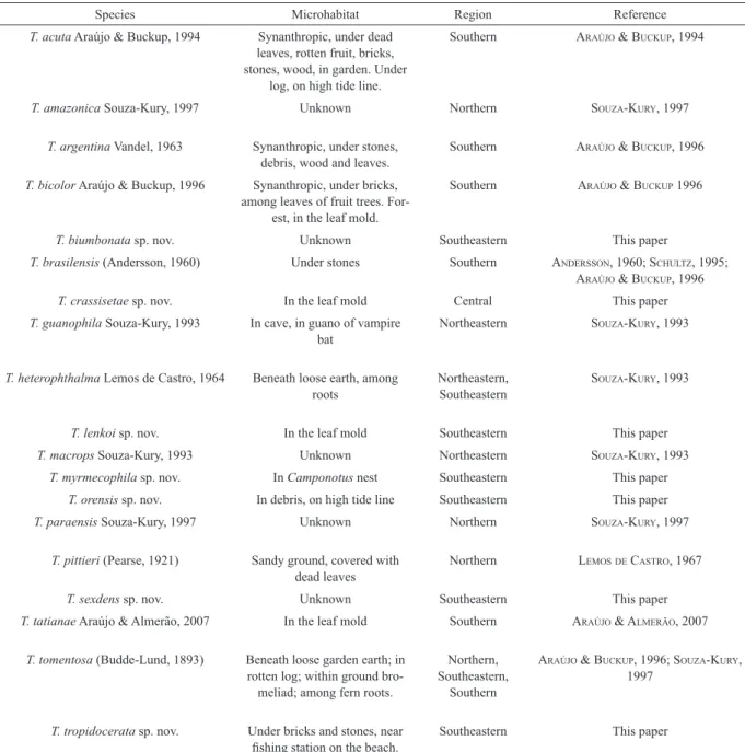
Tab. I. Brazilian records of species of Trichorhina by microhabitats, regions and reference.
Species Microhabitat Region Reference
T. acuta Araújo & Buckup, 1994 Synanthropic, under dead leaves, rotten fruit, bricks, stones, wood, in garden. Under
log, on high tide line.
Southern araújo & Buckup, 1994
T. amazonica Souza-Kury, 1997 Unknown Northern Souza-kury, 1997
T. argentina Vandel, 1963 Synanthropic, under stones, debris, wood and leaves.
Southern araújo & Buckup, 1996
T. bicolor Araújo & Buckup, 1996 Synanthropic, under bricks, among leaves of fruit trees.
For-est, in the leaf mold.
Southern araújo & Buckup 1996
T. biumbonata sp. nov. Unknown Southeastern This paper
T. brasilensis (Andersson, 1960) Under stones Southern anderSSon, 1960; Schultz, 1995;
araújo & Buckup, 1996
T. crassisetae sp. nov. In the leaf mold Central This paper
T. guanophila Souza-Kury, 1993 In cave, in guano of vampire bat
Northeastern Souza-kury, 1993
T. heterophthalma Lemos de Castro, 1964 Beneath loose earth, among roots
Northeastern, Southeastern
Souza-kury, 1993
T. lenkoi sp. nov. In the leaf mold Southeastern This paper
T. macrops Souza-Kury, 1993 Unknown Northeastern Souza-kury, 1993
T. myrmecophila sp. nov. In Camponotus nest Southeastern This paper
T. orensis sp. nov. In debris, on high tide line Southeastern This paper
T. paraensis Souza-Kury, 1997 Unknown Northern Souza-kury, 1997
T. pittieri (Pearse, 1921) Sandy ground, covered with dead leaves
Northern lemoSde caStro, 1967
T. sexdens sp. nov. Unknown Southeastern This paper
T. tatianae Araújo & Almerão, 2007 In the leaf mold Southern araújo & almerão, 2007
T. tomentosa (Budde-Lund, 1893) Beneath loose garden earth; in rotten log; within ground
bro-meliad; among fern roots.
Northern, Southeastern,
Southern
araújo & Buckup, 1996; Souza-kury,
1997
T. tropidocerata sp. nov. Under bricks and stones, near
242
Figs 1–7. Ratio b/c and d/c of noduli laterales on pereonites I–VII: 1, Trichorhina biumbonata sp. nov.; 2, Trichorhinacrassisetae sp. nov.; 3,
Trichorhinalenkoi sp. nov.; 4, Trichorhinamyrmecophila sp. nov.; 5, Trichorhinaorensis sp. nov.; 6, Trichorhinasexdens sp. nov.; 7, Trichorhina tropidocerata sp. nov.
Figs. 8–18. Trichorhinabiumbonata sp. nov. Female, habitus: 8, dorsal; 9, cephalothorax and two first pereonites, lateral view; 10, cephalothorax,
frontal view. Male: 11, antennula; 12, antenna; 13, left mandible; 14, right mandible; 15, fan-shaped scale-seta of pereonite; 16, exite of maxillula;
244
Trichorhina crassisetae sp. nov. (Figs 2; 25–43)
Type material. Holotype ♂, BRAZIL, Mato Grosso do Sul: Três Lagoas (Sucuriú river, Corredeira Chupão), in the forest along the river, in the leaf mold, VI.1964, without collector (MNRJ 4113).
Paratypes: 8♂, 8♀, same data as holotype (MNRJ 4114).
Diagnosis. Pigmentation of body dark brown. Eyes with eight black ommatidia. Peduncle of antenna with conspicuous bristles. Exite of maxillulae with four teeth in the outer group, one very small. Pereopod I of male with setae bifurcated in carpus. Endopod of pleopod I of male with row of small setae in inner border. Measurements. Male, length: 3.18 mm, width: 1.19 mm; female, length: 2.52 mm, width: 1.06 mm.
Description. Pigmentation of body dark brown; lighter in head (with small dark brown spots) (Fig. 25), antennae, sides of pereon and of pleon and uropods. Eyes black with eight ommatidia. Pereonite I with anterior margins reaching the eyes. Cephalic lateral lobes small, shorter than median lobe which has rounded apex and straight sides (Fig. 26). Eyes with eight ommatidia. Pleon outline continuous with pereon; pleonites III–V with well developed epimera. Pereon, pleon and telson covered with fan-shaped scale-setae (Fig. 27). In the posterior margins of pereonites, scale-setae large and wide intercalate with small and narrow, and in the lateral margins small. Fan-shaped scale-setae more or less rounded and with four shafted frame.
Noduli laterales with featureless base. Pereonite VII
with only one nodulus lateralis on each side. Position
of noduli laterales as illustrated (Fig. 2). Distal joint of
antennula with five aesthetascs (Fig. 28). Antenna (Fig.
29) stretched reaches posterior margin of pereonite II. Second joint of antennal peduncle without dorsal keel nor crest in outer border. Second joint of antennal
flagellum without groove. Left mandible (Fig. 30)
without rods between molar and incisory processes. Six
penicils in molar process of left mandibles (Fig. 30), right four (Fig. 31). Outer group of exite of maxillulae with four teeth, one much smaller. Inner group of exite
of maxillulae with four teeth: two bifid (Fig. 32). Maxilla
with inner lobe narrower than outer lobe. Sensilla on the inner lobe (Fig. 33). Endite of maxilliped without small teeth in outer distal border, and with inner distal border smooth (Fig. 34). Pleopods without respiratory areas.
Male. Pereopod I (Fig. 35) with bifurcate setae in carpus, contrasting to that of female (Fig. 37); pereopod VII (Fig. 36) without prominent sexual dimorphism, compared to that of female (Fig. 38). Pleopod I with exopod subrectangular (Fig. 39); endopod with a row of small setae parallel to the inner border (Fig. 40). Pleopod II with triangular exopod (Fig. 41); endopod with distal half slender (Fig. 42). Pleopod V with exopod subrectangular (Fig. 43).
Remarks. Trichorhina crassisetae sp. nov.
is distinguished from species with eight to twelve
ommatidia as follows: from T. amazonica Souza-Kury,
1997 by 1) molar process of left mandible with six penicils (four in T. amazonica); 2) maxillulae with eight
teeth, two bifid (in T. amazonica seven, two bifid). From T. atlasi by 1) absence of frontal line (frontal line very
thin in T. atlasi); 2) sexual dimorphism in pereopod I
of male: carpus with remarkable bifurcate setae (males
without particular features, see Vandel 1959:101, for
T. atlasi); 3) pleonites three to five with well developed
points (blunt in T. atlasi). From T. kribensis Ferrara & Schmalfuss, 1983 by 1) antenna without aesthetascs;
2) exite of maxillulae with eight teeth, two bifid (nine
undivided in T. kribensis); 3) pereopod VII of male
without dimorphism. From T. marianii Arcangeli, 1930
by 1) tegument smooth (granulous in T. marianii); 2)
sexual dimorphism in pereopod I of male: carpus with bifurcate setae (sexual dimorphism only in length and
width of merus and carpus in T. marianii). From T.
pubescens (Dollfus, 1893) by 1) antenna with joints
Figs. 19–24. Trichorhina biumbonata sp. nov. Male: 19, pereopod I; 20, pereopod VII; 21, exopod of pleopod I; 22, endopod of pleopod I; 23, exopod of pleopod II; 24, endopod of pleopod II. Scale bars: 0.1 mm.
246
Figs. 35–43. Trichorhinacrassisetae sp. nov. Male: 35, pereopod I; 36, pereopod VII. Female: 37, pereopod I; 38, pereopod VII. Scale bars: Figs 35–38, 0.1 mm; arrowed details, 0.01 mm; 39, exopod of pleopod I; 40, endopod of pleopod I; 41, exopod of pleopod II; 42, endopod of pleopod II; 43, exopod of pleopod V. Scale bars: Figs 39–43, 0.1 mm; arrowed details, 0.01 mm.
of maxillulae with six undivided teeth and one bifid (two of inner group bifid in T. acuta). From T. argentina
by 1) antenna shorter; 2) eyes light brown (black in T.
argentina); 3) coxal plates without glandular pores.
From T. australiensis by 1) exite of maxillulae with
six undivided teeth and one bifid (in T. australiensis
nine, inner group with all bifid); 2) presence of frontal
lobe. From T. barbouri by second joint of antennal
flagellum without groove and tegument smooth. From
T. biumbonata sp. nov. by 1) second joint of antennal peduncle without crest in outer border; 2) molar process
of mandibles with one penicil (five to seven in T. biumbonata). From T. hospes by 1) exite of maxillulae
with six undivided teeth and one bifid (in T. hospes
nine, two bifid); 2) endite of maxilliped with two teeth
in outer distal border (without teeth in T. hospes). From
T. pallida by 1) molar process of mandibles with one penicil (two in T. pallida); 2) exite of maxillulae with six
undivided teeth and one bifid (two of inner group bifid
in T. pallida). From T. papillosa by 1) molar process
of mandibles with one penicil (three in T. papillosa);
2) exite of maxillulae with six undivided teeth and one
bifid (two bifid in T. papillosa); 3) endite of maxilliped with two teeth in outer distal border and one in inner distal border. From T. paraensis by 1) exite of maxillulae
with six undivided teeth and one bifid (in T. paraensis
eight, two bifid); 2) endite of maxilliped with two teeth
in distal outer border (with one tooth in distal outer
border and one tooth in inner border in T. paraensis).
From T. silvestrii by 1) molar process of both mandibles
with one penicil (with three penicils in T. silvestrii); 2) exite of maxillulae with six undivided teeth and one
bifid (in T. silvestrii eight, two bifid). From T. vandeli by
1) antennula with distal joint with about five aesthetascs
(with seven to eight aesthetascs in T. vandeli); 2) exite
of maxillulae with six undivided teeth and one bifid (in
T. vandeli eight, two bifid).
Etymology. Species named after Karol Lenko, who collected the type series, and many other Brazilian Oniscidea.
Trichorhina myrmecophila sp. nov. (Figs 4, 58–78)
Type material. Holotype ♂, BRAZIL, São Paulo: Barueri, in nest of Camponotus rufipes (Fabricius, 1775) (Hymenoptera,
Formicidae), 14.X.1967, K. Lenko col. (MNRJ 4117). Paratypes: 3♂, 18♀, same data as holotype (MNRJ 4118).
Diagnosis. Pigmentation of body pale yellow.
Eyes with five black ommatidia. Pleopod I of male with
exopod slightly rounded; endopod with apex with row of micro-setae.
Measurements. Male, length: 3.00 mm, width: 1.00 mm; female, length: 2.30 mm, width: 0.92 mm.
Description. Pigmentation of body faint, pale yellow, with small brown spots in head. Eyes black
with five ommatidia. Head partly sunken in pereonite
II–V of peduncle without inferior groove; 2) exite of
maxillulae with eight teeth, of which two bifid (all teeth
undivided in T. pubescens). Promptly distinguishable
from all other species of the genus by eyes with eight ommatidia.
Etymology. From Latin crassus = thick + seta =
bristle, due to the stout bristles in antenna.
Trichorhina lenkoi sp. nov. (Figs 3, 44–57)
Type material. Holotype ♂, BRAZIL, São Paulo: São Roque, Araçariguama, in the leaf mold, 11.IV.1970,
K. Lenko col. (MNRJ 4115). Paratypes: 4♂, 2♀, same
data as holotype (MNRJ 4116).
Diagnosis. Pigmentation of body brown. Eyes
with five ommatidia light brown. Exite of maxillulae with six undivided teeth plus one bifid tooth.
Measurements. Male, length: 3.03 mm, width: 1.10 mm; female, length: 2.34 mm, width: 0.96 mm.
Description. Pigmentation of body brown, with small dark brown spots in head. Eyes light brown with
five ommatidia, not contiguous. Pereonite I with anterior
margins reaching the eyes. Cephalic lateral lobes small, shorter than median lobe which has apex rounded and straight sides (Fig. 44). Pleon outline continuous with pereon; telson subtriangular with sides slightly convex. Surface with hexagonal plates. Pereon, pleon and telson covered with fan-shaped scale-setae (Fig. 46), with four
axial rods. Noduli laterales (Fig. 45) with featureless
base. Pereonite VII with only one nodulus lateralis on
each side. Position of noduli laterales as illustrated (Fig.
3). Antennula with distal joint with about five aesthetascs.
Antenna (Fig. 47) stretched reaches posterior margin of pereonite II. Second joint of antennal peduncle without crest in outer border and without dorsal keel.
Second joint of antennal flagellum without groove.
Left mandible (Fig. 48) without rods between molar and incisory processes. Number of penicils in molar process of left mandibles one (Fig. 48), right one (Fig. 49). Outer group of exite of maxillulae with three teeth. Inner group of exite of maxillulae with four teeth: one
bifid (Fig. 50). Maxilla with inner lobe narrower than
outer lobe. Sensilla on the inner lobe. (Fig. 51). Endite of maxilliped with two small teeth in distal outer border, and with inner distal border smooth (Fig. 52). Pleopods without respiratory areas.
Male. Pereopods I (Fig. 53) and VII (Fig. 54) without apparent sexual dimorphism. Endopod of pleopod I with distal half slightly turned to the outside, apex simple (Fig. 55). Pleopod II with exopod triangular (Fig. 57); endopod with distal half slender (Fig. 56).
Remarks. Trichorhina lenkoi sp. nov. is
distinguished from other species with four to six
ommatidia as follows: from T. acuta by 1) molar process
248
Figs. 44–57. Trichorhina lenkoi sp. nov., male: 44, cephalothorax, frontal view; 45, nodulus lateralis; 46, fan-shaped scale-seta of pereonite; 47, antenna; 48, left mandible; 49, right mandible; 50, exite of maxillula; 51, maxilla; 52, maxilliped. Scale bars: Fig. 44, 0.5 mm; Figs 45–46, 0.01 mm; Figs 47–52, 0.1 mm; 53, pereopod I; 54, pereopod VII; 55, endopod of pleopod I, detail; 56, endopod of pleopod II; 57, exopod of pleopod II. Scale bars: 0.1 mm.
I, anterior margins of pereonite I reaching the eyes. Cephalic lateral lobes slightly ahead of median lobe which has apex rounded and straight sides (Figs 59, 60). Pleon slightly narrower than pereon, pleonites III–V with well developed points (Fig. 58). Body surface with hexagonal plates. Pereon, pleon and telson covered with fan-shaped scale-setae bearing two axial rods (Fig.
63). Noduli laterales with featureless base (Fig. 62).
Pereonite VII with only one nodulus lateralis on each
side. Position of noduli laterales as illustrated (Fig. 4). Antennula with middle joint much smaller than others,
distal joint with five aesthetascs (Fig. 61). Antenna (Fig.
64) stretched reaches posterior margin of pereonite II. Second joint of antennal peduncle without crest in outer border and without dorsal keel. Second joint of
antennal flagellum with a slightly transversal groove.
Left mandible (Fig. 65) without rods between molar and incisory processes. Number of penicils in molar process of left mandibles one (Fig. 65), right one (Fig. 66). Outer group of exite of maxillulae with three teeth. Inner group of exite of maxillulae with four simple teeth (Fig. 67). Maxilla with inner lobe narrower than outer lobe wich is apically truncate. Sensilla on the inner lobe (Fig. 68). Endite of maxilliped with two small teeth in outer distal border, and irregular inner distal border (Fig. 69). Pleopods without respiratory areas.
Male. Pereopods I (Fig. 70) and VII (Fig. 71) without apparent sexual dimorphism (see pereopods I
and VII of female in figures 73 and 72, respectively). Carpus of pereopod I of male without bristle field.
Pleopod I with exopod rounded (Fig. 74); endopod (Fig. 75) with distal half slightly turned to the outside and apex armed with a short row of micro-setae parallel to the inner border. Pleopod II with triangular exopod (Fig. 77); endopod with distal half gradually tapered (Fig. 76). Pleopod V with sub-rectangular exopod (Fig. 78).
Remarks. Trichorhina myrmecophila sp. nov.is
distinguished from other species with eye with four to six
ommatidia as follows: from T. acuta by 1) molar process
of mandibles with one penicil (six in T. acuta); 2) exite of maxillulae with seven undivided teeth (two of inner
group bifid in T. acuta). From T. argentina by 1) exopod
of pleopod I of male rounded (ovoid in T. argentina);
2) endopods of uropods relatively longer. From T.
australiensis by 1) antennula with joints of different proportions; 2) exite of maxillulae with seven undivided teeth (in T. australiensis nine, five of which bifid); 3)
exopod of pleopod I of male rounded (narrowed in inner side in T. australiensis). From T. barbouri by 1) second
joint of antennal flagellum without groove and tegument
smooth. From T. biumbonata sp. nov. by 1) second joint
of antennal peduncle without crest in outer border; 2) molar process of mandibles with one penicil (with about
seven in T. biumbonata); 3) endite of maxilliped with
two teeth in upper outer margin (one in T. biumbonata);
4) pereopod I of male with setae undivided (with up
to four points in T. biumbonata). From T. hospes by 1)
antennula with distal joint with five aesthetascs (six in T. hospes); 2) exite of maxillulae with seven undivided teeth (in T. hospes nine, two bifid). From T. lenkoi sp. nov. by
1) pigmentation of body pale yellow (light brown in T.
lenkoi); 2) eyes black (light brown in T. lenkoi); 3) pleon slightly narrower than pleon (continuous with pereon in
T. lenkoi); 4) endopod of pleopod I of male with row of
micro-setae in apex. From T. pallida by 1) molar process
of mandibles with one penicil (two in T. pallida); 2)
exite of maxillulae with seven undivided teeth (in T.
pallida seven, two bifid); 3) exopod of pleopod I of male
rounded (elliptic in T. pallida). From T. papillosa by 1)
molar process of mandibles with one penicil (three in T.
papillosa); 2) exite of maxillulae with seven undivided teeth (in T. papillosa seven, two bifid); 3) endite of
maxilliped with two teeth in distal outer border. From T.
paraensis by 1) pereon, pleon and telson with fan-shaped
scale-setae (with ovoid scale-setae in T. paraensis); 2)
endite of maxilliped with two teeth in outer distal border (with one tooth in T. paraensis). From T. silvestrii by 1)
distal joint of antennula with five aesthetascs (four in T. silvestrii); 2) exite of maxillulae with seven undivided teeth (in T. silvestrii eight, two bifid). From T. vandeli by
1) distal joint of antennula with five aesthetascs (seven
to eight in T. vandeli); 2) molar process of mandibles
with one penicil (with a group of many penicils in T.
vandeli).
Etymology. Species name stems from Greek and refers to the collecting of the type series in nest of ants.
Trichorhina orensis sp. nov. (Figs 5, 79–94)
Type material. Holotype ♂, BRAZIL, Rio de Janeiro: Ilha de Itacuruçá, Praia dos Mocambos (supra tidal line, in garbage), 15.XII.1973, A. L. Castro & B. Prazeres col. (MNRJ 4119). Paratypes:
12♂, 41♀, same data as holotype (MNRJ 4120).
Diagnosis. Body unpigmented. Eyes with five
brown ommatidia. Molar process of mandible with one
penicil. Exite of maxillulae with seven teeth, two bifid.
Measurements. Male, length: 2.48 mm, width: 0.83 mm; female, length: 2.89 mm, width: 0.99 mm.
Description. Body unpigmented. Eyes brown with
five ommatidia. Pleon outline continuous with pereon
(Fig. 79); telson subtriangular with rounded apex of sides slightly concave. Pereon, pleon and telson covered with fan-shaped scale-setae. In the posterior margins of pereonites they are large and wide, alternating with small and narrow ones, and in the lateral margins they are small. Both median axes of fan-shaped scale-setae evident, as well as the secondary supporting axioles
(Fig. 83). Noduli laterales with simple base (Fig.
84). Pereonite VII with one nodulus lateralis on each
side. Position of noduli laterales as illustrated (Fig.
250
Figs. 58–69. Trichorhinamyrmecophila sp. nov. Female, habitus: 58, dorsal; 59, cephalothorax and two first pereonites, lateral view; 60, cepha -lothorax, frontal view. Male: 61, antennula; 62, nodulus lateralis; 63, fan-shaped scale-seta of pereonite; 64, antenna; 65, left mandible; 66, right mandible; 67, exite of maxillula; 68, maxilla; 69, maxilliped. Scale bars: Fig. 58, 1 mm; Figs 59–60, 0.5 mm; Figs 62–63, 0.01 mm; Figs 61, 64–69, 0.1 mm.
252
crest in outer border and without dorsal keel. Second
joint of antennal flagellum without groove (Fig. 82).
Left mandible without rods between molar and incisory processes. Number of penicils in molar process of left mandibles one, right one (Fig. 86). Outer group of exite of maxillulae with three teeth. Inner group of exite of
maxillulae with four teeth: two bifid (Fig. 87). Maxilla
with inner lobe narrower than outer lobe. Sensilla on the inner lobe, with a sinuous median margin (Fig. 88). Endite of maxilliped with two small teeth in distal outer border, and with inner distal border smooth (Fig. 89). Pleopods without respiratory areas.
Male. Carpus of pereopod I of male (Fig. 90)
with bristle field. Pereopod VII (Fig. 91) without
sexual dimorphism. Pleopod I with endopod with distal half turned to the outside and apex slightly crenulate (Fig. 92). Pleopod II (Fig. 93) with triangular exopod; endopod with distal half strongly narrowed. Pleopod V with subtriangular exopod (Fig. 94).
Remarks. Trichorhina orensis sp. nov. is
distinguished from other species with four to six
ommatidia, as follows: from T. acuta by molar process
of mandibles with one penicil (six in T. acuta). From
T. argentina by 1) body unpigmented (purplish uniform
pigmentation in T. argentina); 2) brown ommatidia
(black in T. argentina); 3) coxal plates without glandular
pores. From T. australiensis by exite of maxillulae with
seven teeth, of which two bifid (in T. australiensis nine,
five bifid), and endite of maxilliped without tooth in
distal inner border. From T. barbouri by second joint
of antennal flagellum without groove and tegument
smooth. From T. biumbonata sp. nov. by 1) second joint
of antennal peduncle without crest in outer border; 2) molar process of mandibles with one penicil (with about
seven in T. biumbonata); 3) endite of maxilliped with
two teeth in distal outer border (one in T. biumbonata).
From T. hospes by exite of maxillulae with seven teeth,
of which two bifid (in T. hospes nine, two bifid). From T. lenkoi sp. nov. by endite of maxilliped without teeth in inner distal border, and dactylus of pereopod I of male with inner seta larger. From T. myrmecophila sp. nov. by
the inner group of exite of maxillulae with two bifid teeth
(four undivided teeth in T. myrmecophila), and endopod
of pleopod II of male without row of scale-setae. From
T. pallida by the molar process of mandibles with one
penicil (two in T. pallida), and endopod of pleopod I of
male with distal half turned to the outside (straight in
T. pallida). From T. papillosa by the molar process of
mandibles with one penicil (three in T. papillosa), and
endite of maxilliped with two teeth in outer distal border. From T.paraensis by exite of maxillulae with seven teeth,
of which two bifid (in T. paraensis eight, two bifid), and
endite of maxilliped without teeth in inner distal border
(with one tooth in inner distal border in T. paraensis).
From T. silvestrii by distal joint of antennula with about eight aesthetascs (four in T. silvestrii), and molar process
of mandibles with one penicil (three in T. silvestrii).
From T. vandeli by molar process of mandibles with one
penicil (with a group of many penicils in T. vandeli), and
exite of maxillulae with seven teeth, of which two bifid
(in T. vandeli eight, two bifid).
Etymology. From Latin ora = beach, referring to
microhabitat where the individuals were collected.
Trichorhina sexdens sp. nov. (Figs 6, 95–107)
Type material. Holotype ♂, BRAZIL, São Paulo: Ilha dos Búzios, 16.X to 04.XI.1963, K. Lenko col. (MNRJ 4121). Paratypes:
3♂, 6♀, same data as holotype (MNRJ 4122).
Diagnosis. Body brown. Eyes with four dark brown ommatidia. Molar process of mandibles with one penicil. Exite of maxillulae with six simple teeth. Measurements. Male, length: 2.13 mm, width: 0.65 mm; female, length: 2.13 mm, width: 0.71 mm.
Description. Body brown, with small dark brown spots in head. Eyes dark brown with four ommatidia. Head narrowed in the anterior half and not sunken in pereonite I. Cephalic lateral lobes very small, shorter than the median lobe, which has rounded apex (Figs 95, 96). Body surface with plates. Pereon, pleon and
telson covered with fan-shaped scale-setae. Noduli
laterales with simple base. Each side of pereonite VII
with one nodulus lateralis. Position of noduli laterales
as illustrated (Fig. 6). Second joint of antennal peduncle without crest in outer border and without keel in dorsal
side. Second joint of antennal flagellum entire (Fig. 97).
Left mandible (Fig. 98) without rods between molar and incisory processes. Number of penicils in molar process of left mandibles one (Fig. 98), right one. Outer group of exite of maxillulae with three teeth. Inner group of exite of maxillulae with three undivided teeth (Fig. 99). Endite of maxilliped (Fig. 100) with two small teeth in distal outer border, and one small tooth in distal inner border. Pleopods without respiratory areas.
Male. Pereopods I (Fig. 101) and VII (Fig. 102) without sexual dimorphism. Pleopod I with subrectangular exopod (Fig. 103); endopod with distal half slightly turned to the outside and apex armed with a short row of scale-setae parallel to the inner border (Fig. 104). Pleopod II with triangular exopod (Fig. 106); endopod with distal half slender (Fig. 105). Pleopod V
with subtriangular exopod with five large bristles (Fig.
107).
Remarks. Trichorhina sexdens sp. nov. is
distinguished from other species with four to five
ommatidia as follows: from T. acuta by 1) exite of
maxillulae with six undivided teeth (in T. acuta seven,
with two bifid); 2) endite of maxilliped with two teeth
in inner distal border and one tooth in outer distal border
(without teeth in outer distal border in T. acuta). From
T. barbouri by 1) tegument smooth (granulous in T. barbouri); 2) second joint of antennal flagellum without
groove. From T. hospes by 1) exite of maxillulae with
Figs. 79–89. Trichorhina orensis sp. nov. Female, habitus: 79, dorsal; 80, cephalothorax and two first pereonites, lateral view; 81, cephalothorax,
254
Figs. 90–94. Trichorhinaorensis sp. nov., male: 90, pereopod I; 91, pereopod VII; 92, endopod of pleopod I; 93, pleopod II; 94, exopod of pleopod V. Scale bars: Figs 90–94, 0.1 mm; arrowed details, 0.01 mm.
six undivided teeth (in T. hospes nine, with two bifid);
2) endite of maxilliped with two teeth in inner distal
border and one tooth in outer distal border. From T.
paraensis by 1) eyes with dark brown ommatidia (light
brown in T. paraensis); 2) exite of maxillulae with six
undivided teeth (in T. paraensis eight, two bifid). From
T. silvestrii by 1) absence of rods between molar and incisory processes of left mandible; 2) molar process
of mandible with one penicil (three in T. silvestrii);
3) exite of maxillulae with six undivided teeth (in T.
silvestrii eight, of which two bifid). From T. vandeli by 1) molar process of mandibles with one penicil (many in T. vandeli); 2) exite of maxillulae with six undivided teeth (in T. vandeli eight, of which two bifid); 3) exopod
I of male subrectangular (subtriangular in T. vandeli).
From T. biumbonata sp. nov., T. lenkoi sp. nov., T.
myrmecophila sp. nov. and T. orensis sp. nov. mainly by
the eyes with four ommatidia (five in these species).
The species with three ommatidia [e.g. T.
triocellata Ferrara & Taiti, 1985 and T. squamata
(Verhoeff, 1933)] have eyes with a conspicuous triangular conformation of ommatidia.
Etymology. Species name originates from Latin, meaning six teeth, due to dentition of maxillular exite.
Trichorhina tropidocerata sp. nov. (Figs 7, 108–124)
Type material. Holotype ♂, BRAZIL, São Paulo: Santos
(Ponta da Praia), under bricks and stones 100 m from fishing station, 27.XI.1963, P. S. Moreira col. (MNRJ 4125). Paratypes: 23♂, 38♀,
same data as holotype (MNRJ 4126).
Diagnosis. Body pale yellow, with small brown spots in head. Eyes with 15 black ommatidia. Molar process of mandibles with seven penicils. Exite of maxillulae with eight simple teeth.
Measurements. Male, length: 3.58 mm, width: 1.33 mm; female, length: 5.04 mm, width: 1.99 mm. Description. Pigmentation of body faint, pale yellow, with small brown spots in head. Eyes black with 15 ommatidia. Pereonite I with anterior margins reaching the line of the eyes, and lateral lobes slightly ahead of median lobe which has apex rounded and straight sides (Figs 109, 110). Pleon narrower than pereon (Fig. 108), pleonites III–V with well developed epimera; telson triangular and depressed in the middle. Body surface with semi-circular scales. Pereon, pleon and telson covered with fan-shaped scale-setae and two axial rods (Fig. 114). In posterior margins of pereonites they are large and wide, intercalate with small and narrow ones,
and in lateral margins they are small. Nodulus lateralis
with featureless base (Fig. 115). Pereonite VII with only one nodulus lateralis on each side. Position of noduli laterales as illustrated (Fig. 7). Projected tubercles apically in merus and carpus of pereopods II–VII (Fig. 118). Antenna (Fig. 111) stretched reaches half of pereonite III. Second joint of antennal peduncle with
dorsal keel, projecting over third join. Second joint of
antennal flagellum with distal groove. Left mandible
(Fig. 112) without rods between molar and incisory processes. Number of penicils in molar process of left mandibles seven (Fig. 112), right seven (Fig. 113). Outer group of exite of maxillulae with four teeth, one smaller. Inner group of exite of maxillulae with four simple teeth (Fig. 116). Endite of maxilliped without small teeth in distal outer border; distal border irregular (Fig. 117). Pleopods without respiratory areas.
Male. Pereopods I (Fig. 119) and VII (Fig. 118) without sexual dimorphism. Pleopod I with subtriangular exopod (Fig. 120); endopod with distal half slightly turned to the outside, apex simple (Fig. 121). Pleopod II with exopod subtriangular with rounded apex (Fig. 123); endopod narrowed in second half (Fig. 122). Pleopod V with subtriangular exopod (Fig. 124).
Remarks. Trichorhina tropidocerata sp. nov. is
distinguished from Trichorhina lobata Verhoeff, 1946
which has a similar number of ommatidia (13 to 14) by 1) second joint of antennal peduncle with a keel; 2) endite of maxilliped without teeth (with three stout teeth in T. lobata); 3) ischium of pereopod VII of male without keel; 4) scales in pereopods II–VII. Distinguished from all other species of the genus by the eyes composed by 15 ommatidia.
Etymology. Species name comes from Greek
tropis = keel and ceras = feeler, due to the strong keel in antennal peduncle.
Trichorhina acuta Araújo & Buckup, 1994
Trichorhina acutaaraújo & Buckup, 1994: 130, figs 1-12.
Material examined. BRAZIL, Mato Grosso do Sul: Bonito
(Toca do Tamanduá), ♂, 10♀, 16.IV.1998, E. Trajano & J. J. Geoffroy
col. (UECE).
Distribution. BRAZIL, Santa Catarina and Rio Grande do Sul; new record for the state of Mato Grosso do Sul.
Key to the species of Trichorhina from Brazil
1. Ischium of pereopod VII of the male with conspicuous expansion. . . T. tatianae
Ischium without expansion. . . .2 2. Eyeless or with only one diminute ommatidium;
unpigmented . . . . . . 3 At least one well developed ommatidium;
color variable . . . 4
3. Telson acutely pointed; antennary tubercles
prominent . . . T. brasiliensis
Telson subtriangular broadly rounded; antennary tubercles not prominent . . . T. pittieri
4. Pereonite VII with two noduli laterales on each
side . . . 5
Pereonite VII with only one nodulus lateralis
256
Figs. 95–100. Trichorhina sexdens sp. nov., male: 95, cephalothorax and four first pereonites, dorsal view; 96, cephalothorax, frontal view; 97,
antenna; 98, left mandible; 99, exite of maxillula; 100, maxilliped. Scale bars: Fig. 95, 1 mm; Fig. 96, 0.5 mm; Figs 97–100, 0.1 mm.
258
Figs. 108–117. Trichorhinatropidocerata sp. nov. Female, habitus: 108, dorsal; 109, cephalothorax and first pereonite, lateral view; 110, cephalo -thorax, frontal view. Male: 111, antenna; 112, left mandible; 113, right mandible; 114, fan-shaped scale-seta of pereonite; 115, nodulus lateralis; 116, exite of maxillula; 117, maxilliped. Scale bars: Figs 108–109, 1 mm; Fig. 110, 0.5 mm; Figs 114–115, 0.01 mm; Figs 111–113, 116–117, 0.1 mm.
260
5. Left mandible with rods between molar and
incisory processes; eye with five ommatidia;
noduli laterales with featureless base . . . . . . T. guanophila
Left mandible without rods between molar and incisory processes; eye with one or two
ommatidia; noduli laterales with base provided
with two lateral processes . . . 6 6. Eye formed by two unequally sized . . .
. . . .T. heterophthalma
Eye formed by only one ommatidium . . . . . . .T. tomentosa
7. Second article of antennal peduncle ridged or crested . . . .8 Second article of antennal peduncle . . . . . . .9 8. Eye formed by 15 ommatidia; second article of
antennal peduncle with dorsal keel (Fig. 111) . . . .T. tropidocerata sp. nov.
Eye formed by five ommatidia; second article of
antennal peduncle with outer crenulate (Fig. 12) . . . .T. biumbonata sp. nov. 9. Eye formed by a single very large ommatidium
. . . .T. macrops
Eye formed by at least four ommatidia . . . . 10 10. Eyes with four ommatidia . . . 11
Eyes with five to ten ommatidia . . . 13
11. Noduli laterales very close to the posterior border of pereonites I–VII (ratio b/c smaller than 0.1). . . .12
Noduli laterales located more far of the posterior border of pereonites I–VII (ratio b/c larger than 0.2, larger than 0.1 in P VII) (Fig. 6) . . . .T. sexdens sp. nov. 12. Telson clearly acute . . . T. acuta
Telson subtriangular, with rounded apex . .T. paraensis
13. Eyes formed by eight to ten ommatidia . . . 14
Eyes formed by five to six ommatidia . . . .15
14. Noduli laterales with steep increase from P I to P II in d/c (Fig. 2). . . .T. crassisetae sp. nov.
Noduli laterales without steep increase from
P I to P II in d/c . . . T. amazonica
15. Exite of maxillula with 4+4 (two cleft)
teeth . . . .T. argentina
Exite of maxillula with 3+3–4 (zero to two cleft) teeth . . . .16
16. Pereon yellow to brown, pleon unpigmented;
only six teeth at exite of maxillula, none cleft; molar process of left mandible with three penicils . . . .T. bicolor
Body background of pereon and pleon uniform; seven teeth in exite of maxillula, one or two cleft; molar process of left mandible with only one penicil . . . 17 17. Body background brown with darker spots in head;
eyes lighter; one cleft tooth in exite of maxillula (Fig. 50) . . . T. lenkoi sp. nov. Body pale yellow to unpigmented; eyes dark
brown to black; two cleft teeth in exite of maxillula . . . .18
18. Carpus of pereopod I of male without bristle field
(Fig. 70) . . . T. myrmecophila sp. nov.
Carpus of pereopod I of male with bristle field
(Fig. 90) . . . .T. orensis sp. nov.
Acknowledgments. To Dr. Ana Maria S. P. Vanin (USP) for making material available for study; Dr. Eleonora Trajano (USP) for the material from cave of Mato Grosso do Sul and for providing encouragement along the years; Dr. Célia Maria de S. Sampaio (UECE) for laboratorial facilities; Claudia Melo (MZSP) for helping with the bibliography; Dr. Adriano B. Kury (MNRJ) for his great assistance in the early studies of this work and suggestions for the names of the species; Dr. Franco Ferrara (Centro di Studio per la Faunistica ed Ecologia Tropicali del C.N.R., Firenze) and the anonymous referees of Iheringia for the critical review of the manuscript; Catherine Ehret for the English review. Thanks are also due to Fundação Cearense de Apoio à Pesquisa (FUNCAP) and Conselho Nacional de Desenvolvimento
Científico e Tecnológico (CNPq), for the financial support.
REFERENCES
anderSSon, Å. 1960. South American terrestrial isopods in the
collection of the Swedish State Museum of Natural History. Arkiv för Zoologi 12:537-570.
araujo, p. B. & almerão, m. 2007. Nova espécie de Trichorhina
(Isopoda, Oniscidea, Platyarthrydae) do Brasil. Iheringia, Série Zoologia 97(2):219-222.
araújo, p. B. & Buckup, l. 1994. Nova espécie de Trichorhina
Budde-Lund, 1908 (Crustacea, Isopoda, Platyarthridae) do sul do Brasil. Iheringia, Série Zoologia 77:129-134.
_____. 1996. Novos registros e uma espécie nova de Trichorhina
Budde-Lund, (Isopoda, Oniscidea, Platyarthridae) do sul do Brasil. Revista Brasileira de Zoologia 13:799-810.
Budde-lund, g. 1908. Isopoda von Madagaskar und Ostafrika
mit Diagnosen verwandter Arten. In: Voeltzkow, A. ed. Reise
in Ostafrika in den Jahren 1903–1905. Wissenschaftliche Ergebnisse, v.2. p. 265-308.
leiStikow, a. & wägele, j. 1999. Checklist of the terrestrial isopods
of the new world (Crustacea, Isopoda, Oniscidea). Revista Brasileira de Zoologia 16:1-72.
lemoSde caStro, a. 1967. Isópodos terrestres da Amazônia brasileira
(Isopoda, Oniscoidea). In: lent, h. ed. Atas do Simpósio sôbre
a Biota Amazônica. Rio de Janeiro, Conselho Nacional de Pesquisas, v.5. p. 311-336.
SchmalfuSS, h. 2003. World catalog of terrestrial isopods (Isopoda:
Oniscidea). Stuttgarter Beiträge zur Naturkunde, Serie A, 654:1-341.
Schultz, G. 1995. Terrestrial isopod crustaceans (Oniscidea) from
Paraguay with definition of a new family. Revue Suisse de Zoologie 102:387-424.
Souza-kury, l. 1993. Notes on Trichorhina I. Two new species from
northeastern Brazil (Isopoda, Oniscidea, Platyarthridae). Revue Suisse de Zoologie 100:197-210.
_____. 1997. Two new species of Trichorhina from Brazilian Amazonia (Isopoda, Oniscidea, Platyarthridae). Crustaceana 70:180-190.
taiti, S. & ferrara, f. 1987. Contributions to the knowledge of the
mountain fauna of Malawi. 6. Terrestrial isopods (Crustacea). Revue de Zoologie Africaine 101:69-102.
Vandel, a. 1952. Etude des isopodes terrestres recoltes au Venezuela
par le Dr. G. Marcuzzi. Memorie del Museo Civico di Storia Naurale di Verona 3: 59-203.
_____. 1959. Sur la présence du genre Trichorhina au Maroc et sur les
affinités de ce genre (Crustacés, Isopodes terrestres). Bulletin du Muséum national d’Histoire Naturelle 31:100-104.
_____. 1962. Isopodes Terrestres (Deuxieme partie). In: Fédération Française des Sociétes de Sciences Naturelles. ed. Faune de France. Paris, P. Lechevalier. p.417-931.
Verhoeff, k. 1946. Über Land–Isopoden der Seychellen und aus
Burma. Arkiv för Zoologi 37 A:1-18.
1