153
Integrated biomarker response index using a Neotropical fish
to assess the water quality in agricultural areas
Carlos Eduardo Delfino Vieira
1,2, Mayara da Silva Almeida
1,2, Bruno Ambrósio Galindo
2,
Lindalva Pereira
2and Claudia Bueno dos Reis Martinez
1Aquatic ecosystems in areas with intense agricultural activity are subject to pesticide contamination, which may compromise the health of the fish. In order to verify the quality of the water and the possible effects of pesticides on fish, a method that combines different biomarker responses into an index named “integrated biomarker response” (IBR) was applied using the biological alterations in the Neotropical fish Astyanax altiparanae. Fish were maintained in situ at five sites along a stream that
runs in an agricultural area and in a stream within a forest fragment, considered a reference site. After seven days of exposure the following alterations were observed in fish confined at experimental sites: increased activity of glutathione-S-transferase (GST) and catalase (CAT) and increase in the content of reduced glutathione (GSH) in liver and gills, reduction of acetylcho-linesterase (AChE) activity in the brain and muscle, increase in the occurrence of DNA strand breaks and in the frequency of micronuclei (MN) and nuclear abnormalities (ENA) in erythrocytes. The IBR highlighted three sites as the most affected, as the animals confined at these sites showed greater variations in biological responses. The biomarkers most important for the IBR results were GST, AChE, DNA breaks and ENA.
Ecossistemas aquáticos inseridos em áreas com intensa atividade agrícola estão susceptíveis à contaminação por pesticidas, os quais podem comprometer a saúde dos peixes. A fim de verificar a qualidade da água e os possíveis efeitos de pesticidas sobre peixes, um método integrando o uso de diferentes biomarcadores, denominado índice integrado de respostas de biomarcadores (IBR), foi aplicado utilizando-se as alterações biológicas avaliadas em vários órgãos do peixe neotropical Astyanax altiparanae.
Os peixes foram confinados in situ em cinco pontos ao longo de um ribeirão localizado em área de produção agrícola e em um
córrego dentro de um fragmento florestal, considerado um local de referência. Após sete dias foram observadas as seguintes alterações nos peixes confinados nos pontos experimentais: aumento da atividade da glutationa-S-transferase (GST) e catalase (CAT) e aumento do conteúdo de glutationa-reduzida (GSH) em fígado e brânquias, redução da atividade da acetilcolinesterase (AChE) no músculo e cérebro, aumento de quebras no DNA e na frequência de micronúcleos (MN) e alterações nucleares (ENA) em eritrócitos. O IBR destacou três dos pontos estudados como os mais afetados pela contaminação agrícola, uma vez que foram observados nos animais confinados nestes locais maiores variações nas respostas biológicas. Os biomarcadores mais significativos para os resultados de IBR foram a GST, AChE, quebras no DNA e ENA.
Key words: Astyanax altiparanae, Biomonitoring, Biotransformation, Genotoxicity, Pesticides.
1Universidade Estadual de Londrina, Centro de Ciências Biológicas, Departamento de Ciências Fisiológicas. Rodovia Celso Garcia Cid,
PR-445, Km 380, Câmpus Universitário, Caixa Postal 10.011, 86057-970 Londrina, PR, Brazil. [email protected] (CEDV), [email protected] (MAS), [email protected] (CBRM, correspondent author)
2Universidade Estadual do Norte do Paraná - Câmpus Cornélio Procópio, Colegiado de Ciências Biológicas, PR-160, Km 0, 86300-000
Cornélio Procópio, PR, [email protected] (BAG), [email protected] (LP)
Introduction
The agricultural expansion in Brazil was characterized by lack of planning and consequent destruction of natural resources, particularly forests. Throughout the history of the country the original forest cover has been giving more space
(ANVISA & UFPR, 2012). In turn the Paraná State stands out in the Brazilian agricultural scenario, being in the third place in the national ranking of pesticide consumption, equivalent to 13% of the total sold in Brazil (ANVISA & UFPR, 2012). Environmental pollution caused by these pesticides, especially in aquatic ecosystems has been documented worldwide and is a major problem both at local and global scales (Cerejeira et al.,2003; Spalding et al.,2003). However
field studies that demonstrate the effect of these contaminants on aquatic fauna are lacking as well as the effects of the reduction of riparian vegetation and the use of the adjacent landscape, the quality of water resources and the health of aquatic biota, have been little investigated.
In order to monitor the effects of pesticides in the environment, in situ tests have proven useful, especially
because they combine ecological relevance to toxicity testing, by incorporating field fluctuations under semi-controlled conditions. Fish have been extensively employed in in situ
tests, because containment can be a useful strategy for monitoring of toxic agents in the aquatic environment (Barbee
et al., 2008; Schlenk et al., 2008; Klobucar et al., 2010)
through the analysis of different parameters.
Among these parameters, biomarkers are defined as variations induced by toxic agents in molecular or cellular components, processes, structures and functions, measurable in biological systems or samples (Depledge et al.,1995). These
biomarkers may be able to provide an early warning signal well before severe environmental degradation has already occurred (Mouneyrac & Amiard-Triquet, 2013).
The application of biomarkers in environmental assessment is limited without an integrated system to overcome difficulties in relating information and in categorizing sites according to pollutant-induced changes in the health status of the organisms (Cravo et al., 2012). The use of a methodology that
integrates the responses of different biomarkers into a single value or graph has proven advantageous, as it allows a better understanding of the results and large-scale deployment of these tools in environmental monitoring (Sanchez et al., 2011).
Among these indices, the Integrated Biomaker Response (IBR), described by Beliaeff & Burgeot (2002), is one of the most used in field and laboratory studies (Arzate-Cardenas & Martinez-Jeronimo, 2011; Serafim et al., 2012). Recently,
Sanchez et al. (2013) proposed a second version for the index
(IBRv2) to eliminate some drawbacks of the first version, and achieved satisfactory results.
Species of Astyanax have been pointed out as useful in
environmental monitoring in several field studies (Schultz & Martins-Junior, 2000; Winkaler et al.,2001; Silva & Martinez,
2007; Lemos et al.,2008) and in laboratory (Akaishi et al.,
2004; Rossi et al.,2011). The fish Astyanax altiparanae
Garutti & Britski, 2000,is important in fishing and as food and is found in the microbasin of the Água das Araras Stream.
The region where this microbasin is inserted, in northern Paraná State, consists of a complex of water springs that form important water bodies, placed into a mosaic of agricultural areas and forest fragments. The intensive use of pesticides, along with the reduction of riparian vegetation makes these water bodies even more susceptible to chemical contamination from agricultural activities. In this context this study aimed at evaluating the water quality of the Água das Araras Stream through biochemical and genotoxic biomarkers on the fish species A. altiparanae subjected to in situ tests at different
sites along the stream.
Material and Methods Study area
The Água das Araras stream is located in Paraná State, Southern Brazil, and runs about 18 km from its source to its mouth, on the left bank of the Laranjinha River. The region of the stream is characterized by intensive agricultural activity, with predominance of crops of corn, wheat and soybean. Along its course, besides crossing agricultural areas, the stream passes through a conservation unit, the Parque Estadual Mata São Francisco (PEMSF). For this study, we selected five experimental sites along this stream (Fig. 1).
Site 1 (23°09’39.2”S 50°35’52.4”W): artificial impoundment used for aquaculture, near the main source of the stream, with banks devoid of riparian vegetation, surrounded by corn monoculture.
Site 2 (23°10’05.2”S 50°33’18.3”W): at the southern limit of the PEMSF, close to wheat and corn fields. Although this stretch is still protected by the vegetation of the park, proximity to these crops may be interfering with the water quality.
Site 3 (23°09’38.4”S 50°31’27”W): in the middle portion of the stream, with banks lacking riparian vegetation, predominance of grass, surrounded by cornfields. At this location, there is a station for water abstraction that supplies the municipality of Santa Mariana, Paraná State.
Site 4 (23°09’48.5”S 50°30’08.9”W): in the upper-middle portion of the stream, with banks also devoid of riparian vegetation, and proximity to wheat and corn crops.
Site 5 (23°09’59.0”S 50°28’57.0”W): in the upper portion of the stream, with total absence of riparian vegetation. Among all the sites evaluated, this is the one that may be receiving the largest load of agricultural contaminants, since monocultures in the surroundings reach a few meters from the bed of the stream.
In situ tests
Specimens of A. altiparanae (N = 48) weighing 8.09 ± 0.35
g and total length of 8.43 ± 0.09 cm (mean ± SE), obtained from a fish farm, were kept in tanks containing dechlorinated water with constant aeration for at least seven days before the start of the in situ tests, which were conducted during
the winter (July and August 2011). Fish were transported to the field in plastic bags containing water and oxygen and confined (N = 8 per site) in cages (50 x 50 x 50 cm) in the different experimental sites, where they remained for seven days. Voucher specimen of A. altiparanae (MZUEL 6465)
was deposited in the fish coleção do Museu de Zoologia da Universidade Estadual de Londrina (MZUEL), Brazil.
Water parameters (pH, dissolved oxygen, temperature, and conductivity) were determined on the first and last day of exposure, with a multiparameter probe (HANNA- HI 9828) in all the sites. It was also conducted a survey with farmers in the study area in order to check the most used pesticides.
Sampling
After the exposure period, fish were removed from the cages, anaesthetized with benzocaine (0.1g L-1) and the blood
was taken from the caudal vein, using heparinized syringes. Blood samples (10 µL per fish) were preserved in microtubes containing fetal bovine serum (Gibco®) which were kept cool until the comet assay. After blood collection, the animals were killed by medullar section and samples of gills, liver, muscle and brain were taken and maintained in dry ice. In
the laboratory, samples were stored in ultrafreezer (-80°C) until biochemical analyses. These procedures were performed according to the protocol approved by the Animal Experiments Committee of Londrina State University.
Biochemical analysis
Samples of liver, gill, muscle and brain were weighed, homogenized (1:10 w/v) in potassium phosphate buffer (0.1M, pH 7.0) and centrifuged (10000 g, 20 min, 4°C). The supernatant was used for biochemical analyses.
The activity of glutathione S-transferase (GST) was determined by monitoring the complexation of reduced glutathione (GSH) with the substrate 1-chloro-2,4-dinitrobenzene (CDNB) in a spectrophotometer at 340 nm (Keen et al.,1976). The enzyme activity was expressed as
nmol CDNB conjugates. min-1. mg protein-1.
The activity of catalase (CAT) was determined from the rate of decomposition of H2O2 by the enzyme, based on the
decrease in absorbance at 240 nm (Beutler, 1975). The enzyme activity was expressed as µmol H2O2.min
-1.mg protein-1.
The concentration of reduced glutathione (GSH) was determined according to the method of Beutler et al. (1963), by
the reaction of glutathione with the color reagent 5,5-dithiobis-2-nitrobenzoic acid (DTNB), forming a thiolate anion (TNB), which was measured at 412 nm. The GSH concentration was expressed in µg GSH.mg protein-1.
The activity of acetylcholinesterase (AChE) was determined according to the method described by Ellman et al. (1961) adapted to microplate by Alves Costa et al. (2007),
Fig. 1. Maps are showing South America (A), Paraná State (B) and Água das Araras stream (C) indicating the five experimental
diameter; class 2 = tail length corresponding to once or twice the diameter of the nucleoid; class 3 = tail length greater than twice the diameter of the nucleoid. The DNA damage score was obtained by multiplying the number of cells in each class by the class value. The results of DNA damage was expressed by the mean of scores of damages for each group at each exposure site.
The micronucleus test (MN) was performed with fish erythrocytes according to the technique described by Heddle (1973) and Schmid (1975) and the occurrence of erythrocytic nuclear abnormalities (ENA) was analyzed according to Carrasco et al. (1990). The ENA were classified according to
Monteiro et al. (2011) in three categories: segmented nucleus,
lobulated nucleus, and kidney-shaped nucleus. Immediately after sampling, a small amount of blood withdrawn from each animal was smeared over two clean glass slides, dried at room temperature overnight, fixed with methanol for 10 min and stained with Giemsa (10%). A total of 1000 erythrocytes per fish were examined on an Olympus microscope (1000x magnification). The mean frequency of micronuclei (MN) and erythrocytic nuclear abnormalities (ENA) of each site was calculated and expressed per 1000 cells (‰).
Integrated Biomarker Response (IBR)
The biomarkers results were applied into the “integrated biomaker response” (IBR) index, described by Beliaeff & Burgeot (2002) and modified by Sanchez et al. (2013). For
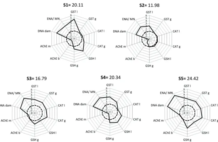
IBR calculation, the ratio between the experimental sites individually and the reference site for each biomarker was log-transformed (Yi) and then the overall mean (μ) and standard deviation (s) was calculated. Then, Yi values were standardized by the formula: Zi = (Yi - µ)/s and the difference
between Zi and Z0 (reference) determined A values. IBR value was calculated for each exposure site by the sum of A values. For each site data were represented in a radar chart indicating the deviation of biomarker investigated in relation to the reference site (0). The area above 0 reflects induction of the biomarker, and below 0 indicates reduction of the biomarker. using the substrate acetylcholine iodide and the color regent
5,5-dithiobis-2-nitrobenzoic acid (DTNB) at 415 nm. AChE activity was expressed in nmol min-1.mg protein-1.
The total protein concentration was determined in a spectrophotometer at 700 nm according to the method of Lowry et al. (1951) using a standard curve of bovine serum
albumin (BSA).
Genotoxic analysis
The alkaline comet assay with erythrocytes was performed according to Singh et al. (1988), with some modifications
described by Ramsdorf et al. (2009). Only blood samples
with cell viability above 80%, determined by the Trypan blue exclusion method, were used in the comet assay.
After sampling, an aliquot of blood mixed with fetal bovine serum was added to the low melting point agarose. This mixture was placed on a glass slide previously covered with standard agarose, covered with coverslip, and remained in the refrigerator for 30 min. Then coverslips were removed and the slides were subjected to: a) lysis: 1h at 4°C, protected from light, in lysis solution (2.5 M NaCl, 100 mM EDTA, 10 mM Tris, 10% DMSO, 1 mL Triton X-100, pH 10.0); b) DNA denaturation: 30 min in the dark in an electrophoresis buffer (0.3 N NaOH, 1 mM EDTA, pH>13); c) electrophoresis: 20 min, 300 mA , 25 V, 1 V cm-1; and d) neutralization: three
rinses for 5 min each with buffer (0.4 M Tris, pH 7.5). The slides were then fixed with absolute ethanol for 10 min and placed in the refrigerator until analysis.
Subsequently, the slides were stained with gelRed (Uniscience®) and analyzed on a Leica microscope (DM 2500)
adapted for fluorescence/epifluorescence, equipped with blue excitation filter (450-490 nm), and a 515 nm barrier filter with a magnification of 1000X. All slides were analyzed in blind test, being evaluated 100 nucleoids per fish. The extent of DNA damage was quantified by the length of the tail formed by the migration of DNA fragments and were classified into four classes according to Kobayashi et al. (1995): class 0 =
no apparent damage; class 1 = tail shorter than the nucleoid
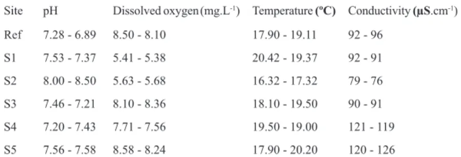
Site pH Dissolved oxygen(mg.L-1) Temperature (ºC) Conductivity (µS.cm-1)
Ref 7.28 - 6.89 8.50 - 8.10 17.90 - 19.11 92 - 96
S1 7.53 - 7.37 5.41 - 5.38 20.42 - 19.37 92 - 91
S2 8.00 - 8.50 5.63 - 5.68 16.32 - 17.32 79 - 76
S3 7.46 - 7.21 8.10 - 8.36 18.10 - 19.50 90 - 91
S4 7.20 - 7.43 7.71 - 7.56 19.50 - 19.00 121 - 119
S5 7.56 - 7.58 8.58 - 8.24 17.90 - 20.20 120 - 126
Table 1. Physical and chemical parameters of the water from the reference site (Ref) and from the sites along Água das Araras
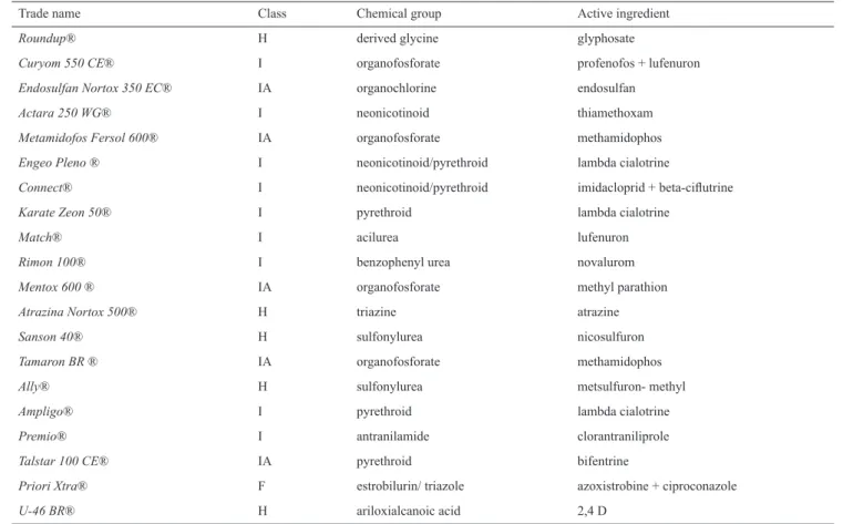
in the study area (Table 2). Among the pesticides used in the region, 45% correspond only to insecticides, 25% are herbicides, standing out glyphosate and atrazine as the most widely used in the region, and the rest are insecticides/ acaricide and fungicides.
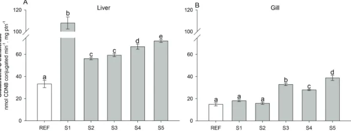
The data obtained from analyses with biomarkers showed a significant increase in GST activity in the liver of fish exposed in all experimental sites with respect to the reference site (Fig. 2A). In the site 1, it was observed a significant increase in enzyme activity with respect to all other sites. In the gills, there was a significant increase in GST activity in animals kept in sites 3, 4, and 5 compared to the reference site (Fig. 2B).
The activity of liver CAT was significantly increased in sites 3 and 5 in relation to the others (Fig. 3A), and in the gills was also observed a significant increase in enzyme activity in sites 1, 3, 4, and 5 (Fig. 3B). It should be emphasized that the CAT activity determined in the gills was very low and in fish from the reference site the enzyme activity was about 50 times lower than in the liver.
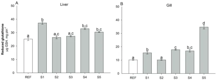
The concentration of GSH increased significantly in the liver of fish confined in the sites 1, 4, and 5 (Fig. 4A) and in the gills of fish from sites 1, 3, 4, and 5 (Fig. 4B), with respect to the reference site.
Statistical analysis
The mean values obtained from each biological variable analyzed in fish from different exposure sites were compared with each other by parametric analysis of variance (ANOVA), after checking for normality and homogeneity of variance. When necessary, differences were identified by Student-Newman-Keuls post hoc test. The significance level was
set at P < 0.05.
Results
The results of chemical and physical parameters of the water collected from the different sites at the beginning and at the end of the exposure period of the animals are shown in Table 1. There were no marked differences in evaluated parameters between the first and last day of the experiment. Sites 1 and 2 showed the lowest concentration of dissolved oxygen. This can be explained because these stretches have slower water flow, compared with the other sites. Likewise, the site 1 also presented the highest temperature because it is an impoundment, which receives a higher incidence of solar radiation. The survey with local farmers identified the 20 agrochemical contaminants that could be present
Trade name Class Chemical group Active ingredient
Roundup® H derived glycine glyphosate
Curyom 550 CE® I organofosforate profenofos + lufenuron
Endosulfan Nortox 350 EC® IA organochlorine endosulfan
Actara 250 WG® I neonicotinoid thiamethoxam
Metamidofos Fersol 600® IA organofosforate methamidophos
Engeo Pleno ® I neonicotinoid/pyrethroid lambda cialotrine
Connect® I neonicotinoid/pyrethroid imidacloprid + beta-ciflutrine
Karate Zeon 50® I pyrethroid lambda cialotrine
Match® I acilurea lufenuron
Rimon 100® I benzophenyl urea novalurom
Mentox 600 ® IA organofosforate methyl parathion
Atrazina Nortox 500® H triazine atrazine
Sanson 40® H sulfonylurea nicosulfuron
Tamaron BR ® IA organofosforate methamidophos
Ally® H sulfonylurea metsulfuron- methyl
Ampligo® I pyrethroid lambda cialotrine
Premio® I antranilamide clorantraniliprole
Talstar 100 CE® IA pyrethroid bifentrine
Priori Xtra® F estrobilurin/ triazole azoxistrobine + ciproconazole
U-46 BR® H ariloxialcanoic acid 2,4 D
Table 2. Most used pesticides in the watershed area of the Água das Araras stream. Class: H = herbicide; I = inseticide; IA
Fig. 3. Activity (mean ± SEM, n = 8) of catalase in liver (A) and gills (B) of A. altiparanae exposed in situ for seven days
in five sites along Água das Araras stream (S1, S2, S3, S4, and S5) and in a reference site (Ref). Different letters indicate significant differences between sites (P < 0.05).
Fig. 2. Activity (mean ± SEM, n = 8) of glutathione S-transferase in liver (A) and gills (B) of A. altiparanae exposed in situ
for seven days in five sites along Água das Araras stream (S1, S2, S3, S4, and S5) and in a reference site (Ref). Different letters indicate significant differences between sites (P < 0.05).
Results showed a significant reduction in AChE activity both in muscle (Fig. 5A) and brain (Fig. 5B) of fish confined in sites 1, 2 and 4 compared to the other sites.
The results of the comet assay showed a significant increase in the occurrence of DNA damage in fish erythrocytes in all sites of the Água das Araras stream, with respect to fish kept in the reference site (Fig. 6). Fish from site 5 showed a higher DNA damage score than all other locations. The frequency of MN (Table 3) was significant higher in fish from sites 1, 3, and 5 in relation to animals from the reference site, while ENA frequency was significantly higher only in fish from site 5 in relation to the reference.
IBR values for each location of the in situ tests are shown
Fig. 4. Content (mean ± SEM, n = 8) of glutathione in liver (A) and gills (B) of A. altiparanae exposed in situ for seven days
in five sites along Água das Araras stream (S1, S2, S3, S4, and S5) and in a reference site (Ref). Different letters indicate significant differences between sites (P<0.05).
Fig. 5. Activity (mean ± SEM, n = 8) of acetylcholinesterasein muscle (A) and brain (B) of A. altiparanae exposed in situ for
seven days in five sites along Água das Araras stream (S1, S2, S3, S4, and S5) and in a reference site (Ref). Different letters indicate significant differences between sites (P < 0.05).
Discussion
One of the most investigated biochemical biomarkers in fish are the enzymes involved in the detoxification of toxic agents and their metabolites, such as biotransformation and antioxidant defense enzymes. The family of glutathione-S-transferase (GST) enzymes is essential in the protection against damage from potentially reactive compounds, combining them with endogenous molecules such as reduced glutathione (GSH), to be later eliminated by the body. The activity of this enzyme is considered a good biomarker of exposure to environmental pollutants (Van der Oost et al., 2003).
The increased GST activity in the liver and gills of fish caged at various sites along the Água das Araras stream can indicate the presence of xenobiotics that are metabolized
by conjugation with GSH, to be eliminated from the body. Several authors have reported increased GST activity in different fish species exposed to pesticides, some of which are used in the study region. For example, Dong
et al. (2013) reported increased GST activity in Danio rerio larvae exposed to the organochlorine endosulfan
and Oruc et al. (2004) observed increased activity of this
enzyme in two species of fish exposed to 2,4 D. Paulino et al. (2012) observed an increase in GST activity in gills of
the Neotropical fish Prochilodus lineatus after sub-chronic
exposure to atrazine, a herbicide widely used to control undesirable organisms in corn crops (Mudiam et al., 2012).
Fig. 6. DNA damage scores (mean ± SEM, n = 8) in
erythrocytes of A. altiparanae exposed in situ for seven days
in five sites along Água das Araras stream (S1, S2, S3, S4, and S5) and in a reference site (Ref). Different letters indicate significant differences between sites (P < 0.05).
Fig. 7. Integrated biomarkers response index (IBR) values for each experimental site along Água das Araras stream (S1, S2,
S3, S4, and S5) where in situ tests were performed. Biomarkers results are represented in relation to the reference site (0). The
area above 0 reflects induction of the biomarker, and below 0 indicates reduction of the biomarker.
Site N MN frequency (‰) ENA frequency (‰)
Ref 8 0.50 ± 0.18 a 1.57 ± 0.29 a
S1 8 2.85 ± 0.69 b 3.71 ± 0.83 a,b
S2 8 1.25 ± 0.25 a,b 2.87 ± 0.89 a,b
S3 8 2.37 ± 0.32 b 4.50 ± 0.56 a,b
S4 8 1.66 ± 0.28 a,b 3.57 ± 0.84 a,b
S5 8 3.62 ± 0.37 b 6.14 ± 0.85 b
Table 3. Frequency of micronuclei (MN) and other nuclear
abnormalities (ENA) in erythrocytes of A. altiparanae, taking
of this biotransformation enzyme was also high in these same sites. In the gills, it was also observed an increased concentration of GSH in the sites 1, 3, 4, and 5, where it was found increased gill CAT, indicating a possible adaptation of antioxidant defenses.
In relation to AChE activity, there was an inhibition of this enzyme activity in both brain and muscle of fish exposed in the sites 1, 2, and 4. This enzyme, which occurs in cholinergic synapses and motor end-plates, is responsible for the hydrolysis of the neurotransmitter acetylcholine into choline and acetic acid. Inhibition of AChE is classically associated with the mechanism of toxic action of organophosphates and carbamates insecticides (Payne et al., 1996) and disturbances
on its activity may affect locomotion and balance in fishes, impairing feeding, escape and reproductive behavior (Pessoa
et al., 2011). AChE activity in fish can also be modified by
other classes of pesticides, like organochlorines such as endosulfan (Dutta & Arends, 2003). Other authors have also demonstrated the anticholinesterase effect of glyphosate in different fish species (Glusczak et al.,2006; Glusczak et al.,
2007; Cattaneo et al., 2011) as well as glyphosate
based-products, Roundup® and Roundup Transorb® on Prochilodus
lineatus (Modesto & Martinez, 2010a, 2010b).
In assessing DNA damages, it was observed an increase in damage scores in erythrocytes of fish confined in all experimental sites in relation to the control. There was also an increase in the occurrence of MN in erythrocytes of fish confined in the sites 1, 3, and 5 and increased frequency of ENA in fish of the site 5. Genotoxic effects of various groups of pesticides, such as organophosphates, organochlorines, and pyrethroids have been demonstrated in vivo and in vitro
tests (Bolognesi, 2003; Abdollahi et al., 2004; Kaushik &
Kaushik, 2007).
The effects of atrazine on genetic material have also been described for fish. Santos & Martinez (2012) observed increased occurrence of DNA damage in blood, liver and gill cells in fish exposed to this herbicide. Cavas (2011) observed a significant increase in the occurrence of DNA damage and the frequency of micronuclei in erythrocytes of Carassiusauratus
after exposure to atrazine. Ventura et al. (2008) observed an
increase in DNA strand breaks and in the frequency of MN and ENA in erythrocytes of O. niloticus exposed to different
concentrations of atrazine after only 72 hours of exposure. Likewise, Nwani et al. (2011) observed an increase of MN for Channa punctatus after seven days of exposure to atrazine.
Genotoxic effects of glyphosate on fish have also been reported in the literature. Cavalcante et al. (2008) verified an
increase in DNA strand breaks in blood cells of P. lineatus
exposed to commercial formulation of the herbicide. Similarly, Rossi et al. (2011) reported an increased frequency of MN
and ENA in Astyanax sp. exposed to this pesticide. Ramsdorf et al. (2012) also observed an increase in the frequency of
Another widely used herbicide in the study area is Roundup®, a glyphosate based product. The effects of
this herbicide on aquatic organisms have been addressed, especially in fish (Glusczak et al.,2006; Cattaneo, 2011; Rossi et al., 2011). Modesto & Martinez (2010a) have shown that
this herbicide increases the activity of liver GST and other enzymes involved in antioxidant defense of P. lineatus.
The antioxidant defense system has been increasingly studied given the ability of oxiradicals to promote responses that are used as biomarkers (Di Giulio et al., 1989; Winston
& Di Giulio, 1991). The main components of this defense system are enzymes, including superoxide dismutase (SOD), catalase (CAT) and glutathione peroxidase (GPX). The catalase (CAT) works in eliminating H2O2, producing H2O
and O2. In this study, an increase in the activity of this enzyme
was observed in the liver of fish from sites 3 and 5 and in the gills of animals from sites 1, 3, 4 and 5, suggesting an increase in antioxidant defense to eliminate reactive oxygen species (ROS), mainly formed during the metabolism of chemical compounds. Although it was observed an increase in CAT activity in the gills, it should be borne in mind that this organ has very low activity of this enzyme, compared with the liver, as demonstrated for other fish species, such as P. lineatus (Simonato et al., 2011). Wilhelm Filho et al.
(1994) suggest that gills may have alternative mechanisms to eliminate hydrogen peroxide.
Various pesticides can lead the organism to a state of oxidative stress, causing an increase in ROS generation and changes in antioxidant defense mechanisms. Several studies have shown that organophosphate pesticides promote oxidative changes in fish species, such as in Cyprinus carpio,
Ictalurus nebulosus (Hai et al., 1997) and Anguilla anguilla
(Peña-Llopis et al.,2003) exposed to dichlorvos, in Brycon cephalus exposed to Folisuper BR 600 (methyl parathion)
(Monteiro et al., 2009) and in Oreochromis niloticus exposed
to fenthion (Piner et al., 2007) and trichlorfon (Thomaz et al., 2009). Glyphosate-based herbicides are also reported to
induce oxidative stress in different fish species (Lushchak et al., 2009), including Leporinus obtusidens (Glusczak et al.,
2006), Rhamdia quelen (Glusczak et al., 2007) and P. lineatus
(Modesto & Martinez, 2010a).
Glutathione (GSH), a tripeptide that plays a key role in reactions of oxidation/reduction, amino acid transport and detoxification of many toxic agents, is the first line of defense against cellular damage mediated by oxidants (Van der Oost et al., 2003). The increased concentration of liver
altiparanae was effective to evaluate water quality. New
and ongoing monitoring programs in these sites should be established, combining the use of biomarkers and in situ
exposure with chemical analysis of water, aiming to identify the pesticides present in this mixture and relate them to the observed effects in animals.
Acknowledgments
This work was supported by grants from the Brazilian research funding institutions CNPq (INCT-TA/CNPq: process 573949/2008-5) and Fundação Araucária. Carlos E. D. Vieira thanks Fundação Araucária for the scholarship. C. B. R. Martinez is research fellow from CNPq and member of the Brazilian Institute of Aquatic Toxicology (INCT-TA/CNPq).
Literature Cited
Abdollahi, M., A. Ranjbar, S. Shadnia, S. Nikfar & A. Rezaiee. 2004. Pesticides and oxidative stress: A review. Medical Science Monitor, 10: 144 -147.
Akaishi, F. M., H. C. Silva de Assis, S. C. G. Jakobi, D. R. Eiras-Stofella, S. D. St-Jean, S. C. Courtenay, E. F. Lima, A. L. R. Wagener, A. L. Scofield & C. A. Oliveira Ribeiro. 2004. Morphological and neurotoxicological findings in tropical freshwater fish (Astyanax sp.) after waterborne and acute
exposure to water soluble fraction (WSF) of crude oil. Archives of Environmental Contamination and Toxicology, 46: 244-253. Alves Costa, J. R., M. Mela, H. C. Silva de Assis, E. Pelletier, M.
A. Randi & C. A. Oliveira Ribeiro. 2007. Enzymatic inhibition and morphological changes in Hoplias malabaricus from
dietary exposure to lead (II) or methylmercury. Ecotoxicology Environmental Safety, 67: 82-88.
ANVISA & UFPR. 2012. Seminário de mercado de agrotóxico e regulação. Agência Nacional de Vigilância Sanitária, Brasília, 11 abril de 2012.
Arzate-Cardenas, M. A. & F. Martinéz-Jeronimo. 2011. Age-altered susceptibility in hexavalent chromium-exposed Daphnia schodleri (Anomopoda: Daphniidae): integrated biomarker
response implementation. Aquatic Toxicology, 105: 528-534. Barbee, G. C., J. Barich., B. Duncan., J. W. Bickham., C. W. Matson., C.
J. Hintze., R. L. Autenrieth, G. D. Zhou, T. J. Mcdonald, L. Cizmas, D. Norton & K. C. Donnelly. 2008. In situ biomonitoring of PAH-contaminated sediments using juvenile coho salmon (Oncorhynchus kisutch). Ecotoxicology Environmental Safety, 71: 454-464.
Beliaeff, B. & T. Burgeot. 2002. Integrated biomarker response: a useful tool for ecological risk assessment. Environmental Toxicology Chemistry, 21: 1316-1322.
Beutler, E. 1975. Red Cell Metabolism: a manual of biochemical methods. New York. Grune and Stratton.
Beutler, E., O. Durom & B. M. Kelly. 1963. Improved method for the determination of blood glutathione. Journal of Laboratory and Clinical Medicine, 61: 882-888.
Bolognesi, C. 2003. Genotoxicity of pesticides: a review of human biomonitoring studies. Mutation Research, 543: 251-272. Carrasco, K. R., K. L. Tilbury & M. S. Myers. 1990. Assessment of
the piscine micronucleus test as in situ biological indicator of chemical contaminant effects. Canadian Journal of Fisheries and Aquatic Sciences, 47: 2123-2136.
ENA and MN and DNA damage in species of Astyanax sp.
collected in the area potentially contaminated with pesticides, including Roundup, compared with the amount of changes found in fish collected from a reference site. In the same way, Cavas & Könen (2007) registered an increase in the frequency of MN and ENA in Carassius auratus after four
days of exposure to different concentrations of glyphosate. In addition to these pesticides, increased frequency of ENA and MN and DNA strand breaks have also been described in fish exposed to herbicide endosulfan (Neuparth et al., 2006;
Pandey et al., 2006).
Formation of micronuclei is a short term response to a genotoxic substance, so that their expression depends on the intensity of exposure to contaminants and probably independent of the duration of such exposure (Heddle et al.,
1991). The increased frequency of MN and ENA in some sites evaluated may indicate the presence of pesticides able to promote mutagenic damage in erythrocytes of A. altiparanae.
The integrated biomarker index (IBR) was able to discriminate the sites based on the biomarkers responses. The sites with higher IBR values were 5, 4, and 1, respectively. These results are consistent with the degree of local human interference, as they are the stretches of the stream with the lowest cover of riparian vegetation, which is non-existent in some stretches, and are located in areas with more intensive farming activities, which come very close to the bed of the stream. The site 2 also located within the forest unit, had the lowest IBR value among the evaluated sites. Thus, we can assume the protective effect of this vegetation as a barrier to the runoff and leaching of these contaminants coming from the surrounding monocultures. Nevertheless, important variations observed in some parameters evaluated in fish confined in the site 2 still indicate the presence of contaminants in these waters.
As proposed by Beliaeff & Burgeot (2002) the IBR can be associated with a star or radar chart that shows the specific responses of biomarkers in each site analyzed. In the present study, several biomarkers exhibited a response that was induced or inhibited according to the sampling site and the spatial arrangement of these biomarkers in the star plot allowed visualizing more clearly which biomarkers were the most sensitive in this kind of evaluation. Thus, the comet assay that assesses DNA damage, the enzymatic activity of GST in the liver and the frequency of MN and ENA, besides the activity of AChE were the biomarkers that proved to be more efficient in this study.
Mutation Research, 18: 187-192.
Heddle, J. A., M. C. Cimino, M. Hayashi, F. Romagna, M. D. Shelby, J. D. Tucker, P. Vanparys & J. T. MacGregor. 1991. Micronuclei as an index of cytogenetic damage: past, present, and future. Environmental and Molecular Mutagenesis, 18: 277-291. Kaushik, P. & G. Kaushik. 2007. An assessment of structure and
toxicity correlation in organochlorine pesticides. Journal of Hazardous Materials, 143: 102-111.
Keen, J. H., W. H. Habig & W. B. Jakoby. 1976. Mechanism for several activities of the glutathione-S-transferase. Journal Biology Chemistry, 20: 6183-6188.
Klobucar, G. I. V., A. Stambuk, M. Pavlica, M. S. Peric, B. K. Hackenberger & K. Hylland. 2010. Genotoxicity monitoring of fresh water environments using caged carp (Cyprinus carpio).
Ecotoxicology, 19: 77-84.
Kobayashi, H., C. Sugiyama, Y. Morikawa, M. Hayashi & T. Sofuni.1995. Comparison between manual microscopic analysis and computerized image analysis in the single cell gel electrophoresis. MMS Commun, 3: 103-115.
Lemos, C. T., F. A. Iranço, N. C. Oliveira, G. D. Souza & J. M. Fachel. 2008. Biomonitoring of genotoxicity using micronuclei assay in native population of Astyanax jacuhiensis (Characiformes:
Characidae) at sites under petrochemical influence. Science of the Total Environmental, 406: 337-343.
Lowry, O. H., N. J. Rosebrough, A. L. Farr & R. J. Randall. 1951. Protein measurements with the folin phenol reagent. Journal Biology Chemistry, 193: 265-275.
Lushchak, O. V., O. I. Kubrak, J. M. Storey, K. B. Storey & V. I. Lushchak. 2009. Low toxic herbicide Roundup induces mild oxidative stress in goldfish tissues. Chemosphere, 76: 932-937. Martins, S. V. 2001. Recuperação de matas ciliares. Viçosa: Aprenda Fácil. Modesto, K. A. & C. B. R. Martinez. 2010a. Roundup causes
oxidative stress in liver and inhibits acetylcholinesterase in muscle and brain of the fish Prochilodus lineatus. Chemosphere,
78: 294-299.
Modesto, K. A. & C. B. R. Martinez. 2010b. Effects of Roundup Transorb on fish: hematology, antioxidant defenses and acetylcholinesterase activity. Chemosphere, 81: 781-787. Monteiro, D. A., F. T. Rantin & A. L. Kalinin. 2009. The effects
of selenium on oxidative stress biomarkers in the freshwater characid fish matrinxã, Brycon cephalus (Günther, 1869) exposed
to organophosphate insecticide Folisuper 600 BR (methyl parathion). Comparative Biochemistry Physiology C, 149: 40-49. Monteiro, V., D. G. S. M. Cavalcante, M. B. F. A. Viléla, S. H.
Sofia & C. B. R. Martinez. 2011. In vivo and in vitro exposures for the evaluation of the genotoxic effects of lead on the Neotropical freshwater fish Prochilodus lineatus. Aquatic
Toxicology, 104: 291-298.
Mouneyrac, C. & C. Amiard-Triquet. 2013. Biomarkers of ecological relevance. In: Pp 210-219. Férard, J. & C. Claise (Eds.). Encyclopedia of Aquatic Ecotoxicology. Berlin, Springer. Mudiam, M. K. R., S. P. Pathak, K. Gopal & R. C. Murthy. 2012.
Studies on urban drinking water quality in a tropical zone. Environmental Monitoring and Assessment, 184: 461-469. Neuparth, T., J. W. Bickham, C. W. Theodorakis, F. O. Costa & M.
H. Costa. 2006. Endosulfan-Induced Genotoxicity Detected in the Gilthead Seabream, Sparus aurata L., by Means of Flow
Cytometry and Micronuclei Assays. Bulletin of Environmental Contamination and Toxicology, 76: 242-248.
Nwani, C. D., N. S. Nagpure, R. Kumar, B. Kushwaha, P. Kumar & W. S. Lakra. 2011. Mutagenic and genotoxic assessment of atrazine-based herbicide to freshwater fish Channa punctatus (Bloch)
Cattaneo, R., B. Clasen, V. L. Loro, C. C. Menezes, A. Pretto, B. Baldisserotto, A. Santi & L. A. Avila. 2011. Toxicological Responses of Cyprinus carpio Exposed to a Commercial
Formulation Containing Glyphosate. Bulletin Environmental Contamination Toxicology, 87: 597-602.
Cavalcante, D. G. S. M., C. B. R. Martinez & S. H. Sofia. 2008. Genotoxic effects of roundup on the fish Prochilodus lineatus.
Mutation Research, 655: 41-46.
Cavas, T. 2011. In vivo genotoxicity evaluation of atrazine and atrazine-based herbicide on fish Carassius auratus using
the micronucleus test and the comet assay. Food Chemical Toxicology, 49: 1431-1435.
Cavas, T. & S. Könen. 2007. Detection of cytogenetic and DNA damage in peripheral erythrocytes of goldfish (Carassius auratus)
exposed to a glyphosate formulation using the micronucleus test and the comet assay. Mutagenesis, 22: 263-268.
Cerejeira, M. J., P. Viana, S. Batista, T. Pereira, E. Silva, M. J. Valério, A. Silva, M. Ferreira & A. M. Silva-Fernandes. 2003. Pesticides in Portuguese surface and ground waters. Water Research, 37: 1055-1063.
Cravo, A., C. Pereira, T. Gomes, C. Cardoso, A. Serafim, C. Almeida, T. Rocha, B. Lopes, R. Company, A. Medeiros, R. Norberto, R. Pereira, O. Araújo & M. J. Bebianno. 2012. A multibiomarker approach in the clam Ruditapes decussatus to assess the impact
of pollution in the Ria Formosa lagoon, South Coast of Portugal. Marine Environmental Research, 75: 23-34.
Depledge, M. H., A. Aagaard & P. Gyorkost. 1995. Assessment of trace metal toxicity using molecular, physiological and behavioral biomarkers. Marine Pollution Bulletin, 31: 19-27.
Di Giulio, R. T., P. C. Washburn, R. J. Wenning, G. W. Winston & C. S. Jewell. 1989. Biochemical responses in aquatic animals: a review of determinants of oxidative stress. Environmental Toxicology and Chemistry, 8: 1103-1123.
Dong, M., L. Zhu, B. Shao, S. Zhu, J. Wang, H. Xie, J. Wang & F. Wang. 2013. The effects of endosulfan on cytochrome P450 enzymes and glutathione S-transferases in zebrafish (Danio rerio) livers. Ecotoxicology and Environmental Safety, 92: 1-9.
Dutta, H. M. & D. A. Arends. 2003. Effects of endosulfan on brain acetylcholinesterase activity in juvenile bluegill sunfish. Environmental Research, 91: 157-162.
Ellman, G. L., K. D. Courtney, V. Andres Jr. & R. M. Featherstone. 1961. A new and rapid colorimetric determination of acetylcholinesterase activity. Biochemistry Pharmacology, 7: 88-90.
Garutti, V. & H. A. Britski. 2000. Descrição de uma nova espécie de Astyanax (Teleostei: Characidae) na bacia do alto do rio
Paraná e considerações sobre as demais espécies do gênero da bacia. Comunicações do Museu de Ciências e Tecnologia, Série Zoologia, 13: 65-88.
Glusczak, L., D. S. Miron, M. Crestani, M. B. Fonseca, F. A. Pedron, M. F. Duarte & V. L. P. Vieira. 2006. Effect of glyphosate herbicide on acetylcholinesterase activity and metabolic and hematological parameters in piava (Leporinus obtusidens).
Ecotoxicology Environmental Safety, 65: 237-241.
Glusczak, L., D. S. Miron, B. S. Moraes, R. R. Simões, M. R. Schetinger, V. M. Morsch & V. L. Loro. 2007. Acute effects of glyphosate herbicide on metabolic and enzymatic parameters of silver catfish (Rhamdia quelen). Comparative Biochemistry
Physiology C, 146: 519-524.
Hai, D. Q., S. I. Varga & B. Matkovics. 1997. Organophosphate effects on antioxidant system on carp (Cyprinus carpio) and catfish (Ictalurus nebulosus). Comparative Biochemistry Physiology C, 117: 83-88.
Schmid, W. 1975. The micronucleus test. Mutation Research, 31: 9-15. Schultz, U. H. & H. Martins-Junior. 2000. Astyanax fasciatus as
bioindicator of water pollution of Rio dos Sinos, RS, Brazil. Brazilian Journal of Biology, 61: 615-622.
Serafim, A., R. Company, B. Lopes, V. F. Fonseca, S. França, R. P. Vasconcelos, M. J. Bebianno & H. N. Cabral. 2012. Application of an integrated biomarker response index (IBR) to assess temporal variation of environmental quality in two Portuguese aquatic systems. Ecological Indicators, 19: 215-225.
Silva, A. G. & C. B. R. Martinez. 2007. Morphological changes in the kidney of a fish living in an urban stream. Environmental Toxicology and Pharmacology, 23: 185-192.
Simonato, J. D., M. N. Fernandes & C. B. R. Martinez. 2011. Gasoline effects on biotransformation and antioxidant defenses of the freshwater fish Prochilodus lineatus. Ecotoxicology, 20:
1400-1410.
SINDAG (Sindicato Nacional da Indústria de Produtos para Defesa Agrícola), 2009. Available from: http://www.sindag.com.br/ noticia.php?News_ID=1399.
Singh, N. P., M. T. McCoy, R. R. Tice & E. L. Schneider. 1988. A simple technique for quantification of low levels of DNA damage in individual cells. Experimental Cell Research, 175: 184-191. Spalding, R. F., M. E. Exner, D. D. Snow, D. A. Cassada, M. E.
Burbach & S. J. Monson. 2003. Herbicides in ground water beneath Nebraska’s management systems evaluation area. Journal of Environmental Quality, 32: 92-98.
Thomaz, J. M., N. D. Martins, D. A. Monteiro, F. T. Rantin & A. L. Kalinin. 2009. Cardio- respiratory function and oxidative stress biomarkers in Nile tilapia exposed to the organophosphate insecticide trichlorfon (Neguvon). Ecotoxicology Environmental Safety, 72: 1413-1424.
Van der Oost, R., J. Beyer & N. P. E. Vermeulen. 2003. Fish bioaccumulation and biomarkers in environmental risk assessment: a review. Environmental Toxicology and Pharmacology, 13: 57-149.
Ventura, B. C., D. F. Angelis, M. A. Marin-Morales. 2008. Mutagenic and genotoxic effects of the atrazine herbicide in Oreochromis niloticus (Perciformes, Cichlidae) detected by micronuclei test and
the comet assay. Pesticides Biochemical and Physiology, 90: 42-51. Wilhelm Filho, D., B. Gonzalez-Flecha & A. Boveris. 1994. Gill
diffusion as a physiological mechanism for hydrogen peroxide elimination by fish. Brazilian Journal of Medical and Biological Research, 27: 2879-2882.
Winkaler, E. U., A. G. Silva, H. C. Galindo & C. B. R. Martinez. 2001. Biomarcadores histológicos e fisiológicos para o monitoramento da saúde de peixes de ribeirões de Londrina, Estado do Paraná. Acta Scientiarum, 23: 507-514.
Winston, G. W. & R. T. Di Giulio. 1991. Pro-oxidant and antioxidant mechanisms in aquatic organisms. Aquatic Toxicology, 19: 137-161.
Submitted May 21, 2013 Accepted November 7, 2013 by Bernardo Baldisserott Published March 31, 2014
using micronucleus test and single cell gel electrophoresis. Environmental Toxicology and Pharmacology, 31: 314-322. Oruc, E. O., Y. Sevgiler & N. Uner. 2004. Tissue-specific oxidative
stress responses in fish exposed to 2,4-D and azinphosmethyl. Comparative Biochemistry and Physiology, 137: 43-51. Pandey, S., N. S. Nagpure, R. Kumar, S. Sharma, S. K. Srivastava
& M. S. Verma. 2006. Genotoxicity evaluation of acute doses of endosulfan to freshwater teleost Channa punctatus (Bloch)
by alkaline single-cell gel electrophoresis. Ecotoxicology and Environmental Safety, 65: 56-61.
Paulino, M. G., N. E. S. Souza & M. N. Fernandes. 2012. Subchronic exposure to atrazine induces biochemical and histopathological changes in the gills of a Neotropical freshwater fish, Prochilodus lineatus. Ecotoxicology and Environmental Safety, 80: 6-13.
Payne, J. F., A. Mathieu, W. Melvin & L. L. Fancey. 1996. Acetylcholinesterase, an old biomarker with a new future? Field trials in association with two urban rivers and a paper mill in Newfoundland. Marine Pollution Bulletin, 32: 225-231. Peña-Llopis, S., M. D. Ferrando & J. B. Peña. 2003. Increased
recovery of brain acetylcholinesterase activity in dichlorvos-intoxicated European eels Anguilla anguilla by bath treatment with
N-acetylcysteine. Diseases of Aquatic Organisms, 55: 237-245. Pessoa, P. C., K. H. Luchmann, A. B. Ribeiro, M. M. Veras, J. R. M.
B. Correa, A. J. Nogueira, A. C. D. Bainy & P. S. M. Carvalho. 2011. Cholinesterase inhibition and behavioral toxicity of carbofuran on Oreochromis niloticus early life stages. Aquatic
Toxicology, 105: 312-320.
Piner, P., Y. Sevgiler & N. Uner. 2007. In vivo effects of fenthion on oxidative processes by the modulation of glutathione metabolism in the brain of Oreochromis niloticus. Environmental Toxicology,
22: 605-612.
Ramsdorf, W. A., F. S. F. Guimarães, M. V. M. Ferraro, J. Gabardo, E. S. Trindade & M. M. Cestari. 2009. Establishment of experimental conditions for preserving samples of fish blood for analysis with both comet assay and flow cytometry. Mutation Research, 673: 78-81.
Ramsdorf, W. A., T. Vicari, M. I. M. Almeida, R. F. Artoni & M. M. Cestari. 2012. Handling of Astyanax sp. for biomonitoring in
Cangüiri Farm within a fountainhead (Iraí River Environment Preservation Area) through the use of genetic biomarkers. Environmental Monitoring and Assessment, 184: 5841-5849. Rossi, S. C., M. D. Silva, L. D. Piancini, C. A. Oliveira Ribeiro,
M. M. Cestari & H. C. Silva de Assis. 2011. Sublethal effects of waterborne herbicides in tropical freshwater fish. Bulletin Environmental Contamination Toxicology, 87: 603-607. Sanchez, W., T. Burgeot & O. Perceval. 2011. Perspectives from the
French workshop on the development and validation of biomarkers and bioassays for the monitoring of aquatic environments. Environmental Science Pollution Research, 19: 1345-1347. Sanchez, W., T. Burgeot & J. Porcher. 2013. A novel “Integrated
Biomarker Response” calculation based on reference deviation concept. Environmental Science Pollution Research, 20: 2721-2725. Santos, T. G. & C. B. R. Martinez. 2012. Atrazine promotes
biochemical changes and DNA damage in a Neotropical fish species. Chemosphere, 89: 1118-1125.