Isolation of endophytic bacteria from arboreal species of the Amazon
and identification by sequencing of the 16S rRNA encoding gene
Mariza M. Coêlho
1, Monica S. Ferreira-Nozawa
2, Sérgio R. Nozawa
2,3and André L.W. Santos
1 1Laboratório de Biotecnologia Vegetal, Universidade Nilton Lins, Manaus, AM, Brazil.
2
Laboratório de Expressão Gênica, Universidade Nilton Lins, Manaus, AM, Brazil.
3
Laboratório de Genética Química e Bioinformática, Universidade Nilton Lins, Manaus, AM, Brazil.
Abstract
Endophytic bacteria from three arboreal species native to the Amazon (Carapa guianenses, Ceiba pentandra, and Swietenia macrophylla), were isolated and identified, through partial sequencing of the 16S rRNA encoding gene. From these, 16 isolates were obtained, although, when compared to sequences deposited in GenBank, only seven had produced identifiable fragments.Bacillus, Pantoea and two non-culturable samples were identified. Results ob-tained through sequence analysis revealed low genetic diversity across the isolates, even when analyzing different species and plant structures. This is the first report concerning the isolation and identification of endophytic bacteria in these plant species.
Key words:isolation, sequencing,Bacillus,Pantoea.
Received: February 13, 2011; Accepted: July 17, 2011.
Endophytic microorganisms inhabit the inner organs and tissues of plants, such as leaves, stems, seeds and roots, during at least one period of their life-cycles, without caus-ing diseases or produccaus-ing visible external manifestation
(Azevedo et al., 2000). Endophytic communities are
formed mainly by fungi and bacteria. It is estimated that ev-ery plant-species constitutes a possible host for endophytic microorganisms, which, in the vast majority and despite their biotechnological potential (Ezraet al., 2004), remain unidentified. Although the interaction between these mi-croorganisms and their respective host-plants is not, as yet, fully understood, over recent years they have been progres-sively more extenprogres-sively employed, either in agriculture (Ryanet al., 2008), or in the production of compounds with therapeutic application, such as taxol (Stierleet al., 1993) and leucinostatin A (Strobel and Hess, 1999).
The origin, entrance pathway, colonization and trans-mission of endophytic bacteria have been the object of con-siderable research efforts (Azevedo et al., 2002). These bacteria may proliferate in seeds, the rhizosphere, the phyl-loplane as well as the material that results from vegetative propagation (Stierleet al., 1993; Kuskeet al., 1997). Pene-tration into the host plant may occur via stomata, wounds, or areas of lateral root development, or may even be facili-tated by the production of hydrolytic enzymes capable of
degrading the cell wall (Souzaet al., 2004). Once inside, the endophytic microorganism may lodge in specific tis-sues, or even systemically colonize the plant, thereby estab-lishing symbiotic, mutualistic, commensal and tropobiotic relationships (Ulrichet al., 2008).
Worldwide, the highest plant diversity is found in the Amazon biome (Strobel and Hess, 1999). Concurrently, it is not surprising that biomes characterized as extremely biodiverse are also believed to harbor significant richness and variety of microorganism populations (Figueiredo et al., 2009). Notwithstanding, there are few reports on endo-phytic microorganisms isolated from Amazonian plant spe-cies. Most studies on native plants have been addressed,
either to economically relevant species, as Euterpe
oleracea(Strobel and Daisy, 2003),Paullinia cupanavar.
sorbilis(Hallmannet al., 1997),Theobroma gradiflorum
(Ribeiroet al., 1999) andBactris gasipes(Downinget al.,
2000), or to anthropotoxic forms, such as Paulicourea
longifloraandStrychnos cogens(Souzaet al., 2004).
From a conservation view-point, the devastation ob-served in recent decades in the Amazon Rainforest has very likely caused the extinction of not only plant species, but also the endophytic microorganisms they host (Strobel and Daisy, 2003). Thus, more in-depth knowledge of this microbiota, as well as the interactions it maintains with host-plants and the environment, is an essential variable in the development of conservation strategies directed to sus-taining environmental balance, thereby preserving bio-diversity as a whole, in efforts that may pave the way for its Send correspondence to André L.W. Santos. Laboratório de
Bio-tecnologia Vegetal, UniNilton Lins, Bloco Unicenter, Av. Prof. Nilton Lins n° 3259, Parque das Laranjeiras, 69058-030 Manaus, AM, Brazil. E-mail: alwsantos@yahoo.com.br.
biotechnological application (Azevedoet al., 2002). More specifically, such knowledge gains increased relevance in the context of Amazonian forest plant species, already un-dergoing intensive exploitation for timber or the production of essential oils.
In this scenario, the present work used the 16S rRNA gene region for identifying endophytic bacteria in three
tree-species, native to the Amazon rainforest: Carapa
guianensesAublet (andiroba),Ceiba pentandra(L.) Gaertn
(kapok tree, locally known as sumauma) and Swietenia
macrophyllaKing (big-leaf mahogany). The plant species chosen have already been intensively exploited by the tim-ber industry and manufacturers of aromatherapy products, thereby causing a significant decrease in populations in those areas where they are native.
Plant material (leaves, apices, stems and seeds) was collected from five seedlings each ofCarapa guianenses
Aublet (andiroba), Ceiba pentandra (L.) Gaertn
(sumaúma), andSwietenia macrophyllaKing (big-leaf ma-hogany), originally from the forest nursery of UniNilton Lins, Manaus, AM, Brazil. The material was first disin-fected by treatment with 70% (v/v) ethanol for 5 min, fol-lowed by sodium hypochlorite (NaClO) 10% (v/v) for 10 min and then rinsed three times with distilled autoclaved water. To confirm disinfection success, 300-mL aliquots were taken from the final autoclaved water wash-offs, and transferred onto a 2xYT culture medium (16 g/L tryptone, 10 g/L yeast extract, 5 g/L NaCl) in Petri dishes, and incu-bated for 7 days at 37±2 °C.
After asepsis, 8 x 8 mm leaf fragments, seeds, apices and 50 mm stem sections were inoculated in Petri dishes containing 2xYT medium supplemented with 0.3 g/L Ben-late® (DuPont), to inhibit growth of fungal colonies. The material was then incubated for 7 days at 37±2 °C. Plant material presenting bacteria colonies was transferred to liq-uid culture medium (2xYT), and cultivated for 14 h in the dark at 37±2 °C, with 220 rpm orbital shaking. After three days, 1.5 mL aliquots were separated from the cultures and centrifuged at 12,000 xg, for 15 min at 4 °C. The super-natant was disposed of and the pellet stored in glycerol 70% (v/v) at -80 °C, awaiting DNA extraction.
DNA extraction was in accordance with the protocol described in the Wizzard Genomic DNA Purification kit (Promega Co.). Extracted DNA was electrophoresed in 1% (w|v) agarosegels, stained with ethidium bromide, and quantitatively analyzed in a micro-volume spectrometer (NanoDrop 1000, V3.6.0, Thermo Scientific, Waltham, MA, USA). PCR was amplifications were carried out using specific primers for the 16S rRNA encoding gene in a 25-mL final volume containing 200 ng/mL of bacterial DNA, 12.5mL of Green Master Mix (Promega Co.), 8.5mL of sterile milli-Q water, and 1mL of each primer. The prim-ers were the same as those used by Kuskeet al.(1997): primer 8F (forward, 5’-AGA GTT TGA TCC TGG CTC
AG-3’), and primer 1100R (reverse, 5’-GGG TTG CGC TCG TTG-3’). DNA was amplified in a thermal cycler (Techne TC-412, Barloworld Scientific Ltd, UK), accord-ing to a 35-cycle program: 30 s at 92 °C; 45 s at 44 °C; 1 min at 72 °C; and a final 5 min extension cycle at 72 °C. PCR
products were cloned in a pGEM-T easy vector system
(Promega,), in accordance with manufacturer’s instruc-tions. Plasmid DNA of selected clones was isolated, ac-cording to the miniprep procedure.
Sequencing reactions were carried out in microplates
using the kitDNA Sequencing-Big Dye Terminator Cycle
Sequencing Ready ABI Prismversion 3. Sequencing reac-tions were conducted in a 10-mL final volume of a solution prepared with 1mLBig Dye, 1 mL primer, 3mL plasmid DNA, 1.5mL buffer, and 3.5mL sterile milli-Q water. The primer used was M13/pUC 1211 (forward; 5’-GTA AAA CGA CGG CCA GT-3’). The quality of the sequences was assessed based on electropherograms generated with Se-quencing Analysis 3.5 software, and analyzed with
Phred/Phrap/Consedsoftware (Guimarães VC, 1998, MSc Dissertação Universidade Federal de São Carlos/ Univer-sidade Federal do Amazonas). Appropriate sequences were selected using Blast2go for automated annotation. Initially, sequences were analyzed for nucleotide similarity in com-parison with sequences deposited in GenBank, and then ac-cessed via the National Center for Biotechnology Informa-tion (NCBI) website using the BlastN tool (Altschulet al., 1997).
Molecular phylogenetic analysis by Maximum Parsi-mony (MP) was done using the Close-Neighbor-Inter-change Algorithm (Tamura and Nei, 1993) with search level 0. Initial trees were obtained through the random addi-tion of sequences (10 replicates). Analysis involved all the nucleotide sequences. Included codon positions were 1st+2nd+3rd+Noncoding. All positions containing gaps and missing data were eliminated.
For estimating the Maximum Likelihood (ML) values, a user-specified toplogy was used. The evolutionary history was inferred by using the Maximum Likelihood method based on the Tamura-Nei model (Tamura and Nei, 1993). Initial tree(s) for the heuristic search were obtained automat-ically as follows. When the number of common sites was < 100 or less than one fourth of the total number of sites, the maximum parsimony method was used; otherwise, the BIONJ method with MCL distance matrix was used. Evolu-tionary analysis was with MEGA5 (Tamuraet al., 2011).
As a rule, plants may be simultaneously colonized by a large variety of endophytic bacteria. This bacterial diversity is affected by a number of factors, such as specificity and age of the host-plant, season of the year, ecological niche, and type of tissue (Azevedoet al., 2002). The present work ana-lyzed the diversity of endophytic bacteria isolated from vari-ous plant tissues, as caulinar apices, foliar discs, stems and mature seeds, from three arboreal species, native to the
Ceiba pentandra (sumauma) and Swietenia macrophylla
(mahogany). The isolation and growth of bacteria in the cul-ture medium used (2xYT), and the subsequent analysis of partial sequencing in nucleotides of the 16S rRNA encoding gene, lead to the identification of 16 bacterial strains. As to the plant structures used, 18,75% of the isolates were ob-tained from the apices and stems ofC. guianiensis, 25% from the seeds and apices ofC. pentandra, and 56.25% from the stems and leaves ofS. macrophylla.
The isolation of endophytic bacteria in a culture-medium is considered one of the simplest methods, when assessing bacterial communities (Andreote et al., 2009). However, in terms of diversity, and when considering the total population, this can lead to underestimation, since identification is restricted to those actually capable of growing in the specific culture-medium chosen. Further-more, differences in growth-rate duringin vitroincubation,
i.e., fast or slow, or, recalcitrance to the cell-lysis process, as observed in Actinobacteria, may also affect the number of isolates obtained (Glare and O’Callaghan, 2000). Most studies based on isolation in a culture medium are faced with such limitations, although the results obtained do in-deed shed more light on the way such bacterial populations are structured (Andreoteet al., 2009).
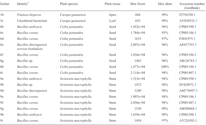
In the present work, results were obtained for only seven samples, in which fragments with approximately 1,200 bp were observed (Figure S1). The identification of endophytic bacteria based on the comparison between the
sequences obtained and those deposited in GenBank using the BlastN revealed the prevalence ofBacillus. Of the total number of samples analyzed, 57.14% belonged to the ge-nusBacillusand 14.28% toPantoea, whereas 28.57% were non-culturable bacteria. A phylogenetic analysis of isolates is shown in Supplementary Figure S2. Gram positive and Gram negative endophytic bacteria have already been iso-lated from many tissue types in numerous plant-species (Glare and O’Callaghan, 2000). Recently, the endophytic microbiota of several plant-species have been studied, the most prevalent genera isolated including Pseudomonas,
Erwinia, Bacillus, Burkholderia, Xanthomonas and
Enterobacter(Kuske et al., 1997). Bacteria belonging to
the genus Pantoea have likewise been observed in
cit-rus-plant species, as well as clover and sugarcane (Araújo
et al., 2002; Polanczyk and Alves, 2003).
On identifying isolates at the species level, incongrui-ties were observed. When sequences obtained in the present analysis were compared to those deposited in GenBank us-ing the BlastN, it was observed that one same given se-quence was actually similar to those sese-quences of more than one species (Table 1). The genetically related species were Bacillus thuringiensis, B. cereus, B. subtilis, B. amyloliquefaciens, B. polyfermenticus, B. anthracis, B. velezensisandPantoea dispersa. According to Polanczyk and Alves (2003), though the termBacillus thuringiensisis used for one single species (based on taxonomic traits), the bacterium belongs to a complex formed by several species
Table 1- Identification of endophytic bacteria based on the 16S rRNA region sequence compared to sequences deposited in GenBank, using Blastn.
Isolate Identity1 Plant species Plant tissue Max Score Max ident Accession number (GenBank)
1b Pantoea dispersa Carapa guianensis Apex 684 99% FJ756350.1
5c Uncultured bacterium Carapa guianensis Leaf 652 99% AY838532.1
6b Bacillus anthracis Ceiba pentandra Seed 1.452e+04 96% CP001598.1
6c Bacillus cereus Ceiba pentandra Seed 1.784e+04 95% CP001186.1
6d Bacillus cereus Ceiba pentandra Seed 1635 97% FJ841975.1
6e Bacillus thuringiensis serovar konkukian
Ceiba pentandra Seed 2.007e+04 96% AE017355.1
6f Bacillus cereus Ceiba pentandra Seed 1.856e+04 96% CP001186.1
6g Bacillussp. Ceiba pentandra Seed 1443 96% AB126763.1
6h Bacillus cereus Ceiba pentandra Seed 1.477e+04 100% CP001186.1
6i Bacillus cereus Ceiba pentandra Seed 2.114e+04 98% CP001407.1
9a Bacillus anthracis Swietenia macrophylla Stem 1.315e+04 94% CP001598.1
9b Bacillussp. Swietenia macrophylla Stem 1472 98% EF428972.1
9d Bacillus thuringiensis Swietenia macrophylla Stem 1249 98% AM778997.1
9e Bacillus cereus Swietenia macrophylla Stem 1.907e+04 98% CP001186.1
9f Bacillus cereus Swietenia macrophylla Stem 2.036e+04 98% CP001407.1
9g Bacillus cereus Swietenia macrophylla Stem 1530 99% AB508868.1
9h Bacillus anthracis Swietenia macrophylla Stem 1.639e+04 99% CP001598.1
(B. anthracis,B. cereus,B. mycoides,B. thuringiensis, and
B. weihenstephanensis). This complex is calledB. cereus. Molecular methods, the analysis of fatty acids and phos-pholipids, comparison of the 16S rRNA sequence, among
other analytical approaches, have shown that B.
thuringiensisandB. cereusare, in fact, one and the same species. Therefore, the need for a better distinction between the two has become the central topic of several taxonomy studies (Gordonet al., 1998).
The results obtained on analyzing partial sequences of the 16S rRNA region encoding gene, demonstrate the low genetic divergence between endophytic bacteria isolated from the three arboreal plant-species native to the Amazon. This may be linked to the growth conditions adopted, as well as to the low specificity of the primers used. Notwithstand-ing, the plant species used in the present study showed good promise as a source of sampling material in studies about the isolation and identification of bacterial genera that may have potential biotechnological applications.
Acknowledgments
The authors are indebted to the Post-Graduation Pro-gramStricto Sensu, Academic MSc course in Urban Biol-ogy, UniNilton Lins.
References
Altschul SF, Gish W, Miller W, Myers EW and Lipman DJ (1997) Gapped BLAST and PSI-BLAST: A new generation of pro-tein database search programs. Nucleic Acids Res 25:3389-3402.
Andreote FD, Araújo WL, Azevedo JL, Van Elsas JD, Rocha UN and Van Overbeek LS (2009) Endophytic colonization of potato (Solanum tuberosumL) by a novel competent bacte-rial endophyte,Pseudomonas putidastrain P9, and its effect on associated bacterial communities. Appl Environ Micro-biol 75:3396-3406.
Araújo WL, Marcon J, Maccheroni Júnior W, Van Elsas JD, Van Vuurde JWL and Azevedo JL (2002) Diversity of endo-phytic bacterial populations and their interaction with Xylella fastidiosain citrus plant. Appl Environ Microbiol 68:4906-4914.
Azevedo JL, Maccheroni Jr W, Pereira JO and Araújo WL (2000) Endophytic microorganisms: A review on insect control and recent advances on tropical plants. Electron J Biotechnol 3:e1-4.
Azevedo JL, Maccheroni Júnior W, Araújo WL and Pereira JO (2002) Microrganismos endofíticos e seu papel em plantas tropicais. In: Serafini LA, Barros NM and Azevedo JL (eds) Biotecnologia: Avanços na Agricultura e na Agroindústria. EDUCS, Caxias do Sul, pp 235-268.
Isolate Identity1 Plant species Plant tissue Max Score Max ident Accession number
(GenBank)
9j Bacillus thuringiensis Swietenia macrophylla Stem 1341 99% FJ932761.1
13a UnculturedBacillussp. Swietenia macrophylla Leaf 1158 98% EU371583.1
13b Bacillus subtilis Swietenia macrophylla Leaf 970 100% GQ161967.1
13c Bacillussp. Swietenia macrophylla Leaf 515 99% FJ465166.2
13c Bacillus subtilis Swietenia macrophylla Leaf 1476 98% EU257444.1
13d Bacillus amyloliquefaciens Swietenia macrophylla Leaf 1.352e+04 99% CP000560.1
13e Bacillus velezensis Swietenia macrophylla Leaf 1437 98% EU852930.1
13f Bacillus velezensis Swietenia macrophylla Leaf 1483 99% EU852930.1
13g Bacillus amyloliquefaciens Swietenia macrophylla Leaf 1294 97% FJ685773.1
13h Bacillus amyloliquefaciens Swietenia macrophylla Leaf 1391 99% FJ960508.1
13j Bacillus subtilis Swietenia macrophylla Leaf 1393 97% EU862566.1
14a Bacillus amyloliquefaciens Swietenia macrophylla Leaf 1.342e+04 98% CP000560.1
14b Bacillus polyfermenticus Swietenia macrophylla Leaf 1489 98% AY149473.2
14c Bacillussp. Swietenia macrophylla Leaf 453 97% FJ654441.1
14d Bacillus subtilis Swietenia macrophylla Leaf 1184 98% EF428247.2
14e Bacillussp. Swietenia macrophylla Leaf 749 99% FJ463041.1
14f Bacillus polyfermenticus Swietenia macrophylla Leaf 1520 99% AY149473.2
14h Bacillus polyfermenticus Swietenia macrophylla Leaf 1404 98% AY149473.2
14g Bacillussp. Swietenia macrophylla Leaf 1227 98% FJ465166.2
14i Bacillus polyfermenticus Swietenia macrophylla Leaf 1415 98% AY149473.2
16a Unculturedbacterium Ceiba pentandra Apex 619 99% GQ096960.1
Downing KJ, Leslie G and Thompson JA (2000) Biocontrol of the sugar-cane borer Eldana saccharinaby expression of the Bacillus thuringiensis cry1Ac7 and Serratia marcescens chiAgene in sugar-cane associated bacteria. Appl Environ Microbiol 66:2804-2810.
Ezra D, Castillo UF, Strobel GA, Hess WM, Porter H, Jensen JB, Contron MAM, Teplow DB, Sears J, Maranta M, et al. (2004) Coronamycins, peptide antibiotics produced by a verticullate Streptomyces sp (MSU-2110) endophytic on Monsterasp. Microbiology 150:785-793.
Figueiredo JEF, Gomes EA, Guimarães CT, Lana UGP, Teixeira MA, Lima GVC and Bressan W (2009) Molecular analysis of endophytic bacteria from the genusBacillusisolated from tropical maize (Zea maysL). Braz J Microbiol 40:522-534. Glare TR and O’Callaghan M (2000)Bacillus thuringiensis: Biol-ogy, Ecology and Safety. John Wiley & Sons, Chichester, 350 pp.
Gordon D, Abajian C and Green P (1998) Consed: A graphical tool for sequence finishing. Genome Res 8:195-202. Hallmann J, Quadt-Hallmann A, Mahaffee WF and Klopper JW
(1997) Bacterial endophytes in agricultural crops. Can J Microbiol 43:895-914.
Kuske CR, Barns SM and Busch JD (1997) Diverse uncultivated bacterial groups from soils of the arid Southwestern United States that are present in many geographic regions. Appl En-viron Microbiol 63:3614-3621.
Polanczyk R and Alves S (2003)Bacillus thuringiensis: Uma breve revisão Agrociencia 7:1-10.
Ribeiro JELS, Hopkins MJG, Vicentini A, Sothers CA, Costa MAS, Brito JM, Souza MAD, Martins LHP, Lohmann LG, Assunção PACL, et al.(1999) Flora da Reserva Ducke: Guia de Identificação das Plantas Vasculares de uma Flo-resta de Terra-Firme na Amazônia Central. INPA, Manaus, 816 pp.
Ryan RP, Germaine K, Franks A, Ryan DJ and Dowling DN (2008) Bacterial endophytes: Recent developments and ap-plications. FEMS Microbiol Lett 278:1-9.
Souza AQL, Souza ADL, Astolfi Filho S, Belém Pinheiro ML, Sarquis MIM and Pereira JO (2004) Atividade
antimicro-biana de fungos endofíticos isolados de plantas tóxicas da Amazônia:Palicourea longiflora(aubl) rich andStrychnos cogensbentham. Acta Amazonica 34:185-195.
Stierle A, Strobel G and Stierle D (1993) Taxol and taxane pro-duction byTaxomyces andreanae, an endophytic fungus of Pacific Yew. Science 260:214-216.
Strobel G and Hess WM (1999) Glucosylation of the peptide leucinostatina A, produced by an endophytic fungus of Eu-ropean yew, may protect the host from leucinostatin toxic-ity. Chem Biol 4:529-536.
Strobel G and Daisy B (2003) Bioprospecting for microbial endo-phytes and their natural products. Microbiol Mol Biol Rev 67:491-502.
Tamura K and Nei M (1993) Estimation of the number of nucleo-tide substitutions in the control region of mitochondrial DNA in humans and chimpanzees. Mol Biol Evol 10:512-526.
Tamura K, Peterson D, Peterson N, Stecher G, Nei M and Kumar S (2011) MEGA5: Molecular Evolutionary Genetics Analy-sis using maximum likelihood, evolutionary distance, and maximum parsimony methods. Mol Biol Evol doi: 10.1093/molbev/msr121.
Ulrich K, Ulrich A and Ewald D (2008) Diversity of endophytic bacterial communities in poplar grown under field condi-tions. FEMS Microbiol Ecol 63:169-180.
Supplementary Material
The following online material is available for this ar-ticle:
Figure S1 - Agarose gel showing the PCR products Figure S2 - Molecular phylogenetic analysis
This material is available as part of the online article at http:www.scielo.br/gmb.
Associate Editor: Luís Carlos de Souza Ferreira
2 3
4
5
6
7
8
9
10
11
12
Figure S1 – Agarose gel 1% (w/v) showing the PCR products of the amplification of the
1316S rRNA encoding region. 1: 200-bp ladder; 2-7: isolates.
1415
16
17
18
19
20
21
22
23
24
25
200 bp 1000 bp
1 2 3 4 5 6 7
Bacillus sp.
Bacillus anthracis
Uncultured bacterium2
Uncultured Bacillus
Bacillus amyloliquefaciens
Uncultured bacterium
Pantoea dispersa
Bacillus thuringiensis serovar konkukian
Bacillus cereus
Bacillus subtilis
Bacillus velezensis
33
100
100 75
50
50
50 50
25 25
26
(a)
27Bacillus thuringiensis Bacillus sp.
Bacillus anthracis Uncultured bacterium2 Uncultured Bacillus Bacillus amyloliquefaciens Uncultured bacterium Pantoea dispersa Bacillus subtilis Bacillus velezensis Bacillus polyfermenticus
Bacillus thuringiensis serovar konkukian Bacillus cereus
0 .0 2
0 .0 1
0 .0 0
2 . 3 4
3 . 1 6
0 . 5 3 0 . 0 0
0 .6 8
0 .5 0 0 .0 0
0 .0 0
2 .0 5
0 . 2 2 1 .0 1 0 .0 0
0 . 1 3
1 . 1 6
0 .8 3 0 . 5 4
0 .0 0
0 .0 0
0 . 6 0
0 . 4 3 0 .5 4
28