Bull Pan Am Health Organ 14(l), 1980
DISTRIBUTION OF HEPATITIS B VIRUS (HBV) M.ARKF.RS IN BLOOD DONORS OF 13 WESTERN HEMISPHERE COUNTRIES: PROCEEDINGS OF THE RED CROSS LATIN AMERICAN HEPATITL5 B WORKSHOP
S. Mazzur,’ N. Nath,’ C. Fang,’ M. J. Bastiaans,l J. L. Molinaris,2 M. Balcaser,S S. Beker G.,4 E. A. Brunings, 5 A.R.E. Cameron,6 V. Farrell,” 0. H. Fayfs G. Labrador-Gonz51ez,4 G. Gonz5lez L.,’ A. Gutie’wz D., lo C. Jaramillo T., 1 r R. Katz,lz M. B. Leme I.&ez,l’ E. Ix~vy-K~enig,~ F. Morales Ayala,14 J. Rodriguez Amaya,* H. Rodriguez-Moyado, l5 R. A. de Torres,16 and M. Velascoig
Although hefiatitis B is a significant health problem in the Americas, data about it in many parts of the Hemisphere have tended to be scanty. To help define the situation, a study was made of hepatitis B viral markers in blood samples from 13 Hemisphere countries. This report presents the results of that study.
Introduction
The Red Cross Latin American Hepatitis B Workshop was sponsored by the Puerto Rican chapter of the American Red Cross
‘Until her death, American Red Cross Blood Serv- ices Laboratories, Bethesda, Md., United States.
%uezo Rico Red &o&Blood Service, -San Juan; Puerto Rico.
SInstitutd Dominicano de Seguros Sociales, Santa Domingo, Dominican Republic.
aHospital General de1 Oeste, SAS-Universidad Central de Venezuela, Caracas, Venezuela.
5Prof. dr. Paul C. Flu Institute, Paramaribo. Suriname.
6Academic Hospital, Paramaribo, Suriname. ‘Queen Elizabeth Hospital, St. Michael, Barbados. *Banco Central de Sangre, Rosario, Argentina. 9Banco de Sangre de la Cruz Roja Provincial de1 Guayas, Guayaquil, Ecuador.
lOLouisiana State University-International Center for Medical Research and Training, San Jose, Costa Rica.
llDepartamenta1 de Salud Piiblica de Antioquia, Medellln, Colombia.
lkniversidad de Chile, Santiago, Chile.
13ComissZo National de Hemoterapia. Rio de Ja- neiro, Brazil.
*%Jniversidad National de Trujillo, Trujillo, Peru.
15Instituto Mexican0 de1 Seguro Social, Mexico 7, D.F., Mkxico.
lkniversidad de Buenos Aires, Buenos Aires, Argentina.
and the American Red Cross Blood Services Laboratories on 19-20 May 1977. The workshop, which was held at the University of Puerto Rico, fulfilled two primary ob- jectives. The first was that of demon-
strating and defining various methqds used to detect hepatitis B surface antigen (HBsAg) in blood. The second was that of defining the magnitude of the hepatitis B problem among donor populations of the various participating countries and com- paring different serologic tests’ efficacy in detecting both HBsAg carriers and previous hepatitis B virus (HBV) infections. In con- nection with this work, each participating country submitted about 500 or about 1,000 donor specimens (see Table 1) to the Red Cross Blood Services Laboratories in Be- thesda, Maryland. These specimens were tested for HBsAg, antibody to surface antigen (anti-HBs), antibody to core anti- gen (anti-HBc), HBeAg, antibody to e (anti-HBe), and subtypes of HBsAg.
Hepatitis Research and Testing Section of the American Red Cross Blood Services Laboratories in Bethesda, the results ob- tained with specimens from different coun- tries are strictly comparable. These results, presented in this communication, thus provide a unified picture of the distribution of HBsAg and other HBV markers in a wide range of Latin American blood donor populations.
Materials and Methods Blood Samples
A total of 7,487 blood samples were ob- tained from donors in 13 Western Hemi- sphere countries (Argentina, Barbados, Brazil, Chile, Colombia, Costa Rica, the Dominican Republic, Ecuador, Mexico, Peru, Puerto Rico, Suriname, and Vene- zuela). Aliquots of serum in one-dram vials containing 200 eg sodium azide were shipped by air at ambient temperatures to the American Red Cross Blood Services Laboratories in Bethesda, Maryland.
HBsAg
All samples were tested for HBsAg by means of solid phase radioimmunoassay em- ploying a double antibody sandwich tech- nique (I) and commercially available kits (AUSRIA II @, Abbott Laboratories, North Chicago, Illinois). Those samples found reactive for HBsAg on screening were tested for specificity by inhibition, following the manufacturer’s instructions. Human serum containing high titers of antibody to HBsAg (anti-HBs) was used for this pur- pose. Only those samples that were specifi- cally inhibited by preincubation with anti- HBs were considered positive for HBsAg. All samples positive for HBsAg were retested in three ways-by immunodiffu- sion (ID) using the technique described by Mazzur (2); by counterelectrophoresis (CEP) using Hapindex@ kits supplied by
Ortho Diagnostics (Raritan, New Jersey); and by reverse passive hemagglutination (RPHA) using AUSCELL@ kits produced by Abbott Laboratories.
intibodies to HBsAg
All samples were tested for the presence of anti-HBs with the passive hemagglutination (PHA) microtiter technique (3), using human erythrocytes coated with HBsAg (supplied by Electra-Nucleonics in Be- thesda, Maryland). Samples were screened at 1:4, l:&, and 1:16 dilutions. Uncoated cells were included to determine nonspecific reactions.
Antibodies to HBcAg
All samples were tested for anti-HBc using the RIA test and CORAB@ kits donated by Abbott Laboratories.
HBeAg and anti-HBe
All samples found reactive to HBsAg were screened for the presence of HBeAg/anti- HBe with a rheophoresis technique. Rhe- ophoresis plates were obtained from AmSott Laboratories. Reagents for testing, i.e., sera positive for anti-HBe and HBeAg, were selected from the American Red Cross collection of HBsAg samples. The specific- ity of these samples was confirmed using reagents supplied by Dr. L. 0. Magnius of the Department of Virology, Statens Bak- teriologiska Laboratory, Stockholm, Sweden.
Subty#Cng of HBsAg
46 PAHO BULLETIN l vol. 14, no. 1, 1980
type ad or ay that had been purified by a previously described method (5). Monospe- cific sera used in the passive hemagglutina- tion-inhibition tests were obtained by heteroiogous absorption in the liquid phase.
Results
HBsAg Testing
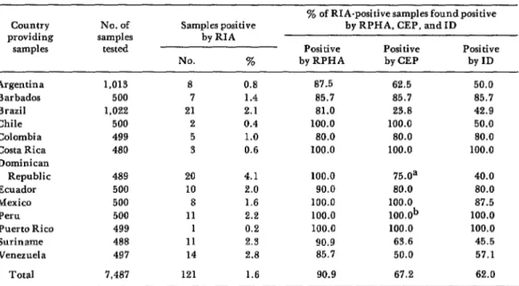
All 7,487 serum samples were tested for HBsAg by radioimmunoassay (RIA), the most sensitive technique. By this method, 121 samples (1.6 per cent) were found to contain HBsAg. Those samples found HBsAg-positive by RIA were subsequently retested by immunodiffusion (ID), counter- electrophoresis (CEP), and reverse passive hemagglutination (RPHA). As indicated in Table 1, RPHA, the most sensitive of these three tests, detected 90.9 per cent of those samples shown by RIA screening to contain HBsAg. Only 11 of the 121 samples found positive (with low levels of HBsAg) by RIA
yielded negative RPHA results. CEP de- tected 67.2 per cent of the positive samples, while ID, the least sensitive method, detected only 62.0 per cent. The HBsAg titers determined by RPHA ranged from 1:8 to at least 1:25,600, with the mode occurring at 1:6,400. As indicated in Figure 1, ID identified all samples with RPHA titers equalling or exceeding 1:25,600, as well as some samples with RPHA titers between 1:16 and 1:12,800. None of the 10 negative controls was judged positive. Similarly, CEP detected HBsAg in all samples with RPHA titers above 1:3,200, and also in some of the samples with titers ranging from 1:8 to 1:1,600.
Anti-HBs and Anti-HBc Testing
As Table 2 shows, the prevalence of anti- body to HBsAg was found to range from 3.8 per cent in the Chilean samples to 55.3 per cent in the samples from the Dominican Republic. This prevalence generally in-
Table 1. Comparison of methods used to detect HEsAg.
Country providing
samples
Argentina Barbados Brazil Chile Colombia Costa Rica Dominican
Republic Ecuador Mexico Peru Puerto Rico Suriname Venezuela
Total
No. of samples
tested
1,013 500 1.022 500 499 480 489 500 500 500 499 488 497 7,487
Samples positive by RIA
No. %
8 0.8
7 1.4
21 2.1
2 0.4
5 1.0
3 0.6
20 4.1
10 2.0
8 1.6
11 2.2
1 0.2
11 2.3
14 2.8
121 1.6
% of RIA-positive samples found positive by RPHA, CEP, and ID
Positive Positive Positive
by RPHA by CEP by ID
87.5 62.5 50.0
85.7 85.7 85.7
81.0 23.8 42.9
100.0 100.0 50.0
80.0 80.0 80.0
100.0 100.0 100.0
100.0 75.0a 40.0
90.0 80.0 80.0
100.0 100.0 87.5
100.0 lOO.Ob 100.0
100.0 100.0 100.0
90.9 63.6 45.5
85.7 50.0 57.1
90.9 67.2 62.0
Figure 1. A total of 121 HBsAg-positive samples detected by radioimmunoassay (RIA) were retested by reveme passive hemagglutination (RPHA),
counterelectrophoresis (CEP), and immunodiffusion (ICI).
titer 3 25,600 12,800 6,400 3,200 1,600 800 400 128 64 32 16 8 4 8
I 62.0%
67.2%
Vertical bars show the number of samples yielding the indicated titers in RPHA tests. The two horizontal bars, marked “ID” and CEP,” pass below certain RPHA titers. Within these ranges (for HBsAg-positive samples with these RPHA titers) ID and CEP tests yielded positive results. Some positive ID and CEP results were also obtained with samples whose RPHA titers fell within the lower ranges indicated by the two horizontal lines.
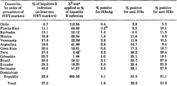
Table 2. Hepatitis B Gnu markem found in serum samples from 13 Western Hcmisphe countries. Countries, Y. of hepa’titis B X2 testa
by order of infection applied to ye prevalence of (at leastone of hepatitis HBV markers H BV markers) B infection
ye positive ye positive y. positive for HBsAg for anti-HBs for anti-HBc
Chile Puerto Rko Barbados Mexico Venezuela Argentina Costa Rica Peru Colombia Brazil Ecuador Suriname Dominican
Republic
6.7 110.56
11.1 66.68
13.1 52.12
16.8 28.34
17.9 22.38
18.6 41.80
20.6 27.3
1o.q 0.02
29.3 144
33.9 28.21
35.3 18.20
40.9 51.27
0.4 0.2b
3.8 9.2
1.4 9.0
1.6 11.6
2.8 11.6
0.8 14.7
0.6 17.3
2.2 20.2
1.0 25.1
2.1 26.7
2.0 29.4
2.3 28.1
82.8 805.38 4.1
Total 27.0 1.6
55.3
20.2
5.3
10.1 11.9 9.0 15.5 9.4
16.7 20.4 18.1 27.6
21.9 37.9 81.1 21.3 aChi-square calculated by comparing positive and negative results for one country with the total positive and ne
48 PAHO BULLETIN l vol. 14, no. 1, 1980
creased in proportion to the number of HBsAg carriers in the population. Testing for anti-HBc showed its prevalence to range from 5.3 per cent in the Chilean samples to 81.1 per cent in the Dominican samples. The prevalence of anti-HBc was shown to increase as the prevalence of other hepatitis B markers rose.
Su btyfiing
Serum samples known to contain HBsAg were tested for antigen subtypes by the pas- sive hemagglutination inhibition reaction and by immunodiffusion. The former test, which is more sensitive than immunodiffu- sion, succeeded in identifying 66 of the 121 samples (54.5 per cent) as HBsAg/ad17 and 5 of the samples (4.1 per cent) as HBsAg/ay. These ay samples came from Argentina, the Dominican Republic, and Suriname. Sub- typing by immunodiffusion confirmed the subtypes of those samples that had suffi- ciently high titers to permit detection by this method; it also further identified the samples involved as HBsAg/adw and HBsAg/ayw. None of these samples con- tained the r determinant.
HBeAg and Anti-HBe
As previously noted, the 121 HBsAg-posi- tive samples were tested by rheophoresis for HBeAg and anti-HBe. Of these, seven samples (5.8 per cent) were found to contain HBeAg. One of these came from Argentina, one from Barbados, one from Suriname, and four from Mexico. The presence of HBeAg in blood donor samples did not correlate with the prevalence of HBsAg in the populations involved.
Antibody to HBeAg was found in 30 of the 121 samples (24.8 per cent); these 30
1’The major antigenic determinants of HBsAg are a, d, y, w, and y. Various combinations of these occur j_n nature, adw, ayw, and adr being the most common.
The antigenic subtypes are curren;iy used as an irn- ,portant marker in epidemiologic studies.
samples came from Argentina, Brazil, the Dominican Republic, Ecuador, Peru, Puerto Rico, and Venezuela.
Overall Test Results
Testing of all the samples from partici- pating Latin American countries showed
1.6 per cent positive for HBsAg, 20.2 per cent positive for anti-HBs, and 21.3 per cent positiv; for anti-HBc. HBsAg was found to occur alone without any of the other HBV markerson occasion, although 94.6 per cent of the HBsAg-positive samples were also found to contain anti-HBc (Figure 2). Anti- HBc was also found in 72.2 per cent of the samples containing anti-HBs, and was detected in 5.3 per cent of the total samples unaccompanied by any other HBV serum marker. These anti-BHc figures cannot be confirmed because no method for confirm- ing the CORAB@ kit RIA test results ob- tained is available at the present time.
The prevalences of HBsAg, anti-HBs, anti-HBc, and HBV infection in the blood donor populations of each of the 13 coun- tries were compared with the prevalences of the indicated markers in all of the countries combined. This was done by applying a chi- square test with one degree of freedom (see Table 2). The results indicated that blood donors of Argentina, Chile, and Puerto Rico had significantly lower HBsAg preva- lences than the overall population tested (p<O.O5), while those of the Dominican Republic and Venezuela had significantly higher H BsAg prevalences (p < 0.005). The prevalence of HBsAg in Costa Rica (0.6 per cent) was not significantly lower (p < 0.1) as it was in Argentina (0.8 per cent). The difference may be due to the different num- ber of samples tested. Similarly, the donors in Argentina, Barbados, Chile, Mexico,
Figure 2. The distribution of HBV serum markers in samples from 7,487 Latin American blood donors.
No HBV Markers
\ 72.98%
Posltrve for HBsAg, but Ant,.HBc unknown 0.09%
HBsAg alone 0.08% HBsAg + Ant!-HBc 1.43%
Ant+HBc 5.30%
AntI-HBs t Anil-HBc
L AntI- HBs 5.61%
All the samples were tested for HBsAg, anti-HBs, anti-HBc, HBeAg, and anti-HBe. A few (0.09 per cent) of the samples could not be tested for anti-HBc because of insuffi- cient sample material.
prevalences (p < 0.005). Observed anti-His prevalences were significantly lower among donors in Argentina, Barbados, Chile, Costa Rica, Mexico, Puerto Rico, and Venezuela (p < 0.025), but were significant- ly higher (p<O.O05) among donors in Brazil, the Dominican Republic, and Suriname.
Prevalences of hepatitis B infection, as indicated by the presence of at least one hepatitis B virus marker, ranged from 6.7 per cent in Chile to 82.8 per cent in the Dominican Republic. The average preva- lence found in the samples from all the participating Latin American countries was 27.0 per cent. Countries that had preva- lences of infection significantly below this average (p<O.O05), as judged by the pre- sence of at least one HBV marker, were Argentina, Barbados, Chile, Costa Rica, Mexico, Puerto Rico, and Venezuela. In contrast, prevalences significantly above the average (p<O.O05) were found for Brazil, the Dominican Republic, Ecuador, and Suriname.
Discussion
This study constitutes the first unified investigation of HBV markers in blood donor populations of 13 Western Hemi- sphere countries. The overall prevalence of HBsAg carriers among 7,487 blood donors in these countries was found to be 1.6 per cent. Respective overall prevalences of anti- HBs and anti-HBc were 20.2 and 21.3 per cent.
50 PAHO BULLETIN l vol. 14, no. 1, 1980
A striking feature of the data was the whether paid or volunteer blood donors extreme variation in the prevalence of HBV were used and the relative socioeconomic markers found in different populations (see status of those donors in the community. Table 2), a prevalence ranging from 6.7 per
cent in Chile to 82.8 per cent in the Domin- ican Republic. This high Dominican Re- public figure confirms earlier work done in that country. That work found HBsAg prevalences of 5.3 per cent by RIA (6) and 2.0 per cent by the immunodiffusion method (7). In addition, a 41 per cent preva- lence of anti-HBs, detected by passive hemagglutination, has been reported among a series of women giving birth at the Salvador B. Gautier Hospital in Santo Domingo (8). Thus the level of HBV infec- tion appears to have been much higher in the Dominican population than among other populations studied.
The contrast between the lowest preva- lence of HBV infection found (6.7 per cent in Chile) and the highest (82.8 per cent in the Dominican Republic) was very dra- matic. However, prevalence levels could not be explained on the basis of geographic con- tiguity. For example, low-prevalence Chile is bordered by Argentina and Peru, where the respective prevalences were 18.6 and 27.3 per cent. Likewise, the prevalences of HBV markers found for the nearby island populations of Barbados, the Dominican Republic, and Puerto Rico were quite dissimilar.
It is difficult to account for these significant variations in HBV prevalence among the various blood donor populations studied; however, some of the variations could be due to local variables-such as
Factors such as enhanced virulence of cer- tain HBV strains, amplification of an existing transmission route, or development of more effective routes could also play some part in these variations.
It was interesting to note that of five HBsAg/ay subtypes identified, three came from the Dominican Republic and one came from Suriname. The Dominican Republic and Suriname populations studied had the two highest prevalences of HBV. It has been suggested that HBsAg/ay is more infectious than other subtypes (9).
Comparative testing of samples reactive for HBsAg by RIA showed that reverse passive hemagglutination (RPHA) detected HBsAg in 90.9 per cent of the RIA-positive samples, while counterelectrophoresis (CEP) and immunodiffusion (ID) respec- tively detected HBsAg in 67.2 and 62.0 per cent of the RIA-positive samples. These data are similar to those obtained by many other investigators (10). Although CEP and ID are much less sensitive than RIA and RPHA, more than half of the HBsAg carriers have sufficient antigen to be detected by these methods; so, while not optimal tests, CEP and ID will detect a sig- nificant number of HBsAg carriers and will permit them to be eliminated from the donor pool. Thus CEP or ID would perhaps be appropriate methods for use by blood programs lacking access to more sensitive technology.
ACKNOWLEDGMENTS
A total of 7,487 donor blood samples from 13 ‘Western~Hemisphere co_untries (Arsntina, Bar-
bados, Brazil, Chiie, Colombia; Costa R&a,-ihe Dominican Republic, Ecuador, Mexico, qeru, Puerto Rico, Suriname, and Venezuela) were tested for various markers of hepatitis B virus (HBV) infection with several different techni- ques. HBsAg was detected in 1.6 per cent of the samples, anti-HBs in 20.2 per cent, and anti-HBs in 21.3 per cent. The incidence of HBsAg varied from 0.2 per cent (in the Puerto Rican samples) to 4.1 per cent (in the samples from the Dominican Republic). Overall, 5.8 per cent of the samples found to contain HBsAg also contained HBeAg, while 24.8 per cent had de- tectable anti-HBe.
Sixty-six (54.5 per cent) of the 121 HBsAg-
positive samples were found to contain subtype HBsAg/ad, and 5 (4.1 per cent) were found to contain subtype HBsAg/ay. Subtypes of the re- maining samples could not be determined because of insufficient antigen. In a comparative study, reverse passive hemagglutination (RPHA), counterelectrophoresis (CEP), and immunodif- fusion (ID) detected HBsAg in 90.9, 67.2, and 62.0 per cent of the samples previously found positive by radioimmunoassay (RIA) screening.
Considerable variation was observed in the prevalence of HBV markers in samples from dif- ferent countries, the highest prevalence being 82.8 per cent in samples from the Dominican Republic. Overall, the findings suggest that exposure to HBV is quite extensive in some Latin American populations.
REFERENCES (I) Ling, C. M., and L. R. Overby. Preva- lence of hepatitis B virus antigen as revealed by direct radioimmuneitssa$ with 1251-antibody. J Immunol 109:834-841, 1972.
(2) Mazzur, S. The detection of Australia antigen by immunodiffusion and counterelec- trophoresis. Am J Med Technol 38:343-349, 1972.
(3) Vyas, G, N.:, and N. R. &ulman. Hemag- glutination assay for antigen and antibody as- sociatedwith viral hepatitis. Science 170:332-333, 1970.
(4) Prince, A. M., B. Brotman, and H. Ikraim. Hemagglutination assay: subtyping by hemagglutination inhibition, an ultrasensitive identity test for HB antigen. In G. S. Vyas et al. (eds.), He#atitis and BZood Transfusion. New York, Grune and Stratton, 1972, pp.
147-154.
(5) Nath, N., S. Mazzur, R. Ledman, and C. T. Fang. Purification of hepatitis B surface
antigen using polyethylene glycol, pepsin and Tween 80. VOX Sung 31 (Suppl. 1):84-95, 1976.
(6) Brea, A. R., and S. Mazzur. Personal communication.
(7) Brea, A. R., S. Mazzur, M. Balacaser, and B. S. Blumberg. Australia antigen in the Dominican Republic. Paper presented at the XIII Congreso Panamericano de Gastroentero- log&z (Buenos Aires, Argentina, 1973).
(8) Mazzur, S., and A. R. Brea. Personal communication.
(9) Nath, N., C. T. Fang, R. Ledman, S. Mazzur, and R. Y. Dodd. Relationship of sub- types of hepatitis B surface (antigen with e antigen and its corresponding antibody. J infect Dis 138:252-256, 1978.
(IO) Nath, N., and R. Y. Dodd. Hepatitis testing. l% T. J. Greenwalt and E. A. Steane teds.), CRC Handbook on Blood Banking (vol.