1 . Specialization in Cardiovascular Surgery, Associate Member of BSCVS, Master’s Degree in Surgery at Federal University of Ceará (UFC), Fortaleza, CE, Brazil.
2 . Medicine Student at UFC, Extension Pro-Rector Scholarship at UFC, Fortaleza, CE, Brazil.
3 . Medicine Student at UFC, FUNCAP scholarship, Fortaleza, CE, Brazil. 4 . Medicine Graduation at UFC, Generalist Physician, Fortaleza,
CE, Brazil.
5 . PhD in Pharmacology at UFC, Adjunct Professor of the Dental Clinics Department at UFC, Fortaleza, CE, Brazil.
6 . Titular Member of BSCVS, Adjunct Professor of the Surgery Department at UFC, Fortaleza, CE, Brazil.
Heraldo Guedis Lobo Filho
1, Nestor Lemos Ferreira
2,
Rafael Bezerra de Sousa
3,
Eduardo Rebouças
de Carvalho
4, Patrícia Leal Dantas Lobo
5,
José Glauco Lobo Filho
6Modelo experimental de infarto do miocárdio induzido por isoproterenol em ratos
Experimental model of myocardial infarction
induced by isoproterenol in rats
Abstract
Objective: To evaluate and validate, in our laboratory,
the essay of myocardial infarction induced by isoproterenol in rats by means of analysis of hematological, biochemical, oxidative stress markers and histopathological parameters. Methods: Thirty young, male, Wistar rats (145 to 230 g) were randomly allocated in two groups: Sham group, which underwent a virtual myocardial infarction induction, and the Infarction group, which underwent a myocardial infarction induction with isoproterenol. The administrations for the infarction induction were performed during two consecutive days and a 24-hour interval between them. Twenty-four hours after the last administration, rats from both groups were anesthetized and sacrificed for blood sample collection to evaluate complete blood count (CBC) and biochemical parameters (SGOT, SGPT, troponin I, urea and creatinin), obtain myocardial fragments for oxidative stress markers analyses (catalase activity and glutathione concentrations) as well as histopathological examinations.
Results: There were no death cases in the Sham group,
while the mortality rate in the Infarction group was 25%. Myocardial infarction induction with isoproterenol raised leukocytes and neutrophils counts, SGOT, troponin I and
urea concentrations, reduced catalase enzyme activity and glutathione concentrations in the myocardium and let to histopathological concentrations as well. It did not exert alterations in terms of hemoglobin, SGPT and creatinin concentrations.
Conclusion: The isoproterenol-induced myocardial
infarction essay in rats was adequately reproduced in our laboratory, causing alterations in hematological, biochemical, oxidative stress markers and histopathological parameters.
Descriptors: Myocardial infarction. Isoproterenol. Rats,
Wistar.
Resumo
Objetivo: Avaliar e validar, em nosso meio, o modelo de
infarto do miocárdio induzido por isoproterenol em ratos por meio de análises de parâmetros hematológicos, bioquímicos, de marcadores do estresse oxidativo e histopatológicos.
Métodos: Trinta ratos jovens, machos, da linhagem
Wistar (145 a 230 g), foram alocados aleatoriamente em dois grupos: grupo Simulado, submetido à falsa indução de infarto do miocárdio, e grupo Infarto, submetido à indução
This study was carried out at Surgery Department of the Faculty of Medicine, Federal University of Ceará, Fortaleza, CE, Brazil.
Correspondence address: Heraldo Guedis Lobo Filho
Rua Dr. José Lourenço, 777 – Aldeota – Fortaleza, CE Brazil – ZIP Code: 60115-280.
E-mail: [email protected]
Article received on April 21st, 2011
In the literature, there is a series of studies using this model, evaluating the effect of different substances on biochemical, oxidative stress markers and histopathological damage resulting from myocardial in rats [8,9].
This study aims to evaluate and validate, in our environment, the model of isoproterenol induced-myocardial infarction in rats, given the scarcity of scientific publications using this model in our country
METHODS
This study, whose data are from a Master’s Degree dissertation presented to the Strictu Sensu Post-graduation Program in Surgery of the Department of Surgery at Faculty of Medicine, Federal University of Ceará, has been developed at the Laboratory of Experimental Surgery, Faculty of Medicine, Federal University of Ceará.
Ethical aspects
The experimental procedures were performed in accordance with the ethical standards established by the Ethics Committee on Animal Research (CEPA), Federal University of Ceará, under protocol (69/10). The norms were established by the Council for International Organization of Medical Sciences (CIOMS) and the precepts of the Brazilian College of Animal Experimentation (COBEA).
Sample
We used 30 young rats, males, weighing between 145 and 230g (mean: 185.12 ± 18.58), Wistar, from the Central INTRODUCTION
Acute myocardial infarction (AMI), defined as death of the cardiac muscle due to ischemia, is one of the most usual diagnoses in hospitalized patients in industrialized nations. In the USA, for example, each year the average of 650,000 individuals present a first-time AMI episode while 450,000 people suffer from a recurrent one [1]. The ever growing rates of such condition in developing countries account as one of the most relevant public health issues nowadays [2].
To study possible protective effects of drugs on the myocardial injury from AMI, a widely used experimental model is the induction of infarction by means of the administration of isoproterenol in rats, since this substance causes a myocardial damage similar to the one observed in AMI in humans [3]. Amidst several mechanisms proposed to explain the isoproterenol-induced myocardial harm, one might say: an unbalance between oxygen supply to and demand from cardiomyocytes inwardly, which is related to myocardial hyperfunction due to increase both in chronotropism and inotropism as well as to hypotension in the coronary bed [4]. Secondly, it is also claimed that there is an elevation of Ca++ overcharge inside the cell [5]. In addition, that ion is related to the activation of the adenylate cyclase enzyme and the depletion of ATP levels on the course of the events [6]. Eventually, there is an oxidative stress augmentation because of several metabolic products originated from isoproterenol, not to mention free radicals genesis [7].
do infarto do miocárdio com isoproterenol. As aplicações, para indução do infarto, foram realizadas durante dois dias consecutivos, com intervalo de 24 horas entre elas. Após 24 horas da última aplicação, os ratos de ambos os grupos foram anestesiados e sacrificados para realização de coleta de sangue para hemograma e análise bioquímica (TGO, TGP, troponina I, ureia e creatinina) e coleta de fragmento do miocárdio para avaliação de marcadores do estresse oxidativo (atividade da catalase e concentração de glutationa) e exame histopatológico.
Resultados: Não houve mortalidade no grupo Simulado,
enquanto a mortalidade no grupo Infarto foi de 25%. A indução do infarto do miocárdio com isoproterenol causou
elevação das contagens de leucócitos e neutrófilos, dos níveis de TGO, troponina I e ureia, reduziu a atividade da catalase e os níveis teciduais de glutationa e causou alterações histopatológicas. Não acarretou alterações nas concentrações de hemoglobina, TGP e creatinina.
Conclusão: O modelo de infarto do miocárdio induzido
por isoproterenol em ratos foi adequadamente reproduzido em nosso laboratório, acarretando alterações em parâmetros hematológicos, bioquímicos, de marcadores de estresse oxidativo e histopatológicos.
Descritores: Infarto do miocárdio. Isoproterenol. Ratos
Bioterium of the Federal University of Ceará, created and maintained under similar environmental conditions and food.
Experimentation Environment
The animals were kept in polypropylene cages, housed in refrigerated facilities (24 ± 2°C). We observed an alternating light/dark cycle every 12 hours. The rats were fed a diet specific for industrial use in laboratories. The water offered was drinking. The cleaning of animal cages and examination were performed daily by the researcher and the technician in charge, respectively. The animals were fasted and no water, from 12 hours prior to the application of anesthesia, which preceded the sacrifice, blood collection, whose aim was to perform complete blood count and serum urea, creatinine, aspartate aminotransferase, aminotransferase and troponin I, removing the apical segment of the left ventricle for histopathology and fragments of the left ventricle to evaluate the activity of catalase and glutathione concentrations.
The rats were first acclimated for a period of seven days prior to the experiments. Surgical procedures were performed in a refrigerated environment at the Laboratory of Experimental Surgery, Department of Surgery, Federal University of Ceará.
Infarction Induction
The induction of AMI was performed in the Infarcted g r o u p , t h r o u g h s u b c u t a n e o u s a d m i n i s t r a t i o n o f isoproterenol at a dose of 150 mg/kg/day diluted in 2 ml of saline on two consecutive days with an interval of 24 hours between applications. The false induction of AMI in the Simulated group was performed by subcutaneous administration of 2 ml of saline on two consecutive days, also with an interval of 24 hours between applications.
Isoproterenol hydrochloride was purchased from Sigma company in the form of vials containing in each one 1 gr of pure powder of the active ingredient. The doses administered in the infarction group were calculated based on the weights of the animals. To measure the exact amount of isoproterenol (150 mg / kg animal) it was used a precision scale, made up a solution with 2 mL of saline solution subcutaneously.
Anesthesia
Twenty-four hours after the last subcutaneous injection, anesthesia was proceeded to perform the sacrifice of animals. The anesthetic technique involved the use of ketamine hydrochloride at a dose of 50 mg/kg of the animal and xilasina hydrochloride at a dose of 10 mg/kg of the animal, both applied intraperitoneally.
Collection of Samples
After adequate anesthesia, an incision was performed in an inverted T extending from neck to pubis.
Collection of blood
We obtained access to the retroperitoneum, the abdominal aorta was dissected in its infrarenal portion. The blood collection was then performed through a puncture of the abdominal aorta with a disposable Jelco number 24 (24 gauge). Using a 5 mL disposable syringe, coupled with Jelco 5 mL of blood were collected. Of this volume, 2 mL were stored in a tube for complete blood count and 3 mL in a tube for biochemical dosages (urea, creatinine, SGOT, SGPT, and troponin I). The tubes, properly identified, were then placed in a cooler with ice.
Collection of the cardiac apex region for histopathologic study
After collection phase of the blood, it was proceeded immediately to the opening of the chest and removing the heart. The heart was then washed with saline solution apical region of the left ventricle was put into a bottle, properly identified, containing a 10% buffered formalin solution.
Collection of fragments of the myocardium to measure the activity of the enzyme catalase and glutathione levels After the withdrawal phase of the cardiac apex, we proceeded immediately to the resection of two fragments of the left ventricle muscle. These fragments were weighed on a precision balance, and placed separately inside cryotubes properly identified and put into container of liquid nitrogen. The cryovials were then stored in a freezer at a temperature of -70°C until the analysis in a period of up to two weeks.
Histopathological evaluation
The apical regions of hearts, fixed in a solution of 10% buffered formalin, were embedded in paraffin in a period of about 24 hours after the onset of fixation. Cuts measuring four micrometers sections were stained with hematoxylin-eosin (HE) and observed microscopically. The severity and extent of MI were observed for each case. The pathologist did not know which group each slide corresponded. The findings were classified into the following degrees, to compose a range of histologic myocardial injury: (0) No change: (1) Mild - focal myocyte damage or small multifocal degeneration with slight degree of inflammation, (2) Moderate - extensive myofibrillar degeneration and/or diffuse inflammatory process, (3) Severe - necrosis with diffuse inflammatory process [10].
Evaluation of activity of the enzyme catalase in the myocardium
mM H2O2. The catalase activity was defined as the amount of enzyme required to decompose 1 nmol of H2O2 per minute at 25°C and pH 7. The absorbance was read in spectrophotometer at 230 nm. The results were expressed as millimoles per minute per gram of tissue (mmol/min.g tissue) [11].
Evaluation of glutathione concentrations in the myocardium
The tissue was homogenized with a solution of 0.02 M EDTA frozen for preparation of the homogenate at 10%. They were then added at a rate of 0.5 mL of homogenate, 0.4 mL of distilled water and 0.1 mL of 50% trichloroacetic acid, and then the samples were centrifuged at 3000 rpm for 15 min. A volume of 0.5 mL of the supernatant was removed and added 1 mL of 0.4 M Tris pH 8.9 and 25 mL of 0.01 M DTNB. The absorbance was measured within 5 min at 412 nm. The concentration of non-protein sulfhydryl groups (NP-SH) was calculated using a standard curve of GSH and the results expressed as mg of NP-SH/g of tissue [12].
Statistical Analysis
Statistical analysis was performed using the statistical software SPSS® for Windows (v.16, SPSS Inc. Chicago, IL, USA). Quantitative variables with normal distribution using the Kolmogorov-Smirnov test were presented as mean and
standard deviation. The range of histological myocardial damage was the only variable that did not show normal distribution, being represented by the median and interquartile range.
To compare the groups in relation to the scale of histologic myocardial injury non-parametric Wilcoxon-Mann-Whitney test was used. The Student’s t test was performed to compare means between groups for all other variables.
The results were expressed in tables and graphs, which were prepared using GraphPad ® software (v. 5, GraphPad Software, San Diego, CA).
In all tests, it was established 0.05 as á probability of type I error, and it was considered statistically significant when P <0.05 (95%).
RESULTS
Regarding mortality, there were no deaths in the Simulated group, while mortality in the Infaction group was 25%.
The induction of myocardial infarction with isoproterenol caused an increase of leukocyte and neutrophil counts, the levels of SGOT, Troponin I and urea, reduced the activity of catalase and tissue levels of glutathione and caused histopathological changes. It did not cause changes in hemoglobin concentrations, ALT and creatinine.
All results for hematological, biochemical and oxidative stress parameters are shown in Tables 1, 2 and 3.
The histopathological changes in the myocardial infarction group had higher scores on the scale of histologic myocardial injury compared to Simulated group, this difference was statistically significant (P = 0.00) (Table 4).
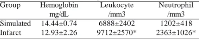
Table 1. Isoprotenerol effect on hemoglobin concentrations and
leukocyte and neutrophil counts Group Simulated Infarct Hemoglobin mg/dL 14.44±0.74 12.93±2.26 Leukocyte /mm3 6888±2402 9712±2570* Neutrophil /mm3 1202±418 2363±1026* *P<0.05
Table 2. Isoprotenerol effect on serum concentration of SGOT, ALT, troponin I, creatinine and urea
Group Simulated Infarct **P<0.01 *P<0.05 SGOT (U/L) 93.57±21.73 240.22±66.66** ALT (U/L) 42.88±11.13 51.72±10.31 Troponin I ng/mL 0.004±0.06 1.98±0.71** Creatinine mg/dL 0.71±0.01 0.72±0.10 Urea mg/dL 46.44±8.98 68.63±11.89*
Table 3. Isoprotenerol effect on the activity of catalase and
glutathione concentrations in the myocardium Groups
Simulated Infarct
Catalase activity (mmol/min.g)
2.51 ± 0.63 1.42 ± 0.22**
**P < 0.01
Glutathione (µg) 292 ± 27.21 192.86 ± 37.46**
Table 4. Comparison of Simulated and Infarction groups,
regarding degree of histologic myocardial injury Groups Simulated Infarct Median 0 3**
**P < 0.01
Fig. 1 - Histopathological study showing myocardial tissue with no histological changes (Grade 0)
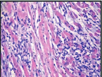
Fig. 2 - Histopathological study showing myocardial tissue with mild histological changes (Grade 1)
Fig. 4 - Histopathological study showing marked histological changes (Grade 3)
Fig. 3 - Histopathological study showing myocardial tissue with moderate histological changes (Grade 2)
In Figures 1 to 4, generated by this study, we present the histological classification of myocardial injury based on the study of Acikel et al. [10].
DISCUSSION
A model widely used for induction of MI in rats is a model of coronary ligation. The heart is exposed through a left thoracotomy incision and ligation of the proximal coronary artery is performed with 7-0 yarn [13,14]. Thus, it is an experimental model more complex, that beyond the need for thoracotomy is associated with ventilatory support and postoperative analgesia. It has a higher incidence of morbidity and mortality not related to MI, such as infection and pneumothorax, reaching a mortality of about 50% [15].
Considering that we are starting this line of research to evaluate the protective effect of substances in cases of myocardial injury, we adopted the model of induction of MI by isoproterenol in rats, given its effectiveness, practicality, reliability and lower morbidity and mortality of animals. This method is performed continuously in international studies [16,17].
myocardial tissue and induced histopathological changes such as inflammation, myofibril degeneration and necrosis. Regarding mortality, the resulting data from the study showed a mortality of 25% in the Infarction group. This value is consistent with data found in literature, pointing to the isoproterenol-induced myocardial infarction as a cause of mortality in this experimental model. Acikel et al. [10] reported a mortality rate of 33.33% in rats undergone administration of isoproterenol, the approximate value found in this research.
With regard to hematological parameters, there was no difference between the levels of hemoglobin in the comparison groups. The influence of isoproterenol administration on the levels of hemoglobin is underreported in the literature. Sangeetha & Quine [18] observed an increase in erythrocyte count and hemoglobin concentrations in rats undergone isoproterenol-induced myocardial infarction.
It was observed that the administration of isoproterenol caused an increase in leukocyte counts and neutrophils in the Infarcted group, when compared to Simulated group. High counts of leukocytes and neutrophils were observed in rats with isoproterenol-induced myocardial infarction [18] and are considered at risk for the development of AMI [19]. Friedman et al. [20] were the first to observe the relationship between neutrophil count and the subsequent development of AMI. In MI, neutrophils may contribute to myocardial injury due to the release of leukotrienes, ROS and hydrolytic enzymes. The degree of leukocytosis may also be associated with the magnitude of myocardial necrosis [18].
Literature data show that administration of isoproterenol subcutaneously as a result has a significant increase in serum markers of myocardial injury, such as SGOT, SGPT, CPK, CK-MB, LDH and troponin [10,21,22]. It is reported that plasma levels of these markers are directly proportional to the degree of necrotic lesions present in the myocardium and thus are markers of myocardial damage [10.23]. Previous studies have shown that kits for troponin T and I for humans can be used in rats [24,25].
In this study, administration of isoproterenol caused increased concentrations of AST and troponin I, when compared the Simulated and Infarction groups, corroborating the results expressed in the literature. Regarding the assessment of markers of renal function, it was found that isoproterenol caused an increase in concentrations of urea. Studies in the literature showing about changes in markers of renal function in this type of experimental model were not found. This observation can open windows to perform further research on renal protection in cases of low cardiac output due to AMI. The administration of isoproterenol caused no changes in serum creatinine. The significant increase in urea levels without elevation of creatinine levels in rats undergone induction
of MI with isoproterenol indicates that the kidney damage is pre-renal in nature, and not intrinsic to renal damage. This is probably related to low cardiac output state of ventricular dysfunction.
The induction of MI led to a reduction of catalase activity in myocardial tissue, as evidenced when comparing the Simulated and Infarction groups. This result is in agreement with literature data [26]. Free radical scavenger enzymes such as catalase, are the first line of defense against oxidative injury, decomposing H2O2 and O2 before their interactions to form the hydroxyl radical (OH). During AMI, superoxide radicals modulate the activity of catalase, resulting in reduced activity of this enzyme and accumulation of superoxide radicals, with consequent damage to the myocardium [27].
The concentrations of glutathione in the myocardial tissue were reduced in the Infarcted group compared to the Simulated group, which is also observed in the literature [21]. ROS are generated in the early stages of AMI, and glutathione is involved in reducing free radical hydrogen peroxide, with decreased levels of glutathione in this period. Glutathione is important in protecting the myocardium against damage by free radicals and a reduction in its levels can compromise recovery after periods of ischemia [28].
The administration of isoproterenol caused moderate to severe histological changes in the Infarction group, while no alterations were observed in the Simulated group, which also agrees with the literature [10,21].
One of the limitations of this study was not evaluation of electrocardiographic, echocardiographic and hemodynamic variables, which certainly would provide more information about effects of myocardial injury. Gelape et al. [29] in their study, assessed echocardiographic parameters and found that the measurement of left atrial volume may be a good parameter for the diagnosis of ventricular dysfunction in rats, 1 to 3 weeks after AMI.
From the 1970s until the present time, several studies have been published using the model of isoproterenol-induced myocardial infarction in rats, in order to evaluate the effects of different drugs in this context of myocardial injury, although only one study has been developed in Brazil [30]. Taking into account the practicality and effectiveness of this model, it is expected that it can be further explored in our midst, especially by researchers related to the injury and myocardial protection themes.
CONCLUSION
activity and levels of glutathione and histopathological changes.
ACKNOWLEDGEMENTS
To Full Professor Paulo Roberto Leitão de Vasconcelos, Full Professor Manoel Odorico de Moraes Filho and Full Professor José Glauco Lobo Filho, all from UFC, by the orientation on this study. To Full Professor Francisco Vagnaldo Fechine Jamacaru by teaching about the care and handling techniques for laboratory animals, and important considerations. To Full Professor Flavia Almeida Santos, from UFC and PhD Student in Pharmacology Talita Cavalcante Morais, for performing the analysis related to markers of oxidative stress. To Full Professor José Telmo Valença Junior, from UFC, by preparation and interpretation of the histopathological studies. To Doctor Eduardo Rebouças Carvalho by statistical analysis. To student of medicine at UFC Nestor Lemos Ferreira for translating the manuscript into English.
REFERENCES
1. Antman EM, Braunwald E. ST-segment elevation myocardial infarction. In: Kasper DL, Fauci AS, Longo DL, Braunwald E, Hauser SL, Jameson JL, eds. Harrison’s principles of internal medicine. 16th ed. New York: McGraw-Hill;2005. p.1448-59.
2. Whelton PK, Brancati FL, Appel LJ, Klag MJ. The challenge of hypertension and atherosclerotic cardiovascular disease in economically developing countries. High Blood Press Cardiovasc Prev. 1995;4:36-45.
3. Ithayarasi AP, Devi CS. Effect of alpha-tocopherol on lipid peroxidation in isoproterenol induced myocardial infarction in rats. Indian J Physiol Pharmacol. 1997;41(4):369-76.
4. Yeager JC, Whitehurst ME. Verapamil prevents isoproterenol-induced cardiac failure in the rat. Life Sci. 1982;30(3):299-306.
5. Bloom S, Davis DL. Calcium as mediator of isoproterenol-induced myocardial necrosis. Am J Pathol. 1972;69(3):459-70.
6. Bhagat B, Sullivan JM, Fischer VW, Nadel EM, Dhalla NS. cAMP activity and isoproterenol-induced myocardial injury in rats. Recent Adv Stud Card Struct Metab. 1976;12:465-70.
7. Singal PK, Kapur N, Dhillon KS, Beamish RE, Dhalla NS. Role of free radicals in catecholamine-induced cardiomyopathy. Can J Physiol Pharmacol. 1982;60(11):1390-7.
8. Engle SK, Jordan WH, Pritt ML, Chiang AY, Davis MA, Zimmermann JL, et al. Qualification of cardiac troponin I concentration in mouse serum using isoproterenol and implementation in pharmacology studies to accelerate drug development. Toxicol Pathol. 2009;37(5):617-28.
9. George JC, Liner A, Hoit BD. Isoproterenol-induced myocardial injury: a systematic comparison of subcutaneous versus intraperitoneal delivery in a rat model. Echocardiography. 2010;27(6):716-21.
10. Acikel M, Buyukokuroglu ME, Erdogan F, Aksoy H, Bozkurt E, Senocak H. Protective effects of dantrolene against myocardial injury induced by isoproterenol in rats: biochemical and histological findings. Int J Cardiol. 2005;98(3):389-94.
11. Aebi H, Bergmeyer HU. Catalase in methods of enzymatic analysis. 2nd ed. New York:Academic Press;1974. p.673-84.
12. Sedlak J, Lindsay RH. Estimation of total, protein-bound, and nonprotein sulfhydryl groups in tissue with Ellman’s reagent. Anal Biochem. 1968;25(1):192-205.
13. Brofman PR, Carvalho KA, Guarita-Souza LC, Rebelatto C, Hansen P, Senegaglia AC, et al. Transplante celular: análise funcional, imunocitoquímica e histopatológica em modelo experimental de miocardiopatia isquêmica utilizando diferentes células. Rev Bras Cir Cardiovasc. 2004;19(3):261-6.
14. Guarita-Souza LC, Carvalho KA, Rebelatto C, Senegaglia AC, Hansen P, Furuta M, et al. A comparação entre o transplante de células tronco mononucleares e mesenquimais no infarto do miocárdio. Rev Bras Cir Cardiovasc. 2004;19(3):261-6.
15. Goldman S, Raya TE. Rat infarct model of myocardial infarction and heart failure. J Card Fail. 1995;1(2):169-77.
16. Prince PS, Dhanasekar K, Rajakumar S. Preventive effects of vanillic acid on lipids, bax, bcl-2 and myocardial infarct size on isoproterenol-induced myocardial infarcted rats: a biochemical and in vitro study. Cardiovasc Toxicol. 2011;11(1):58-66.
17. Thomes P, Rajendran M, Pasanban B, Rengasamy R. Cardioprotective activity of Cladosiphon okamuranus fucoidan against isoproterenol induced myocardial infarction in rats. Phytomedicine. 2010;18(1):52-7.
18. Sangeetha T, Quine SD. Protective effect of S-allyl cysteine sulphoxide (alliin) on glycoproteins and hematology in isoproterenol induced myocardial infarction in male Wistar rats. J Appl Toxicol. 2008;28(5):710-6.
19. Burr ML, Holliday RM, Fehily AM, Whitehead PJ. Hematological prognostic indices after myocardial infarction: evidence from the diet and reinfarction trial (DART). Eur Heart J. 1992;13(2):166-70.
coronary risk factors: the CARDIA study. Int J Epidemiol. 1990;19(4):889-93.
21. Ahmed KK, Rana AC, Dixit VK. Effect of Calotropis procera latex on isoproterenol induced myocardial infarction in albino rats. Phytomedicine. 2004;11(4):327-30.
22. Vimal V, Devaki T. Linear furanocoumarin protects rat myocardium against lipidperoxidation and membrane damage during experimental myocardial injury. Biomed Pharmacother. 2004;58(6-7):393-400.
23. Geetha A, Sankar R, Marar T, Devi CS. Alpha-tocopherol reduces dexorubicin-induced toxicity in rats: histological and biochemical evidences. Indian J Physiol Pharmacol. 1990;34(2):94-100.
24. Chocron S, Alwan K, Toubin G, Kantelip B, Clement F, Kantelip JP, et al. Effects of myocardial ischemia on the release of cardiac troponin I in isolated rat hearts. J Thorac Cardiovasc Surg. 1996;112(2):508-13.
25. O’Brien PJ, Dameron GW, Beck ML, Kang YJ, Erickson BK, Di Battista TH, et al. Cardiac troponin T is a sensitive, specific
biomarker of cardiac injury in laboratory animals. Lab Anim Sci. 1997;47(5):486-95.
26. Sharma M, Kishore K, Gupta SK, Joshi S, Arya DS. Cardioprotective potential of ocimum sanctum in isoproterenol induced myocardial infarction in rats. Mol Cell Biochem. 2001;225(1):75-83.
27. Saravanan G, Prakash J. Effect of garlic (Allium sativum) on lipid peroxidation in experimental myocardial infarction in rats. J Ethnopharmacol. 2004;94(1):155-8.
28. Stanely Mainzen Prince P, Priscilla H, Devika PT. Gallic acid prevents lysosomal damage in isoproterenol induced cardiotoxicity in Wistar rats. Eur J Pharmacol. 2009;615(1-3):139-43.
29. Gelape CL, Sanches MD, Torres RM, Couto CA, Paixão PC, Morales K, et al. Análise ecocardiográfica da função diastólica do ventrículo esquerdo após infarto do miocárdio em ratos. Rev Bras Cir Cardiovasc. 2005;20(1):63-8.