RBCCV 44205-1656 DOI 10.5935/1678-9741.20150012
Cardiac myxoma in pregnancy: a comprehensive
review
Mixoma cardíaco na gravidez: revisão abrangente
Shi-Min Yuan
1, MMed, PhD
1The First Hospital of Putian, Teaching Hospital, Fujian Medical University, Putian, Fujian Province, People’s Republic of China.
This study was carried out at First Hospital of Putian, Teaching Hospital, Fujian Medical University, Putian, Fujian Province, People’s Republic of China.
No inancial support.
Correspondence address: Shi-Min Yuan
Longdejing Street, 389 - Chengxiang District, Putian, Fujian Province, Peo-ple’s Republic of China
E-mail: [email protected]
Article received on September 7th, 2014 Article accepted on February 16th, 2015 Abstract
Objective: Cardiac myxoma in pregnancy is rare and the
clin-ical characteristics of this entity have been insuficiently elucidat -ed. This article aims to describe the treatment options and the risk factors responsible for the maternal and feto-neonatal prognoses.
Methods: A comprehensive search of the literature of cardi-ac myxoma in pregnancy was conducted and 44 articles with 51 patients were included in the present review.
Results: Transthoracic echocardiography was the most com-mon diagnostic tool for the diagnosis of cardiac myxoma during pregnancy. Cardiac myxoma resection was performed in 95.9% (47/49); while no surgical resection was performed in 4.1% (2/49) patients (P=0.000). More patients had an isolated cardiac myxoma resection in comparison to those with a concurrent or staged additional cardiac operation [87.2% (41/47) vs. 12.8% (6/47), P=0.000]. A voluntary termination of the pregnancy was done in 7 (13.7%) cases. In the remaining 31 (60.8%) pregnant patients, cesarean section was the most common delivery mode representing 61.3% and vaginal delivery was more common accounting for 19.4%. Cardiac surgery was performed in the
irst, second and third trimester in 5 (13.9%), 14 (38.9%) and 17
(47.2%) patients, respectively. No patients died. In the delivery group, 20 (76.9%) neonates were event-free survivals, 4 (15.4%) were complicated and 2 (7.7%) died. Neonatal prognoses did not differ between the delivery modes, treatment options,
tim-ing of cardiac surgery and sequence of cardiac myxoma resec-tion in relaresec-tion to delivery.
Conclusion: The diagnosis of cardiac myxoma in pregnan-cy is important. Surgical treatment of cardiac myxoma in the pregnant patients has brought about favorable maternal and feto-neonatal outcomes in the delivery group, which might be attributable to the shorter operation duration and non-emer-gency nature of the surgical intervention. Proper timing of car-diac surgery and improved cardiopulmonary bypass conditions may result in even better maternal and feto-neonatal survivals.
Descriptors: Cesarean Section. Myxoma. Fetal Mortality.
Resumo
Objetivo: Mixoma cardíaco durante a gravidez é raro e as
características clínicas dessa entidade não foram suicientemen -te esclarecidas. Es-te artigo -tem como objetivo descrever as op-ções de tratamento e os fatores de risco responsáveis pelo prog-nóstico materno e fetal-neonatal.
Métodos: Foi realizada uma pesquisa abrangente na lite-ratura sobre mixoma cardíaco durante a gravidez e 44 artigos com 51 pacientes foram incluídos na presente revisão.
INTRODUCTION
Cardiovascular disorders during pregnancy have become a more and more attracting issue concerning both mother and child in terms of their prognoses[1]. Cardiac surgery during
pregnancy remains a tough problem due to the fact that car-diopulmonary bypass jeopardizes fetuses more than mothers[2].
The overall feto-neonatal mortality was 18.6% among the preg-nant patients with a cardiac operation[1]. The fetal deaths were
apparently associated with cardiac surgery during early preg-nancy as well as the use of cardiopulmonary bypass[3]. Cardiac
myxoma in pregnancy is one of the cardiovascular disorders that warrant a surgical resection without delay[1]. However, the
clinical features of cardiac myxomas in the pregnant patients have not been suficiently elaborated, and the risk factors in -luencing the maternal and feto-neonatal outcomes remain un -certain. In order to highlight these aspects, a comprehensive literature review of pregnant cardiac myxoma is conducted.
METHODS
Publications in all languages reporting on cardiac myx-oma during pregnancy until November 2014 were retrieved from MEDLINE, Highwire Press, Google and Yahoo! search engines, Chinese Medical Citation Index (CMCI) and LI-LACs. The search terms “cardiac myxoma” and “pregnancy” were searched. In addition, “left atrial”, “right atrial”, “left ventricular”, “right ventricular”, “mitral valve”, “tricuspid valve” and “aortic valve” were also used in the search strate-gy to ind articles containing cardiac myxomas.
Abbreviations, acronyms & symbols
CMCI Chinese Medical Citation Index QUOROM Quality of Reporting of Meta-Analyses
Primary exclusion criteria included articles describing cardiac myxoma diagnosed postpartum, other types of car-diac tumors, undetermined nature of intracarcar-diac mass, or myxoma of the other organs of the pregnant patients, fetal cardiac myxoma, or cardiac myxoma of animals. Papers with no complete data of the pregnant patients were excluded for the statistical analyses. Table 1 shows the results of literature retrieval. Data were carefully extracted for details of the pa-tient population, demographics, diagnosis, clinical features of cardiac myxomas, associated disorders/comorbidities, car-diac surgical procedures, delivery modes, timing of carcar-diac myxoma and delivery, follow-up length and survival, com-plication and mortality of both mother and baby, etc. This rare condition was only reported in sporadic single case or small series without larger patient population, comparative, or randomized studies. Accordingly, the qualitative analysis of the collective data from the retrieved articles constituted a systematic review, as suggested in the Quality of Reporting of Meta-Analyses (QUOROM) recommendations[4].
Quantitative data were presented as mean ± standard de-viation along with range and median values, and intergroup differences were compared by unpaired t-test. Comparisons of frequencies were made by Fisher’s exact test and P<0.05 was considered statistically signiicant.
RESULTS
Patient information
A total of 47 articles were collected. By excluding 3 duplicate publications[5-7], 44 articles[2,8-50] involving 51 pregnant patients cardíaco durante a gravidez. Ressecção do mixoma cardíaco
foi realizada em 95,9% (47/49); enquanto não foi realizada res-secção cirúrgica em 4,1% (2/49) dos pacientes (P=0,000). Mais pacientes tiveram ressecção isolada do mixoma cardíaco em comparação com aqueles com operação cardíaca concomitante ou adicional [87,2% (41/47) vs. 12,8% (6/47), P=0,000]. A inter-rupção voluntária da gravidez foi feita em 7 (13,7%) casos. Nas restantes 31 (60,8%) pacientes grávidas, a cesariana foi o modo de parto mais comum, representando 61,3% e parto vaginal con-tabilizou 19,4%. A cirurgia cardíaca foi realizada no primeiro, se-gundo e terceiro trimestre em 5 (13,9%), 14 (38,9%) e 17 (47,2%)
pacientes, respectivamente. Nenhuma paciente morreu. No gru-po de parto, 20 (76,9%) recém-nascidos sobreviveram livres de eventos, 4 (15,4%) tiveram complicações e 2 (7,7%) morreram. Os prognósticos neonatais não diferiram entre os modos de parto, opções de tratamento, tempo de cirurgia cardíaca e sequência de ressecção mixoma cardíaco em relação ao parto.
Conclusão: O diagnóstico de mixoma cardíaco durante a gravidez é importante. Tratamento cirúrgico de mixoma cardí-aco em pacientes grávidas trouxe resultados favoráveis para as mães e os neonatos no grupo de parto, o que pode ser atribuído à duração mais curta da operação e à natureza não emergen-cial da intervenção cirúrgica. O momento adequado da cirurgia cardíaca e melhoria das condições de circulação extracorpórea podem resultar em sobrevivência materna e do feto-neonato ainda melhor.
were taken for statistical analysis. Their ages were 29.5±5.1 (20-40; median, 29) (n=39). Their pregnant history was not mentioned in 21 (41.2%) patients, while reported in 30 (58.8%) patients with 8 (26.7%) nulliparous, 8 (26.7%) monoparous and 14 (46.7%) multiparous. The timing of the pregnant patients to be symptomatic was available for 15 (29.4%) patients, with a mean duration of diseased course of cardiac myxoma of 4.0±7.2 months (28 hours-24 months; median, 1 month) (n=10) (no quantitative timing was available in 3 patients[11,19,39].
Clinical features
The symptoms of the cardiac myxoma of the preg-nant patients were described in 39 (76.5%) patients. Eight (20.5%) patients were asymptomatic[12,16,21,35,41,42,45,48]; while
31 (79.5%) manifested one or two of the Goodwin’s triad, namely circulatory, embolic and constitutional symptoms (Table 2). A cardiac murmur or an abnormal heart sound was audible in 18 (35.3%) patients with a systolic murmur [6 (33.3%)][12,15,26,34,35,43] and a diastolic murmur [4 (30.8%)] [8,14,33,45] being the most common one. Twenty-seven (52.9%)
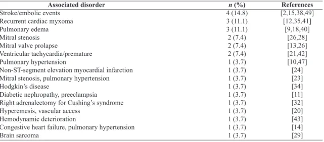
patients had one or more comorbidities or complications of a cardiac myxoma (Table 3).
Diagnosis
The timing of diagnosis of cardiac myxoma was described in 39 (76.5%) individuals. Two (5.1%) patients were diag-nosed with a cardiac myxoma in the 1st and 23rd months before
the current pregnancy, and their gestational ages were recorded as “-4” and “-92” weeks[2,13]. Among the 37 (94.9%) patients,
the cardiac myxoma was diagnosed in the irst, second and third trimesters in 7 (18.9%), 19 (51.4%) and 11 (29.7%) pa-tients, respectively (χ2=9.1, P=0.011) with a mean of 21.7±8.4
(range, 6.7-38; median, 22) weeks (n=35).
The diagnostic techniques for the cardiac myxoma were described in 34 (66.7%) patients, by transthoracic echocar-diography in 26 (70%)[2,8-13,17,20,21,24,27-29,33-35,38,40,42,43,45,47,48,50],
transthoracic and transesophageal echocardiography in 4 (13.3%)[15,27,30,32], transesophageal echocardiography in 2
(6.7%)[36,41], cardiac catheterization[14] and a battery of tests[46]
in 1 (3.3%) patient.
Location of cardiac myxoma was not clearly stated in 5 (9.8%) patients. In the remaining 46 (90.2%) patients, it was located in the left atrium in 37 (80.4%) [intraatrial septum in
11 (29.7%)[8-10,26,29,30,40,42,47,48], anterior mitral lealet in 2 (5.4%) [2,32], free wall[36] in 1 (2.7%) and unknown in 23 (62.2%)
patients[2,13,14,16-18,23,24,26-28,31,33,34,37-39,44-46,50], right atrium in 5
(10.9%)[2,11,15,20,36] (one was multiple[20]), left ventricle in 2
(4.3%)[12,21], right ventricle in 1 (2.2%)[43] and multiple sites
(septal tricuspid lealet, left atrium, intraatrial septum and left atrial appendage) in 1 (2.2%) patient[41]. Three patients
had recurrent cardiac myxomas[12,35,41] and one of them were
recurrent multiple cardiac myxomas[41]. A total of 12 atrial
myxomas including 11 (29.7%, 11/37) left atrial myxomas and 1 (20%, 1/5) right atrial myxoma[20] prolapsed into the
ventricle during diastole.
The attachment of the myxoma was described in 16 (31.4%) patients, it was pedunculated in 15 (93.8%) [8,9,12-15,20,26,29,30,35,36,40,42,48] and sessile in 1 (6.3%) patient[2]. The
dimensions of the cardiac myxomas were available in 25 (49.0%) patients. One of them was described as “egg-sized”[46] and average dimension of the remaining 24
myxo-mas was 55.1±24.5 (range, 22-130; median, 55) mm (n=24)
[2,8,9,11,12-15,20,21,23,26,27,29,30,32,35,39,42,43,47].
The diagnosis of the cardiac myxoma was delayed in 3 (5.9%) patients in the 1st , 3rd and 3rd week after admission[14,33,49].
Treatment
Treatment was not given in 2 (3.9%) patients[22].
Concern-ing the remainConcern-ing 49 patients, surgical operation was not per-formed in 2 (4.1%) patients but with anticoagulant and antibi-otic therapy in one[36] and decline of treatment by another[41].
A surgical resection of cardiac myxoma was performed in 47 (95.9%) patients including a sole cardiac myxoma resection in 41 (87.2%)[2,8-21,23-27,29,31-39,43-46,48-50], concurrent mitral valve
re-pair[40,47] and staged mitral valve replacement[28,29] in 2 (4.3%),
and staged mitral valve repair[30] and concurrent patent fossa
ovalis closure[42] in 1 (2.1%) patient. Cardiac surgery was
per-formed in the irst, second and third trimester in 5 (13.9%), 14 (38.9%) and 17 (47.2%) patients (χ2=9.8, P=0.008) at a mean
of 25.2±9.4 (range, 9-41; median 27) weeks of gestational age (n=36); while timing of cardiac surgery was not given in 15 (29.4%) patients. A cardiac myxoma resection was delayed in 4 (7.8%) patients for a few[43], 7[21], 15[40] and 690 days[13],
respectively. The feto-neonatal fate was not mentioned in 13 (25.5%) cases[15,22,24,27,31,37,38,41,44,47]. Voluntary termination of
pregnancy was done in 7 (13.7%) cases with 6 (85.7%)
early-Table 1. Literature retrieval. Database
PubMed Google Yahoo! Highwire Press
Chinese Medical Citation Index (CMCI) LILACs
Total
58 90 120 662 27
2
Irrelevant
34 34 94 659
23 2
Relevant
24 56 26 3 4 0
Inclusive
(by excluding duplicate indexes) 26
mid-termed[2,9,12,23,42] and 1 (14.3%) late pregnancy
termina-tion[50] with mean gestational age of 17.0±8.3 (range, 11-31;
median, 14) weeks (n=5). Delivery modes of remaining 31 (60.8%) pregnant patients were cesarean section in 19 (61.3%)
[2,10,11,13,16,20,21,28-30,32,35,36,39,40,43,45,46], vaginal delivery in 6 (19.4%)
[8,25,26,48,49], forceps under epidural analgesia in 1 (3.2%)[33] and
unknown in 5 (16.1%) patients[14,17-19,34]. One (3.2%) delivery
timing was not given, 2 (6.5%) deliveries were in second tri-mester and 28 (90.3%) in third tritri-mester with mean gestational age of 35.3±4.6 (range, 22-42; median, 37) weeks (n=30).
Table 2. Presenting symptoms of 31 pregnant patients. Symptom
Circulatory
Dyspnea, palpitation
Chest pain, dyspnea/tachypenia Pulmonary edema
Dyspnea Palpitation
Hemoptysis, dyspnea and cough Dyspnea, pulmonary edema Circulatory + constitutional
Palpitations, dyspnea, fatigue Cough, exhaustion
Palpitation, fatigue, weight loss Palpitation, dyspnea, night sweat
Chest pain, palpitation, cough, fever, chills Embolic
Headache, memory loss
Transient ischemic attack, weakness
Blurred vision
Hemiparesis, optalmoplegia Circulatory + embolic
Dyspnea, orthopnea, dizziness Chest pain, psychomotor restlessness
Chest pain, dyspnea, palpitation, syncope, dizziness Embolic + constitutional
Dizziness, fever
Circulatory + embolic + constitutional Dyspnea, dizziness, fatigue
n (%) 15 (48.4)
5 (33.3) 4 (26.6) 2 (13.3) 1 (6.7) 1 (6.7) 1 (6.7) 1 (6.7) 7 (22.6)
2 (28.6) 2 (28.6) 1 (14.3) 1 (14.3) 1 (14.3) 4 (12.9)
1 (25) 1 (25) 1 (25) 1 (25) 3 (9.7)
1 (33.3) 1 (33.3) 1 (33.3) 1 (3.2)
1 (100) 1 (3.2)
1 (100)
References
[20,26,28,47,50] [10,13,32,34]
[9,18] [26] [29] [27] [40]
[30,39] [19,46] [14] [33] [24]
[49] [38] [2] [29]
[36] [11] [15]
[8]
[43]
Table 3. Associated disorders. Associated disorder
Stroke/embolic events
Recurrent cardiac myxoma Pulmonary edema Mitral stenosis Mitral valve prolapse
Ventricular tachycardia/premature Pulmonary hypertension
Non-ST-segment elevation myocardial infarction Mitral stenosis, pulmonary hypertension
Hodgkin’s disease
Diabetic nephropathy, preeclampsia Right adrenalectomy for Cushing’s syndrome Hyperemesis, vascular access
Hemodynamic deterioration
Congestive heart failure, pulmonary hypertension Brain sarcoma
n (%) 4 (14.8) 3 (11.1) 3 (11.1) 2 (7.4) 2 (7.4) 2 (7.4) 1 (3.7) 1 (3.7) 1 (3.7) 1 (3.7) 1 (3.7) 1 (3.7) 1 (3.7) 1 (3.7) 1 (3.7) 1 (3.7)
References [2,15,38,49] [12,35,41]
All the pregnant patients in the pregnancy termination group received a surgical resection of the cardiac myxoma with an isolated cardiac myxoma resection in 5 (71.4%), myxoma resection with patch repair of the iatrogenic sep-tal defect in 1 (14.3%) and myxoma resection with patent fossa ovalis closure in 1 (14.3%) patient. One (14.3%) patient had pregnancy termination performed before car-diac surgery in 3 (42.9%), after the carcar-diac surgery in 2 (28.6%) and unknown surgical sequence in 2 (28.6%) pa-tients. The indications for pregnancy termination was ma-ternal pulmonary edema in 1 (14.3%)[9], fetal growth
retar-dation (fetal short femur) in 1 (14.3%)[50] and unknown in
5 (71.4%) patients.
In the pregnancy termination group, 6 (85.7%) pregnant patients were event-free and 1 (14.3%) was complicated with postoperative transient acute myocardial infarction[9].
The prognosis of the pregnant patient was not given in 2 pa-tients[22]. In the remaining 42 pregnant patients with a
child-birth, 35 (83.3%) were event-free, 6 (14.3%) were compli-cated, with cerebral involvement (psychomotor agitation and mydriasis anisocoria)[40], heart block[30], pacing dependent
rhythm[28], pulmonary edema[32], uterine contractions[26], and
premature labor[34]), and 1 (2.4%) was recurrent (requiring
reoperation)[29]. No pregnant patients died. Pregnant patients’
event free survival (P=0.680), complication (P=0.686) and recurrence rates (P=0.857) did not differ between pregnancy termination and delivery groups.
Eighteen (42.9%) patients had a delivery before cardiac surgery[8,14,17,19,24,26-30,33,34,38,40,46,47,49], in the irst, second, third and
unknown trimester in 2 (11.1%), 8 (44.4%), 6 (33.3%) and 2 (11.1%) patients with a mean gestational age of 21.6±6.2 (range, 10-28.1; median, 23) weeks (n=16). Thirteen (31.0%) patients has cardiac myxoma resection performed after delivery in sec-ond, third and unknown trimester in 1 (7.7%), 9 (69.2%), and 3 (23.1%) patients at a mean gestational age of 34.2±5.5 (range, 22.2-41; median, 32.7) weeks (n=10)[11,13,16,18,20,21,29,32,35,36,39,41,43].
Two (4.8%) patients received a one-stage delivery and cardiac surgical procedure in the 31st and 32.7th week, respectively.
Sur-gical sequence was unknown in 9 (21.4%) patients.
Prognosis
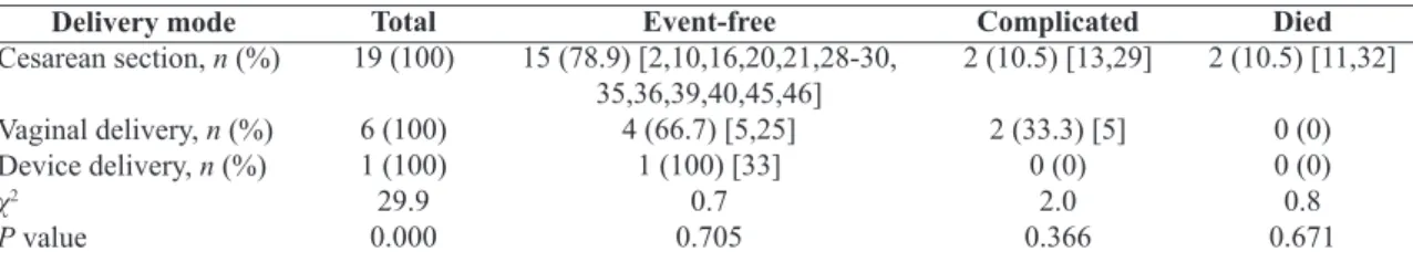
In the delivery group, delivery mode was not given in 16 cases. Among the 26 deliveries with either cesarean section or vaginal delivery, 20 (76.9%) were event-free survivals, 4 (15.4%) were complicated and 2 (7.7%) died. Neonatal prognoses did not differ between delivery modes, treatment options, timing of cardiac surgery and sequence of cardiac myxoma in relation to delivery (Tables 4-7).
Nomimal regression analysis showed that timing of de-livery, delivery mode, surgical resection of the cardiac myx-oma, simple or complex cardiac surgery, timing of cardiac surgery, sequence of cardiac surgery in relation to delivery and maternal complications were not predictive risk factors responsible for fetal outcomes.
Table 5. Neonatal prognosis of different treatment options (Fisher exact test). Treatment
Surgical
Isolated myxoma resection
Myxoma resection with concurrent or staged additional cardiac surgery
Conservative
P value (surgical vs. conservative)
P value (myxoma resection vs. myxoma resection with concurrent or staged additional cardiac surgery)
Total 39 (100) 34 (100)
5 (100)
2 (100) 0.000 0.000
Died 3 (7.7) 3 (8.8) [11,12,32]
0 (0)
0 (0) 0.684 0.489 Event-free
31 (79.5) 27 (79.4) [2,8,10,14,16-21,24-27,29,31,34,35,37-39,44-46,49]
4 (80) [28,30,40,47]
2 (100) [36,41] 0.475 0.976
Complicated 5 (12.8) 4 (11.8) [13,26,33,48]
1 (20) [29]
0 (0) 0.589 0.607 Table 4. Neonatal prognosis subjected to different delivery modes (Fisher exact test).
Delivery mode Cesarean section, n (%)
Vaginal delivery, n (%) Device delivery, n (%) χ2
P value
Total 19 (100)
6 (100) 1 (100) 29.9 0.000
Died 2 (10.5) [11,32]
0 (0) 0 (0) 0.8 0.671 Event-free
15 (78.9) [2,10,16,20,21,28-30, 35,36,39,40,45,46]
4 (66.7) [5,25] 1 (100) [33]
0.7 0.705
Complicated 2 (10.5) [13,29]
2 (33.3) [5] 0 (0)
Table 7. Neonatal prognosis according to sequences of cardiac myxoma resection and delivery (Fisher exact test). Sequence of cardiac myxoma
resection
Before delivery
After delivery
One-stage
χ2
P value
Total
18 (100)
12 (100)
2 (100)
19.5
0.000
Died
0 (0)
3 (25) [11,12,32]
0 (0)
5.5
0.063 Event-free
15 (83.3) [8,14,17,19,24, 26-28,30,34,38,
40,46,47,49]
8 (66.7) [16,18,20,21,29, 35,36,39,41]
2 (100) [10,45]
1.8
0.413
Complicated
3 (16.7) [26,29,33]
1 (8.3) [13]
0 (0)
0.8
0.683 Table 6. Neonatal prognosis according to timing of cardiac surgery (Fisher exact test).
Timing of cardiac surgery 1st trimester
2nd trimester
3rd trimester
χ2
P value
Total 2 (100)
12 (100)
16 (100)
15.6
0.000
Died 0 (0)
0 (0)
2 (12.5) [11,32]
1.9
0.392 Event-free
2 (100) [24,38]
8 (66.7) [8,14,17,19,30,39,40,46]
13 (81.3) [2,10,16,20,21,26,27, 29,34,35,43,45,49]
1.5
0.480
Complicated 0 (0)
4 (33.3) [26,29,33,48]
1 (6.3) [13]
4.1
0.132
DISCUSSION
Cardiac myxoma is rare in pregnant patients. The diagno-sis and management can be challenging in terms of the nature of the intracardiac mass, timing of delivery, necessity of car-diac surgery and risks of subsequent treatment[36]. The clinical
manifestations of a cardiac myxoma can be one or more of the Goodwin’s triad[51]. Patients may present with fatigue and
dyspnea, which, however, can be misinterpreted as asthma or normal fatigue associated with pregnancy[39].
Echocardiogra-phy remains the standard non-invasive diagnostic modality, particularly in the pregnant patient[52]. In some patients, atrial
thrombi may have a stalk and may be mistaken for myxomas, leading to unnecessary and potential harmful surgery[53]. A left
intraatrial mass can be diagnosed as thrombus if associated with atrial ibrillation, dilated left atrium, mitral or tricuspid stenosis, low ejection fraction, prosthetic mitral or tricuspid valves, or spontaneous atrial contrast echoes[54]. Moreover, in
the pregnant patients, cardiovascular magnetic resonance im-aging is indicated for visualizing coarctation, aortitis, aortic dissection and atrial myxoma[52].
Surgical management of cardiac myxoma is similar to that of the valvular disorders in the pregnant patients, even with minimally invasive cardiac surgical techniques[55].
Nevertheless, surgical indications of both conditions can be somehow different from each other. Congestive heart failure as a consequence of rheumatic mitral stenosis is always a contraindication of pregnancy. But it may be curable to per-cutaneous inteventional therapy, however, carrying the risk of fetal teratogenicity by manipulation under X-ray. Or else, an urgent valvular operation is warranted in the presence of infective endocarditis, intramural thrombus, paravalvular leakage, stuck prosthetic valve or thrombus formation. Mean -while, the indications for cardiac myxoma resection are the potential embolic events and sudden death caused by myx-oma-obstructed valve oriice[2]. Wang et al.[2] reported three
pregnant patients with a cardiac myxoma, two of which were complicated with cerebral infarctions and an urgent cardiac myxoma resection with later curettage was performed. Liu et al.[6] described a pregnant patient with a cardiac myxoma
pre-sented with both cerebral infarction and central retinal artery occlusion, and a cardiac surgical resection was performed without delay. As for the potential of cardiogenic embolic events and possible preterm delivery due to hemodynamic changes, a timely surgical resection of cardiac myxoma can be indispensable during pregnancy.
masses who may beneit from non-surgical management[36].
Open heart surgery as well as the use of cardiopulmonary bypass may cause premature labor and endanger the baby[49].
The surgical resection of cardiac myxoma may be associated with a 30% baby loss rate, or postnatal physical or develop-mental disabilities[39]. It is encouraging that maternal survival
rate was 100% in the pregnant patients with a cardiac myx-oma, superior to that of the pregnant patients with infective endocarditis[3]. This might be interpreted as the results of the
advantaged cardiopulmonary bypass techniques including high low rate, high perfusion pressure and pulsatile low ap-plied in cardiac surgery during pregnancy[30]. By comparison,
infective endocarditis and acute aortic dissection might be more dangerous to the pregnant patients than cardiac myx-oma as for the infective nature of the former and the use of profound hypothermic circulatory arrest for the operation of the latter[3,56]. The present study also revealed that timing of
delivery other than the delivery mode (by excluding early termination of pregnancy) and time sequence of cardiac sur-gery and delivery was closely related to feto-neonatal mor-tality. Cardiac surgery under cardiopulmonary bypass should be avoided in the irst trimester, particularly after six weeks, due to the risk of teratogenesis[50]. The pregnant patients may
wait for a few weeks[13], or take weekly thyrotropin-releasing
hormone and ß-methasone therapies for fetal lung matura-tion[35]. Precautions during cardiac operation include using
blood priming solution, normothermic cardiopulmonary by-pass and high perfusion pressure[38].
Possible bias may be generated in present patient setting due to limited data available from the literature for the statis-tical analysis. Therefore, more abundant information of such patients is necessary for further precise results.
CONCLUSIONS
Cardiac myxoma is rare in pregnant patients. In most cas-es, the cardiac myxoma is diagnosed in the second trimester and is resected in the third. Cesarean section was the most frequent delivery mode. The 100% maternal survival of this patient setting is encouraging. A delivery at early gestation was closely related to an increased feto-neonatal mortality. A delivery postponed to late pregnancy until fetal maturity may improve the feto-neonatal survival. In brief, embolic po-tential and hemodynamic deterioration are indications for an urgent cardiac myxoma resection. Otherwise, cardiac surgery should be avoided in the irst trimester and be postponed until fetal pulmonary maturation or after delivery.
Authors’ roles & responsibilities
SMY Study conception and design; analysis and/or interpretation of data; manuscript writing.
REFERENCES
1. Yuan SM. Indications for cardiopulmonary bypass during pregnancy and impact on fetal outcomes. Geburtshilfe
Frauenheilkd. 2014;74(1):55-62.
2. Wang H, Zhang J, Li B, Li Y, Zhang H, Wang Y, et al. Maternal and fetal outcomes in pregnant patients undergoing cardiac surgery with cardiopulmonary bypass. Zhonghua Fu Chan Ke Za Zhi. 2014;49(2):104-8.
3. Yuan SM. Infective endocarditis during pregnancy. J Coll
Physicians Surg Pak. 2015;25(2):134-9.
4. Moher D, Cook DJ, Eastwood S, Olkin I, Rennie D, Stroup DF.
Improving the quality of reports of meta-analyses of randomised controlled trials: the QUOROM statement. Quality of Reporting of Meta-analyses. Lancet. 1999;354(9193):1896-900.
5. John AS, Gurley F, Schaff HV, Warnes CA, Phillips SD, Arendt KW, et al. Cardiopulmonary bypass during pregnancy. Ann Thorac Surg. 2011;91(4):1191-6.
6. Liu GF, Wu Y, Sun L, Meng X, Wang J. Left atrial myxoma and obstruction of central retinal artery: a case report. Chin J Ocul Fundus Dis. 2013;29(6):625-6.
7. Stevens N. Word of mom pregnancy: doctors save pregnant woman and unborn baby after performing open-heart surgery [Accessed Aug 8, 2014]. Available at: http://www.whattoexpect. com/wom/pregnancy/0429/doctors-save-pregnant-woman-and-unborn-baby-after-performing-open-heart-surgery.aspx
8. Agarwal AK, Venugopalan P. Dizziness during pregnancy due to cardiac myxoma. Saudi Med J. 2004;25(6):795-7.
9. Agosti S, Casalino L, Bertero G, Morelloni S, Barsotti A, Brunelli C. Atrial myxoma presenting during pregnancy. G Ital Cardiol (Rome). 2010;11(6):498-500.
10. Arumugam CG, Raju SV, Varma S, Bhaskaran K. Anaesthetic
management of a pregnant patient with left atrial myxoma and myxoma excision of lower segment caesarean. Apollo Med. 2008;5(1):71-3.
11. Berberovic B, Kacila M, Hadzimehmedagic A, Berberovic E. Cardiac myxoma in diabetic pregnancy. Int J Gynaecol Obstet. 2014;125(3):281-2.
12. Bortolotti U, Scioti G, Guglielmi C, Milano A, Nardi C, Tartarini G. Recurrent myxoma of the left ventricle. Case report and review of the literature. J Cardiovasc Surg (Torino). 1999;40(2):233-5.
13. Bryukhina EV, Ishchenko LS, Lomova ES, Ulanova DS. Left
atrial myxoma in a pregnant woman: clinical features, tactics. Sci Pract J Obstet Gynecol. 2014;5(9):114-6.
Thiene G. Surgical removal of a left atrial myxoma during pregnancy. Chest. 1979;75(3):390-2.
15. Christensen M, Tingleff J, Larsen CT, Kjøller SM. Symptoms
debut from a right atrial myxoma during labor. Ugeskr Laeger.
2004;166(47):4267-8.
16. Collins N. Woman and baby saved after emergency surgery[Accessed Aug 8, 2014]. Available at: http://www.
telegraph.co.uk/health/healthnews/8975647/Woman-and-baby-saved-after-emergency-surgery.html
17. Donahoo JS, Weiss JL, Gardner TJ, Fortuin NJ, Brawley RK. Current management of atrial myxoma with emphasis on a new diagnostic technique. Ann Surg. 1979;189(6):763-8.
18. Elston JH. A case report of a myxoma or rhabdomyosarcoma of the left atrium in the third trimester of pregnancy. Nebr Med J. 1984;69(7):225-6.
19. Erikson J. Pregnant woman undergoes rare & risky open-heart
surgery to save her baby [Accessed Aug 21, 2014]. Available at: http://thestir.cafemom.com/pregnancy/171380/pregnant_
woman_undergoes_rare_risky
20. Fang YM, Dean R, Figueroa R. Right atrial myxoma mimicking
an atrial thrombus in the third trimester of pregnancy. J Matern Fetal Neonatal Med. 2007;20(1):77-8.
21. Fiorilli R, Tomasco B, Serino W, Tesler UF. Asymptomatic left ventricular myxoma in pregnancy: echocardiographic diagnosis and surgical treatment. G Ital Cardiol. 1996;26(8):887-90.
22. Gurley FM, Connolly HM, Dearani JA, Warnes CA, Rose CH, Phillips SD, et al. Surgery for valve disease and its sequelae II. Abstract 1806: Cardiac Surgery During Pregnancy: The Mayo Clinic Experience 1976–2005. Circulation. 2006;114:II-356.
23. Han F, Zhao Y, Lu C. Surgery intervention of pregnancy heart disease. J Pract Obstetr Gynecol. 2010;26(3):225-8.
24. Hasan I, Reddy S, Burns T. Myxoma in the mix [abstract]. J Hosp Med. 2014;9(Suppl 2):443 [Accessed Aug 21, 2014]. Available at: http://www.shmabstracts.com/abstract.
asp?MeetingID=800&id=110744
25. Hosseini S, Yaghoubi A, Khamoushi AJ, Raissi K, Kashi F,
Sadeghpour A, et al. Open heart surgery in pregnant women. Iran Heart J. 2004;5(4):34-9.
26. John AS, Connolly HM, Schaff HV, Klarich K. Management of cardiac myxoma during pregnancy: a case series and review of the literature. Int J Cardiol. 2012;155(2):177-80.
27. Kanth P, Vu T, Daroowalla F. Hemoptysis in pregnancy
as the irst sign of left atrial myxoma. Chest. 2014;146(4_
MeetingAbstracts):200A. doi:10.1378/chest.1991271
28. Kither HJ, Stephen G, Vause S. Atrial myxoma in early pregnancy.
Arch Dis Child Fetal Neonatal Ed 2012;97:A52. doi: 10.1136/ fetalneonatal-2012-301809.166
29. Korbel’ M, Kanáliková K, Fischer V, Niznanská Z, Redecha M, Paulíková Z. Management of intracavitary left atrium tumors during pregnancy: two case reports. Zentralbl Gynakol.
2001;123(10):590-2.
30. Koukis I, Velissaris T, Pandian A. Left atrial myxoma associated
with mitral valve pathology in pregnancy. Hellenic J Cardiol. 2013;54(2):138-42.
31. Mahoori A, Farasatkish R, Aghdaie N, Faritus Z, Mollasadeghi G, Kashi F. Outcome of anesthesia and open heart surgery in
pregnant patients. J Teh Univ Heart Ctr. 2007;2(1):21-4.
32. Manfredini R, Calì G, Foresti A. Acute anterior myocardial infarction in a patient with left atrial myxoma during pregnancy. G Ital Cardiol. 1995;25(11):1419-24.
33. Mann MS, Cossham PS, Baker JL, Hurley PA. Left atrial myxoma
in the second trimester of pregnancy. Case report. Br J Obstet Gynaecol. 1987;94(6):592-3.
34. Mercer LJ, Aisenbrey G. Atrial myxoma as a complication of tocolytic therapy. A case report. J Reprod Med. 1985;30(7):561-2.
35. Nakata S, Nakano S, Mitsuda N, Itou N, Takahashi Y, Matsuda
H. Recurrent left atrial myxoma in a patient with a twin fetus pregnancy. Jpn Circ J. 1996;60(2):130-2.
36. Nelson SC, Coleman L, Zakowski MI. Management and cesarean
delivery in a parturient with a right atrial mass. Abstract Number: F-30 [Accessed Aug 21, 2014]. Available at: http://soap.org/ display_2012_abstract.php?id=F-30
37. No authors listed. Interesting cases [Accessed Aug 21, 2014]. Available at: http://www.heartsurgery.in/interesting_cases.htm
38. Obied HY. SHA 21. Surgical removal of left atrial myxoma in
a pregnant lady with recent stroke during 1st trimester. J Saudi
Heart Assoc. 2010;22(2):90 [Accessed Aug 21, 2014]. Available at: http://www.journalofthesaudiheart.com/article/S1016-7315(10)00333-7/fulltext
39. Ogilvie M. ‘I wanted him to have a chance to survive before me’ [Accessed Aug 21, 2014]. Available at: http://www.thestar. com/life/health_wellness/2011/10/22/i_wanted_him_to_have_a_ chance_to_survive_before_me.html
40. Prieto Macías J, Orea Tejeda A, Santiago Bravo M, Vargas Bustos MA, Castillo Razo R. Left atrial myxoma causing severe mitral valve occlusion. Arch Inst Cardiol Mex. 1989;59(6):611-4.
41. Roldán FJ, Vargas-Barrón J, Espinola-Zavaleta N, Keirns C, Romero-Cárdenas A. Recurrent myxoma implanted in the left atrial appendage. Echocardiography. 2000;17(2):169-71.
atrium in pregnancy using 2-dimensional sector visualization.
Vnitr Lek. 1982;28(12):1186-90.
43. Sindjelic R, Vlajkovic G, Djukic P. Management of right
ventricular myxoma diagnosed at full-term pregnancy. Med Sci Monit. 2009;15(10):CS158-61.
44. Siu SC, Sermer M, Harrison DA, Grigoriadis E, Liu G, Sorensen
S, et al. Risk and predictors for pregnancy-related complications
in women with heart disease. Circulation. 1997;96(9):2789-94.
45. Smith A. Asymptomatic atrial myxoma during pregnancy. RCOG World Congress 2013 Liverpool UK, 24-26 June 2013 [Accessed Aug 21, 2014]. Available at: http://www.epostersonline.com/ rcog2013/?q=node/409
46. Stony Brook University. Open-heart surgery performed on patient 27 weeks’ pregnant [Accessed Aug 21, 2014]. Available at: http://
www.sciencedaily.com/releases/2014/04/140421164150.htm
47. Trimakas AP, Maxwell KD, Berkay S, Gardner TJ, Achuff SC.
Fetal Monitoring during cardiopulmonary bypass for removal
of a left atrial myxoma during pregnancy. Johns Hopkins Med
J. 1979;144(5):156-60.
48. Tretina M, Hrdlicka M, Ondrásek J, Nĕmec P, Orban M. Cardiac tumor in a pregnant patient. Vnitr Lek. 2010;56(1):79-81.
49. Wahlberg D. Peaceful Christmas welcome after open heart surgery during pregnancy. Wisconsin State J [Accessed Aug 21, 2014].
Available at: http://obgyn.wisc.edu/news/open-heart-surgery-pregnancy.aspx
50. Witters I, Cannie M, Moerman P, Herijgers P, Rademakers
F, Coudyzer W, et al. Fetal caudal dysgenesis after maternal c a r d i o p u l m o n a r y b y p a ss i n Pr e g n a n c y. Ul t r a so u n d . 2007;15(2):71-2.
51. Maruf MF, Akter T, Islam F, Chowdhury AA, Khan JH, Hassan
K, et al. Left atrial myxoma in a child: an uncommon presentation of a rare tumour in early age. Cardiovasc J. 2012;4(2):171-3.
52. Colletti PM. Guidelines for MRI in the pregnant patient [Accessed Aug 21, 2014]. Available at: cppcongress.com/.../03/Guidelines-for-MRI-in-the-Pregnant-Patient.pdf
53. Kim SE, Park DG. Tentatively diagnosed as myxoma: transit
thrombus entrapped in patent foramen ovale. J Cardiovasc Ultrasound. 2007;15:19-22.
54. Diaconu CC. Left atrial thrombus: a case report [Accessed Aug 21, 2014]. Available at: http://www.medandlife.ro/ medandlife625.html
55. Costa F, Winter G, Ferreira AD, Fernandes TA, Collatusso C, Tremel FT, et al. Initial experience with minimally invasive cardiac operations. Rev Bras Cir Cardiovasc. 2012;27(3):383-91.
56. Yuan SM. Aortic dissection during pregnancy: a dificult clinical