J Bras Pneumol. 2013;39(5):627-629
Epipericardial fat necrosis is a rare, benign, and self-limited cause of acute chest pain, with only 35 cases reported in the literature.(1,2) It presents
as new-onset pleuritic chest pain in otherwise healthy patients, and, therefore, it is an important
differential diagnosis for acute chest pain in this group of individuals. The pathophysiology has been labeled as idiopathic or related to an acute damage of this fat tissue secondary to vascular torsion, trauma, or microvascular bleeding.
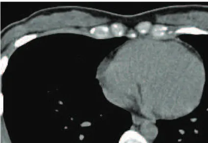
Figure 1 - In A, posteroanterior chest X-ray showing tiny pleural effusion and slight right paracardiac opacity. In B, CT scan of the chest revealing a round encapsulated lesion with fat attenuation and subtle strands in the right epipericardial fat (arrow). In C, coronal and sagittal views of the round encapsulated lesion in the right epipericardial fat (arrows), suggesting epipericardial fat necrosis.
A B
C
Epipericardial fat necrosis: an unusual cause of chest pain
Necrose da gordura epipericárdica: uma causa incomum de dor torácica
Karina de Souza Giassi, André Nathan Costa, André Apanavicius, Guilherme Hipólito Bachion, Rafael Silva Musolino, Ronaldo Adib Kairalla
To the Editor:
628 Giassi KS, Costa AN, Apanavicius A, Bachion GH, Musolino RS, Kairalla RA
J Bras Pneumol. 2013;39(5):627-629
We report the case of a 23-year-old healthy female patient, a chronic oral contraceptive user, who presented to the emergency department with a two-day history of right-sided pleuritic chest pain, associated with mild dyspnea. No fever, cough, wheezing, or other respiratory symptoms were present. Physical examination in the emergency department revealed oxygen saturation on room air of 98%, temperature of 36.8°C, HR of 110 bpm, RR of 24 breaths/min, and blood pressure of 110 × 65 mmHg. Laboratory tests were unremarkable. Electrocardiography showed normal sinus rhythm, whereas chest X-ray showed a slight right paracardiac opacity and tiny right pleural effusion (Figure 1A), and CT revealed a round encapsulated lesion with fat attenuation in the right epipericardial fat with subtle peripheral strands, which are typical abnormalities due to epipericardial fat necrosis (Figures 1B and 1C). Pulmonary embolism was ruled out by a contrast-enhanced protocol. The patient was then treated with nonsteroidal anti-inflammatory drugs, and her symptoms resolved in the next few days. Four weeks later, CT scans of the chest showed complete resolution of all findings (Figure 2).
Epipericardial fat necrosis is a rare entity, being first reported in 1957 by Jackson et al.(3)
Since then, other similar cases have been reported, and the most recent studies have characterized this condition as benign and self-limited.
The epicardial or visceral layer of fat tissue is found in the interventricular groove along the
Figure 2 - CT scan of the chest performed four weeks after admission showing complete resolution of the previous findings.
atria and extends to the right and left pleural surface. It can be more prominent in obese individuals, in whom it can completely cover the epicardial surface.(1)
The physiopathology of necrosis of epipericardial fat is still under debate, but an acute torsion of a vascular pedicle has been proposed.(3) Indeed,
small parts of fat tissue attached to the heart by a pedicle have been found in some patients submitted to cardiac surgery,(4) and an acute torsion of the
vascular pedicle could lead to necrosis. Another explanation would be a pre-existing structural abnormality of the adipose tissue, making this fat vulnerable to trauma caused by the beats of the heart.(5) In addition, it has been postulated
that straining or heavy lifting could trigger abrupt changes in the intravascular pressure associated with the Valsalva maneuver, causing hemorrhage into the adipose tissue that is loosely attached to the pericardium.(1)
The most common presentation of epipericardial fat necrosis is acute pleuritic chest pain. There is no age or gender predilection. The pain can be associated with dizziness, syncope, dyspnea, tachycardia, or diaphoresis. Physical examination is usually unremarkable. In general, the pain lasts only a few days, but it can persist for weeks and recur in intermittent episodes.(6)
Epipericardial fat necrosis: an unusual cause of chest pain
J Bras Pneumol. 2013;39(5):627-629 629
André Apanavicius
Pulmonologist,
Hospital Sírio-Libanês
, São Paulo, BrazilGuilherme Hipólito Bachion Chest Radiologist,
Hospital Sírio-Libanês
,São Paulo, Brazil
Rafael Silva Musolino
Pulmonologist,
Hospital Sírio-Libanês
, São Paulo, BrazilRonaldo Adib Kairalla
Pulmonologist,
Hospital Sírio-Libanês
and Pulmonary Division,
Instituto
do Coração
(InCor, Heart Institute),Hospital das Clínicas
, University of São Paulo Medical School, São Paulo, BrazilReferences
1. Baig A, Campbell B, Russell M, Singh J, Borra S. Epicardial fat necrosis: an uncommon etiology of chest pain. Cardiol J. 2012;19(4):424-8. http://dx.doi.org/10.5603/ CJ.2012.0076 PMid:22825906
2. Runge T, Greganti MA. Epipericardial fat necrosis - a rare cause of pleuritic chest pain: case report and review of the literature. Arch Med Sci. 2011;7(2):337-41. http:// dx.doi.org/10.5114/aoms.2011.22088 PMid:22291777 PMCid:PMC3258729
3. Pineda V, Cáceres J, Andreu J, Vilar J, Domingo ML. Epipericardial fat necrosis: radiologic diagnosis and follow-up. AJR Am J Roentgenol. 2005;185(5):1234-6. http://dx.doi.org/10.2214/AJR.04.1310 PMid:16247140 4. Jackson RC, Clagett OT, Mcdonald JR. Pericardial
fat necrosis; report of three cases. J Thorac Surg. 1957;33(6):723-9. PMid:13429689
5. Lee BY, Song KS. Calcified chronic pericardial fat necrosis in localized lipomatosis of pericardium. AJR Am J Roentgenol. 2007;188(1):W21-4. http://dx.doi. org/10.2214/AJR.04.1989 PMid:17179322
6. Lacasse MC, Prenovault J, Lavoie A, Chartrand-Lefebvre C. Pericardial fat necrosis presenting as acute pleuritic chest pain. J Emerg Med. 2013;44(2):e269-71. http://dx.doi. org/10.1016/j.jemermed.2012.05.032 PMid:22877971 7. Inoue S, Fujino S, Tezuka N, Sawai S, Kontani K, Hanaoka
J, et al. Encapsulated pericardial fat necrosis treated by video-assisted thoracic surgery: report of a case. Surg Today. 2000;30(8):739-43. http://dx.doi.org/10.1007/ s005950070088 PMid:10955740
8. Mazzamuto G, Ghaye B. Epipericardial fat necrosis. JBR-BTR. 2012;95(3):154-5. PMid:22880517
During the first days, chest X-rays can be normal or reveal small pleural effusion. Thereafter, an ill-defined round mass appears near the cardiophrenic angle on the side of the chest pain. These findings are nonspecific and formerly led patients to undergo surgery due to the lack of cross-sectional imaging or the need to rule out malignancies, such as lung cancer and liposarcoma.(7) The pathologic findings have
been described as necrotic fat cells surrounded by macrophages, neutrophils, or fibrous tissue, resembling the features of epiploic appendagitis. Since 2005, when the first case of successful conservative management of epipericardial fat necrosis was described,(3) CT has played an
important role in the diagnosis and follow-up. The typical finding is a round encapsulated fat-containing lesion with strands in the epipericardial fat, which can be mild or marked. Pericardial thickening and ipsilateral pleural effusion can also be present. Once this finding is present in a typical clinical context and other causes of acute chest pain are ruled out, physicians should consider epipericardial fat necrosis as the cause of pain.(8)
Our patient was a young user of oral contraceptives who presented with severe right-sided pleuritic chest pain, which implicates pulmonary embolism as the main diagnosis to be ruled out. Pulmonary embolism was excluded, and epipericardial fat necrosis was visualized by CT scans, which led to the correct diagnosis and allowed the institution of conservative management with excellent results. It is imperative that clinicians and radiologists be aware of this condition in order to manage it properly in emergency departments.
Karina de Souza Giassi Chest Radiology Fellow,
Hospital
Sírio-Libanês
, São Paulo, BrazilAndré Nathan Costa
Pulmonologist,
Hospital Sírio-Libanês
and Pulmonary Division,