Inês Pais Cunha Próteses Endoscópicas para a Paliação de Obstrução Gastrointestinal Intra-abdominal Maligna / Endoscopic Stenting for Palliation of Intra-abdominal Gastrointestinal Malignant Obstruction: Predictive Factors for Clinical Success
Mestrado Integrado em Medicina
Área: Gastroenterologia Tipologia: Dissertação
Trabalho efetuado sob a Orientação de: Professor Doutor Pedro Filipe Vieira Pimentel Nunes E sob a Coorientação de: Professor Doutor Mário Jorge Dinis Ribeiro
Trabalho organizado de acordo com as normas da revista: European Journal of Gastroenterology & Hepathology
Intra-abdominal Maligna / Endoscopic Stenting for Palliation of Intra-abdominal Gastrointestinal Malignant Obstruction: Predictive Factors for Clinical Success
Full Title: Endoscopic Stenting for Palliation of Intra-abdominal
Gastrointestinal Malignant Obstruction: Predictive Factors For
Clinical Success
Short Title: Gastrointestinal Stenting: Success Factors
Pais-Cunha, Inês3; Castro, Rui 1; Libânio, Diogo1,2; Pita, Inês1; Bastos, Rui P.1; Silva, Rui1; Dinis-Ribeiro, Mario1,2; Pimentel-Nunes, Pedro1,2,3 1. Gastroenterology Department, Portuguese Oncology Institute of Porto, 2. Center for research in health technologies and information systems (CINTESIS) Faculty of Medicine, University of Porto 3. Department of Surgery and Physiology, Faculty of Medicine, University of Porto Conflict of Interests and Source of Funding: None declared. Correspondence address: Inês Pais-Cunha Faculty of Medicine, University of Porto Al. Prof. Hernâni Monteiro, 4200 - 319 Porto, PORTUGAL Tel: + 351 933339925 Email: [email protected]
ABSTRACT
Background: Endoscopic stenting proved to be a safe alternative to surgery for malignant intra-abdominal gastrointestinal obstruction. Although high technical success rates are reported, some patients do not experience symptoms relief. Aim: To analyse factors predicting effectiveness of stent placement in patients with gastrointestinal obstruction.
Methods: Retrospective study including 160 patients submitted to palliative stenting for intra-abdominal obstruction in a tertiary centre, from December 2012 to July 2017. Technical and clinical success, stent dysfunction and adverse events were analysed.
Results: Technical success was of 98%. Early clinical success was of 69% and 81% in upper and lower gastrointestinal obstructions, respectively (p=0.107). In the upper tract, obstruction caused by carcinomatosis was the only independent factor predicting early and late clinical failure (OR 9.7, 95%CI 2.4-38.4, p=0.001; OR 7.6, 95%CI 1.8-31.9, p=0.006, respectively) and absence of late clinical benefit (OR 9.2, 95%CI 1.8-47.0, p=0.008). In the colon, ECOG score≥3 was an independent factor for early clinical failure (OR 29.8, 95%CI 1.9-464.9, p=0.002) and obstruction caused by carcinomatosis was an independent factor for late clinical failure (OR 14.4, 95%CI 1.7-119.6, p=0.013) and absence of late clinical benefit (OR 8.3, 95%CI 1.2-57.5, p=0.035). Perforation occurred in 4 patients (2.5%) and stent dysfunction occurred in 15% of patients (4% stent migration; 9% reestenosis). Carcinomatosis was a risk factor for perforation (p=0.039) and migration was higher with shorter 6 cm stents (p=0.044).
Conclusion: Stents are effective and safe for palliating intra-abdominal obstruction. Carcinomatosis predicts an unfavourable clinical outcome. Palliative stenting should be carefully weighed in these patients.
Keywords: stents; intestinal obstruction; carcinoma; stomach neoplasms; colorectal
Introduction
Gastrointestinal (GI) tumors are among the most common malignant diseases worldwide. In fact, colorectal cancer is the second most commonly diagnosed cancer in females and third in males. Similarly, gastric cancer is within the five most commonly diagnosed cancers in both gender.[1] These frequent malignancies often present at advanced unresectable stages requiring palliative care.[2] [3]
Irrespective of GI segment affected, intra-abdominal malignancies can result in intestinal obstruction with dreadful clinical conditions that lead to severe symptom, impaired quality of life and, in more advanced stages, patient’s death.[4,5] In most of these cases, curative treatment is no longer possible and the focus is on symptom control and palliation. Therefore, minimally invasive methods are preferred in this setting.
In the past, the standard treatment for malignant gastric and duodenal outlet obstruction was surgical gastrojejunostomy and for colorectal obstruction was colostomy. However, in recent years, palliative endoscopic stenting has been used as an alternative to surgery to manage the stenosis. [6-9]
Endoscopic stenting is suggested in current practice as a safe and minimally invasive method for re-establishing luminal continuity and relieving GI obstructive symptoms.[10] Although endoscopic stenting has been reported to have a high technical success, the reported clinical success is not as high and some patients still complain of ongoing symptoms after the procedure.[11]
Accordingly, our aim was to determine the overall clinical benefit and predictive factors for unsuccessful clinical outcomes and adverse events of palliative GI stenting for
malignant intra-abdominal obstruction, arguing in favor of a more individualized approach to these patients.
Materials and Methods
Study design and selection of Patients
We conducted a retrospective study of a consecutive series of patients undergoing either gastroduodenal or colorectal stent placement for treatment of malignant intra-abdominal gastroduodenal or colonic obstruction in a tertiary-care medical centre (Instituto Português de Oncologia do Porto Francisco Gentil, E.P.E), between December 2012 and July 2017. The study was approved by the Ethics Committee of Instituto Português de Oncologia do Porto Francisco Gentil, E.P.E
Patients were included if palliative stent placement was indicated for either upper or lower GI tract obstruction and recommended in a multidisciplinary decision (1); had clinically and imagiologically documented unresectable malignant cancers (2); with symptoms of obstruction of the stomach, duodenum or colon, such as vomiting, obstipation, diarrhoea or abdominal pain (3).
Patients were excluded if they had a non-palliative indication for stenting, such as bridge to surgery. In addition, patients without follow-up clinical records were also excluded.
Stent placement
Two fully trained endoscopists with different levels of experience were involved: endoscopist 1 (RS), with more than 10 years of stent placement experience and over 500 intra-abdominal stents placed; and endoscopist 2 (PPN), with more than 5 years of stent placement experience and more than 50 intra-abdominal stents placed.
Diameter of the stents varied from 22 to 27 mm and length varied from 6, 9 or 12 cm
(Boston Wallstent ™ and Cook Evolution™ duodenal or colonic Stents), depending on the patients’ conditions and obstructive causes. All stents were deployed under endoscopic and fluoroscopic guidance and patients were sedated with propofol.
The stenting procedure was always performed with a forward viewing therapeutic endoscope (GIF 2TH 180; Olympus Medical Systems, Tokyo, Japan) or colonoscope (GIF 165, 180 or 190 Olympus Medical Systems, Tokyo, Japan) with a 3.7mm working channel. A catheter with a guidewire was used to allow the endoscope to come closer to the stenosis site. Firstly, the endoscope came close to the stenosis site, after which a guidewire was passed through it. Contrast injected through the catheter was used for
estimation of stenosis length. After that, the appropriate length of the stent was chosen, considering the stent’s shortening after the extension. Finally, the stent was placed under endoscopic and fluoroscopic guidance. Entry point and Follow-up All patients were followed-up from stent placement to endpoint or death. Stent patency was evaluated in every patient with a radiographic examination at 24 hours (generally with contrast for gastroduodenal obstruction). In the absence of any complication, every patient started a liquid diet within 24 hours after the procedure.
Definition of risk factors and endpoints
Clinical data was collected from electronical clinical records. The presence of
carcinomatosis was proven histologically; cytologically or by imaging exams such as MRI scan; CT scan or ultrasound.
Our primary outcome was clinical success and benefit of stent placement. Other endpoints were: technical success; stent dysfunction and adverse events.
Technical success was defined as an adequate placement of the self-expandable metal stent across the stenosis, confirmed by a combination of endoscopy and fluoroscopy.
Early and late clinical success were defined as tolerance for food intake, regular stool canalization and relief of obstructive symptoms, with no procedure-related adverse events and no subsequent intervention at 7 and 30 days (respectively) after the procedure or at time of death if the patient died before evaluation.
Late clinical benefit was considered when the patient was alive and had tolerance for food intake, regular stool canalization and obstructive symptom relief at 30 days, the same as late clinical success but death was not considered as a benefit (even if the patient died with no symptoms of obstruction), since these patients did not benefit of
the stent for more than 1 month.
Stent dysfunction was considered when there was a recurrence of the obstructive symptoms due to stent migration, restenosis or other factors impairing stent function.
Statistical analysis
Statistical analysis was performed using IBM SPSS version 24.0 (IBM Corp., Armonk, N.Y.,
USA). Categorical variables were analysed using Chi-square test or Fisher’s test and continuous variables were expressed as mean and standard deviation, and compared using student t test. Logistic regression was applied to analyse factors associated independently with clinical success. Odds ratio (OR) and 95% confidence intervals (CIs) were reported for failure and not for success. A p-value of < 0.05 was considered statistically significant. Overall survival and symptom-free survival were analysed using Kaplan-Meier analysis. Overall survival was calculated from the time of stent placement until the time of death or endpoint of this study, if the patient remained alive. Censored values were patients who were still alive at the end of the study. Symptom-free survival was defined as the time from initial stent placement until the recurrence of clinical symptoms, stent dysfunction (obstruction, migration or perforation) or death. Censored values were patients who were still symptom-free at the end of the study.
Results
Patient’s Characteristics One hundred and sixty-five intra-abdominal stents were placed during the study period. Three patients were excluded because they had a stent indication for bridge to surgery
(2%). In addition, two patients who didn’t have clinical records for follow-up after the procedure were also excluded (1%).
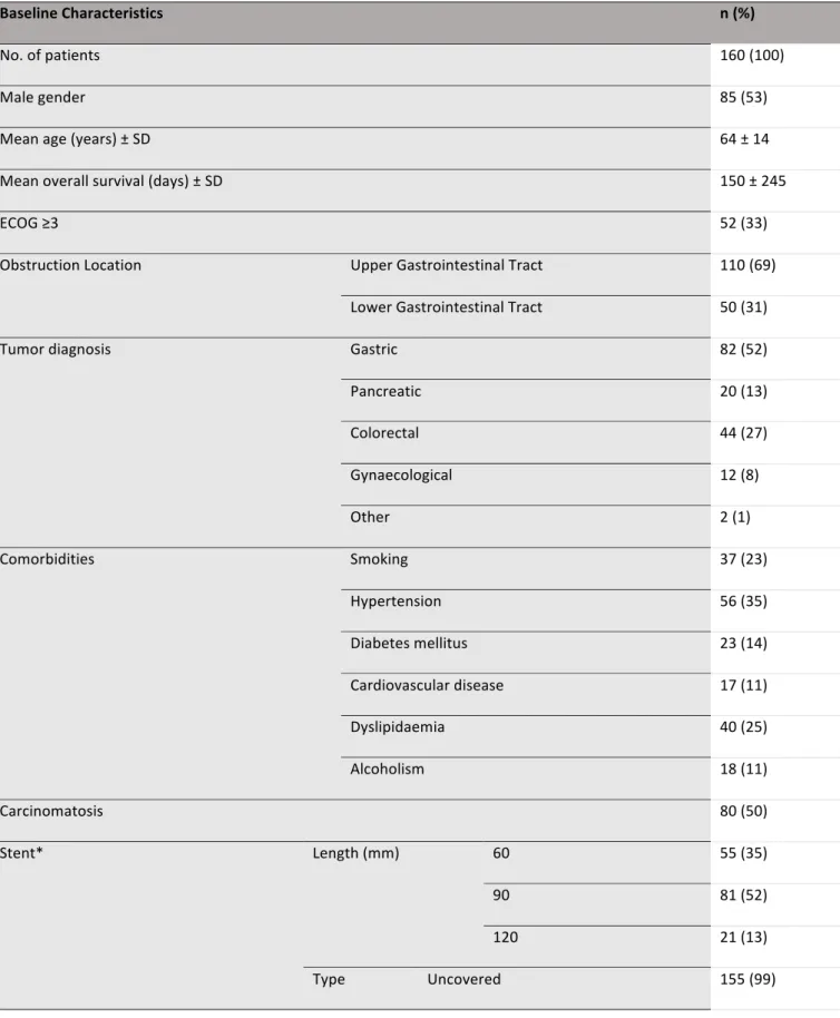
In total, 160 patients had a palliative indication for stent placement and were considered for analysis. The baseline characteristics of these patients are shown in Table 1. The mean age was 64 ± 14 years and 53% of patients were male. The ECOG (Eastern Cooperative Oncology Group) performance status was ≥ 3 in 52 patients (33%). The primary tumour was gastric cancer in 82 patients (51%); pancreatic cancer in 20 patients (13%); colorectal cancer in 44 patients (28%); gynaecological cancer in 12 patients (8%) and others (mesothelioma and unknown) in 1% of patients. Uncovered stents were placed in 99% of the cases. One hundred patients (63%) had been submitted to treatment before stent placement (either chemotherapy, radiotherapy and/or surgery).
Technical success
Technical success was achieved in 157 patients (98%). Three patients only achieved successful stent placement in a second procedure but were considered to have technical success. Technical failure occurred in 3 cases because the guidewire was unable to pass through the stenosis site (one in a gastroduodenal obstruction and 2 in colorectal obstructions). In all cases of technical failure, the obstruction was caused by carcinomatosis. The three patients without technical success were excluded from further analysis.
Factors predicting early and late clinical success
Overall, 114 (73%) patients reached early clinical success: 75 patients with upper GI
obstruction and 39 with lower GI tract obstruction (69%, OR 1.2, 95%CI 0.9-1.5 vs 81%, 0.6 OR, 95%CI 0.3-1.2, p=0.107, respectively) (Table 2).
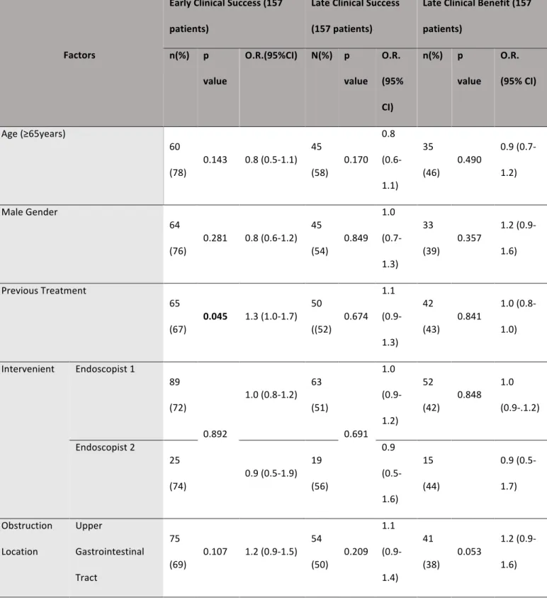
Carcinomatosis and previous treatment were significantly associated with a worse early clinical outcome (OR 1.9, 95%CI 1.4-2.5, p<0.001 and OR 1.3, 95%CI 1.0-1.7, p=0.041, respectively). (Table 2)
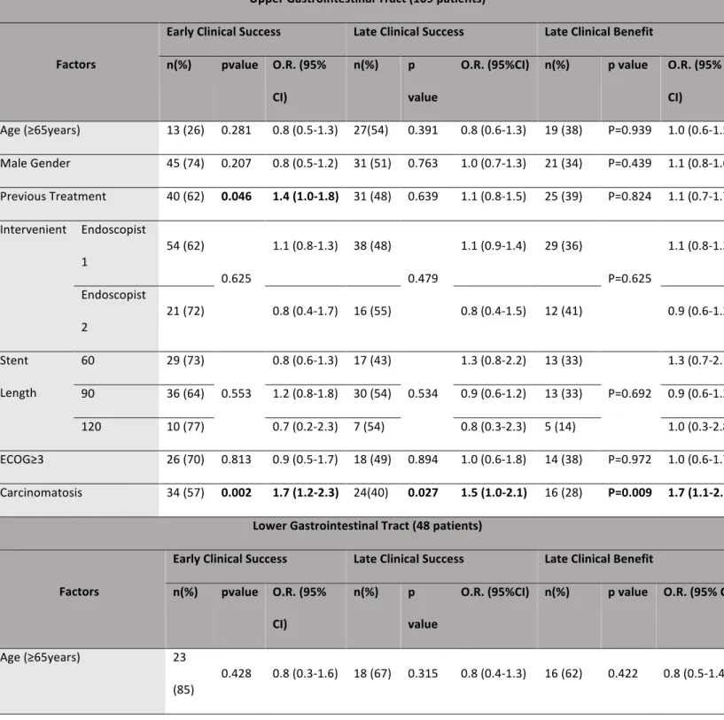
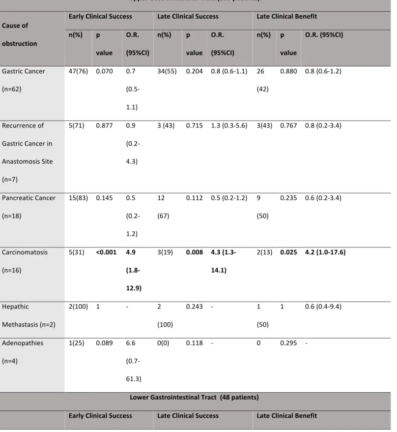
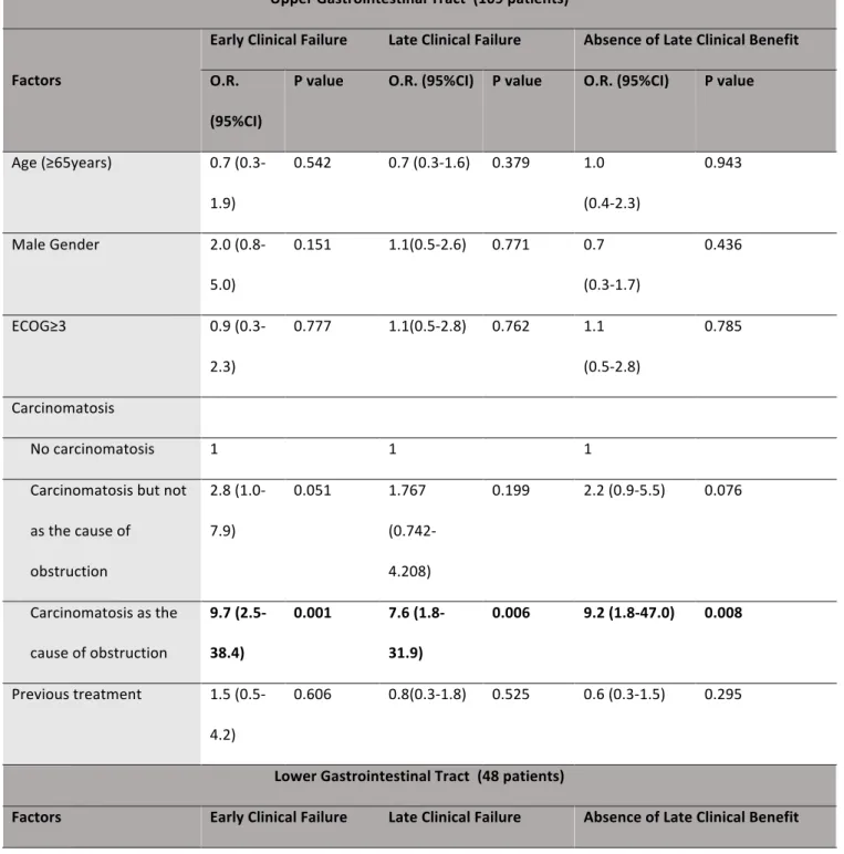
In the subanalysis of the upper and lower GI tract obstruction (Table 3), previous treatment was significantly associated with a worse early clinical outcome in the upper GI tract (OR 1.4, 95%CI 1.0-1.8, p=0.046), ECOG performance score ≥3 was associated with a worse early clinical outcome in the lower GI tract (OR 3.8, 95%CI 1.9-7.7, p=0.001) and carcinomatosis was associated with a worse early clinical outcome in both the upper and lower GI tracts (OR 1.7, 95%CI 1.2-2.3, p=0.002 and OR 2.4, 95%CI 1.2-4.7, p=0.03, respectively). In addition, obstruction caused by carcinomatosis was related to a worse early clinical outcome in both the upper and lower GI tract (OR 4.9, 95%CI 1.8-12.9, p<0.001 and OR 3.5, 95%CI, 1.2-10.4, p=0.028, respectively) (Table 4). In the multivariate analysis of factors related to early clinical failure (Table 5), we found that, in the upper GI tract, carcinomatosis as the cause of obstruction was the only independent factor for early clinical failure (OR 9.7, 95%CI 2.5-38.4, p=0.001). In addition, the presence of carcinomatosis without it being the cause of obstruction showed a tendency for worse early clinical outcome (OR 2.8, 95%CI 1.0-7.9, P=0.051). In the lower GI tract, ECOG performance score ≥3 was the only independent factor for worse early
clinical outcome (OR 29.8, 95%CI 1.9-464.9, p=0.002) and carcinomatosis as the cause of obstruction had a tendency for a worse outcome (OR 8.2, 95%CI 0.8-89.0, p=0.084). Late clinical success was achieved in 83 (53%) patients: 54 patients with upper GI tract obstruction and 29 patients with lower GI tract obstruction (50% OR 1.1, 95%CI 0.9-1.4 vs 60% OR 0.9, 95%CI 0.5-1.6, p=0.209) (Table 2). At 30 days, 54 (34%) patients had died: 43 with upper GI obstruction and 9 with lower GI obstruction (39% vs 19%, p=0.008). Late clinical benefit was achieved in 67 (43%) patients: 26 patients with lower GI obstruction and 41 patients with upper GI obstruction, there was a tendency for better outcome in the first group (57% OR 0.6, 95%CI 0.4-1 vs 38% OR 1.2, 95%CI 0.9-1.7, p=0.053). Carcinomatosis was associated with a worse late clinical success and a worse late clinical benefit (OR 1.7, 95%CI 1.2-2.3, p=0.002 and OR 1.9, 95%CI 1.3-2.7 p<0.001, respectively) (Table 2). In the subanalysis of late clinical success and late clinical benefit, carcinomatosis was associated with a worse late clinical outcome in both the upper and lower GI tract (OR 1.5, 95%CI 1.0-2.1, p=0.027 and OR 2.2, 95%CI 1.0-4.7, p=0.044, respectively) and with a worse late clinical benefit in the upper GI tract (OR 1.7, 95%CI 1.1-2.5, p=0.009) (Table 3). In the lower GI tract, there was a tendency for worse clinical benefit in patients with carcinomatosis (OR 2.2, 95%CI 1.0-4.9, p=0.052). ECOG performance score≥3 had a tendency for late clinical failure in this segment (OR 2.3, 95%CI 0.9-5.4). (Table 3)
Obstruction caused by carcinomatosis, in both the upper and lower GI tract, was related
to a worse late clinical outcome (OR 4.3, 95%CI 1.3-14.1, p=0.008 and OR 5.3 95%CI 1.2-
10.4 p=0.009, respectively) and worse late clinical benefit (OR 4.2, 95%CI 1.0-17.6, p=0.025 and OR 0.7, 95%CI 0.5-1.0, p=0.033, respectively). Obstruction caused by
colorectal cancer was related to a greater late clinical outcome (OR 2.7, 95%CI 0.5-1.0, p=0.027) and late clinical benefit (OR 0.7, 95%CI 0.5-1.0, p=0.006) in the lower GI tract. (Table 4) Multivariate analysis showed that obstruction caused by carcinomatosis was the only independent factor for late clinical failure in both the upper and lower GI tract (OR 7.6, 95%CI 1.8-31.9, p=0.006 and OR 14.4, 95%CI 1.7-119.6, p=0.013, respectively) (Table 5) and also for absence of late clinical benefit in both the upper and lower GI tract (OR 29.8, 95%CI 1.9-464.9, p=0.002 and OR 8.3, 95%CI 1.2-57.5, p=0.035, respectively). In addition, the presence of carcinomatosis without it being the cause of obstruction showed a tendency to absence of late clinical benefit (OR 2.2, 95%CI 0.9-5.5, p=0.076) ( (Table 5).
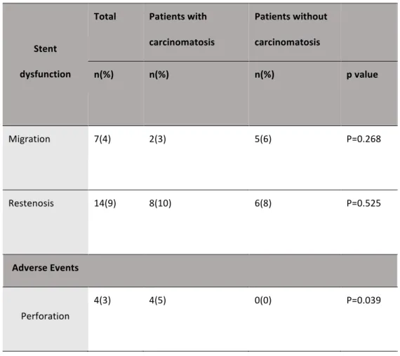
Both overall survival (122 days, 95% CI 83-161 vs. 243, 95% CI 154-333; p=0.02) (Figure 1) and symptom-free survival (66 days, 95% CI 32-100 vs. 183, 95% CI 83-282; p=0.002) (Figure 2) were lower in patients with carcinomatosis compared to patients without carcinomatosis. Factors predicting stent dysfunction and perforation Stent dysfunction and perforation are described in Table 6. Perforation occurred in 4 procedures (2.5%), all occurring in patients with carcinomatosis (p=0.039). One patient had an obstruction caused by a recurrence of gastric cancer in an anastomosis site and the other three had obstruction caused by extraluminal compression by carcinomatosis (1 patient in the jejunum; 1 patient in the antrum and 1 in the sigmoid colon). All of the
4 patients had been submitted to previous treatment (p=0.111), three of them had been submitted to chemotherapy (p=0.454).
Stent dysfunction occurred in 21 patients (13%), 7 patients (15%) who had been submitted to colorectal stent placement and 14 (13%) patients who had been submitted to gastroduodenal stent placement (p=0.731). Of the 7 patients who had stent migration, one had an accidental exit of the rectal stent and did not have an indication for a second stent placement and another patient was submitted to colostomy because of failure to retrieve the stent that migrated from the sigmoid colon. The other 5 patients were submitted to a second stent placement, four gastroduodenal and one colorectal stents. Migration was associated with short 6 cm stents (9% vs 2% p=0.044). Of the 14 patients with stent reestenosis, one patient, whose cause of obstruction was gastric cancer, did not need reintervention because of clinical deterioration. Two patients were submitted to colostomy; one patient’s cause of obstruction was carcinomatosis and the other’s was colorectal cancer. The other 11 patients had to receive a second stent, 9 gastroduodenal and 2 colorectal stents. We did not find any risk factors for stent restenosis even though there seemed to be a tendency for higher rate of reestenosis in patients younger than 65 years old (13% vs 5%, p=0.108), with ECOG≥3 (14% vs 7%, p=0.160) and those submitted to previous treatment (11% vs 5%, p=0.176).
Discussion
Intra-abdominal malignant GI tumors may lead to obstruction, with severe symptoms that greatly affect quality of life, irrespective of GI tract segment affected. Moreover, it can compromise additional treatments such as systemic chemotherapy, radiotherapy or surgery. Palliative stenting has been proven to be a safe alternative to surgery, but not all patients benefit of this treatment.[11] In the present study, we aimed to understand which factors were implicated in patient’s clinical outcome. To our knowledge, this is the first study to analyse factors predicting outcomes of palliative stenting in all causes of intra-abdominal GI obstruction, regardless of the location or cause of obstruction.Technical success rate was 98% (similar for upper and lower GI segment) and in line with that of previous studies [7,8,12-18]. Importantly, with the exception of obstruction caused by carcinomatosis, stent placement was always possible. Perforation is a major adverse event that has to be considered when pondering stent placement. Previous studies have shown iatrogenic perforation rates of 0 to 5%[11,13,18-23]. In our study population, perforation was a rare event, occurring in 4 (2.5%) of our patients. In all patients carcinomatosis was present, being a predictive factor for this adverse event (p=0.039). Three of the 4 patients had been submitted to chemotherapy (p=0.454), a factor that has been considered to be related to this adverse event[24,25]. Additionally, all the 4 perforation cases occurred in patients with previous treatments, suggesting that this may be a risk factor for this complication. In our study this association was not significant because the study was underpowered for detecting this association since we
had very few perforation cases. Therefore, in experienced hands, it looks that intra-abdominal stent placement is a very safe and feasible procedure.
Early clinical success was 81% in lower GI tract obstructions and 69% in upper GI tract obstructions. When looking at the rates of early clinical success in each cause of obstruction (Table 4), they are similar to previous studies. In fact, pancreatic cancer had a high early clinical success rate (83%) which is in accordance with published literature reporting early clinical success rates of 83-88% [26,27]. The same applies to gastric cancer with 76% of patients reaching early clinical success, also comparable to a recent study by Park et al, which compared the outcome of stenting and surgery in obstructions caused by gastric cancer, reporting a clinical success of 77%[28]. Colorectal cancer also had a high rate of early clinical success (87%) similar to previous data, which reported rates of 85-100% [23,29,30].
On the other hand, when the cause of obstruction was recurrence of stenosis in an anastomosis site, patients had a clinical success rate of 71%, comparable to reported early clinical success rate of 70% in a study by Cho et al.[31]. In spite of this, Park MS et al[32] have reported higher success rates (more than 90% in different types of gastric reconstruction) in a large series report of 196 patients, which differs from our results probably because of the characteristics of the baseline population. We had few cases of obstruction caused by gynecological cancer, hepatic metastasis and adenopathies, but we found a high early success rate in the first two groups as all the patients reached symptom relief and a low clinical success rate in the second (25%). The two last groups are seldom reported in literature.
Few studies have reported late clinical outcome at a specific follow-up time and therefore there is no consensual definition of this variable.
We defined two different variables to reflect two different late outcomes: late clinical success and late clinical benefit. The first variable considered that patients who died before 30 days but were free of obstructive symptoms at time of death reached clinical success. The second variable considered death as a negative outcome because, at 30 days follow-up, only patients who were alive and without obstructive symptoms benefited from the stenting procedure. To our knowledge, this is the first study of endoscopic stenting that considers both of these important late clinical outcomes simultaneously.
Patients with upper GI tract obstruction tended to have an earlier death compared to patients with lower GI tract obstruction which reflected in a tendency for worse late clinical benefit (p=0.053) but not for worse late clinical success (p=0.209). This difference shows that there was a group of patients with upper GI tract obstruction who died before the 30 days follow up that were symptom-free at the time of death. Even though these patients improved after the procedure, they died soon after it and from a practical and economical point of view it is arguable if stents should have been placed in these subjects. On the other hand, obstruction caused by colorectal cancer was related to a better late clinical outcome as well as a better late clinical benefit. These findings reflect better prognosis and longer survival of patients with colorectal cancer. Similarly, Keswani et al found that stent palliation of obstruction caused colorectal cancer had a better clinical
outcome than extracolonic obstructive causes.[33] Furthermore, Moon et al also compared these two groups and although they did not find significant early clinical
differences, they found that patients who underwent palliation for obstructions caused by colorectal cancer had a higher overall and stent-patency survival, compared to patients with extracolonic malignancies.[29]
Previous data has reported the presence of carcinomatosis as a predictor of worse clinical outcome in both gastroduodenal and colorectal obstruction. Rademacher et al[34] compared clinical success of gastroduodenal obstruction patients with and without carcinomatosis and found that the first group had a lower rate of clinical success (63%, p=0.036). In a recent study, Faraz et al[19] showed that carcinomatosis was an independent factor for worse clinical outcome in patients submitted to colorectal stenting and Lee et al [17] recognized carcinomatosis as an independent factor for lower obstruction symptom-free survival. Furthermore, Shin et al[13], in a multivariable analysis, found that carcinomatosis was an independent factor for worse clinical outcome in gastroduodenal stenting (p<0.001). In spite of these results, Mendelson et al[18] didn’t find differences in clinical outcomes of patients with or without
carcinomatosis in gastroduodenal stenting. However, in this study, patients with carcinomatosis weren’t considered for stent placement if they had more than one obstruction site or if their lesions were thought not to be amenable for stent placement, which wasn’t the case in our study population, since these factors are not always easy to determine.
related, in the univariate analysis of upper and lower GI tract obstruction, to both early and late clinical failure as well as lower rates of late clinical benefit. Furthermore, we
found that overall survival and especially symptom-free survival seemed to be lower in patients who had carcinomatosis, reflecting a more advanced stage of malignancy and poor symptom relief, respectively. These results can be explained by the fact that carcinomatosis is associated with multiple obstruction sites and decreased bowel movements, which have a negative influence in clinical improvement.
In spite of this, we found that, more important than the sole presence of carcinomatosis were the cases when carcinomatosis itself was the cause of obstruction. In fact, to our knowledge this is the first study analysing the outcome of palliative stenting in patients with carcinomatosis as the cause of obstruction. Our results showed that obstruction caused by carcinomatosis was the only independent factor of early and late clinical failure and absence of clinical benefit in the upper GI tract and late clinical failure and absence of clinical benefit in the lower GI tract. Although it was not an independent factor for early clinical failure in lower GI obstruction, patients showed a tendency for worse outcome. In fact, patients with obstruction caused by carcinomatosis had low early clinical success rates of 31% and 56% in the upper and lower GI tract, respectively. Furthermore, the presence of carcinomatosis without it being the cause of obstruction showed a tendency for a worse early clinical outcome and late clinical benefit in the upper GI tract.
Higher ECOG performance scores were also an independent factor for early clinical failure in the lower GI tract. This could be explained by the fact that patients with poorer performance status are confined to a bed or chair for most of the waking hours. Bowel
movement is improved by low intensity exercise, which is impaired in this group of patients. This impairment justifies their poor rates of stent placement’s clinical success, in spite of being technically successful. [35] This variable also had a tendency for a worse late clinical success but not for worse late clinical benefit, which may be explained by the fact that the patients with lower ECOG performance scores who died before the 30 days follow-up were symptom-free. Therefore, although performance score influenced obstructive symptoms within the first month, it did not reflect on patient’s survival and benefit at 30 days.
Literature reports migration and restenosis rates of 2%-9% and 6%-31% respectively[13,14,20,22,36], which is in agreement with migration and restenosis rates of 4% an 9% in our study. We found stent migration to be more frequent in shorter stenosis sites (6cm). It has been described in previous literature that migration tends to occur with stents too narrow in diameter and/or too short in length for the stricture they are placed in[37]. Although covered stents have been associated with higher rates of migration[38,39], we didn’t find this in our study probably because only a minority of the patients was submitted to covered stent placement (n=2, 1.3%).
This was a nonrandomized, retrospective, single centre study, with its inherent disadvantages and limitations. Due to this study design, more than one physician performed ECOG scores and we cannot rule out interobserver bias, which could explain why this was not a risk factor for patients with upper GI tract obstruction, in contrast to the lower GI tract. In fact, although we did not find this in our study, low performance scores have been associated with clinical success of gastroduodenal stenting in previous
articles[13,16,40]. In addition, we were unable to clearly access the presence and level of ascites in all patients, which could have been a factor that would influence the
multivariate analysis as it was considered a risk factor in other studies[15,18,40]. So, future prospective studies analysing these variables would be of great interest. Furthermore, given that our stent dysfunction and perforation rates were low, our analysis of predictive factors isn’t as strong as it would be if we had a bigger group of patients with these complications. On the other hand, this study had some advantages since a large number of patients and a long follow up period were considered, which enhances our understanding of GI stenting for obstruction caused by intra-abdominal malignancies. When looking at specific scenarios, we can see that patients with obstruction caused by carcinomatosis had a low rate of clinical success, independently of the location of the obstruction (gastroduodenal or colorectal). Only 22% of patients with lower GI tract and 13% of patients with gastroduodenal obstruction were alive at 30 days and were free of obstructive symptoms, respectively. This is a dreadful scenario. Stent placement is an expensive treatment that might not be cost-effective in this specific setting. In addition, 3 of the patients in this group suffered perforation related to the procedure, a major adverse event with deathly consequences.
In conclusion, in this large series of endoscopic stents for palliative intra-abdominal obstruction we showed that stent placement in these situations is feasible and safe, independently of the location of obstruction. However, carcinomatosis not only limits the clinical success of this procedure, both in upper and lower GI tract obstruction, but
also increases the risk of serious complications. Therefore, in the scenario of intra-abdominal malignant obstruction in patients with carcinomatosis, especially in the cases
where carcinomatosis is the cause of obstruction, the decision of stent placement should be fully scrutinized and may not always be an adequate option. Just because we can does not mean we should do it!
References
1. Torre LA, Bray F, Siegel RL, Ferlay J, Lortet-Tieulent J, Jemal A. Global cancer statistics, 2012. CA: A Cancer Journal for Clinicians 2015; 65 (2):87-108.
2. Rothenberger DA. Palliative therapy of rectal cancer. Overview: epidemiology, indications, goals, extent, and nature of work-up. Journal of gastrointestinal surgery : official journal of the Society for Surgery of the Alimentary Tract 2004; 8 (3):259-261. 3. Catalano V, Labianca R, Beretta GD, Gatta G, de Braud F, Van Cutsem E. Gastric cancer. Crit Rev Oncol Hematol 2009; 71 (2):127-164. 4. Kato H, Tsutsumi K, Okada H. Recent advancements in stent therapy in patients with malignant gastroduodenal outlet obstruction. Ann Transl Med 2017; 5 (8):186. 5. Cetinkaya E, Dogrul AB, Tirnaksiz MB. Role of self expandable stents in management of colorectal cancers. World journal of gastrointestinal oncology 2016; 8 (1):113-120. 6. Chandrasegaram MD, Eslick GD, Mansfield CO, Liem H, Richardson M, Ahmed S, et al. Endoscopic stenting versus operative gastrojejunostomy for malignant gastric outlet obstruction. Surg Endosc 2012; 26 (2):323-329. 7. Endo S, Takiguchi S, Miyazaki Y, Nishikawa K, Imamura H, Takachi K, et al. Efficacy of endoscopic gastroduodenal stenting for gastric outlet obstruction due to unresectable advanced gastric cancer: a prospective multicenter study. Journal of surgical oncology 2014; 109 (3):208-212. 8. Varadarajulu S, Roy A, Lopes T, Drelichman ER, Kim M. Endoscopic stenting versus surgical colostomy for the management of malignant colonic obstruction: comparison of hospital costs and clinical outcomes. Surg Endosc 2011; 25 (7):2203-2209.
9. Mergener K, Kozarek RA. Stenting of the gastrointestinal tract. Dig Dis 2002; 20 (2):173-181.
10. Mangiavillano B, Pagano N, Arena M, Miraglia S, Consolo P, Iabichino G, et al. Role of stenting in gastrointestinal benign and malignant diseases. World J Gastrointest Endosc 2015; 7 (5):460-480.
11. Masci E, Viale E, Mangiavillano B, Contin G, Lomazzi A, Buffoli F, et al. Enteral self-expandable metal stent for malignant luminal obstruction of the upper and lower gastrointestinal tract: a prospective multicentric study. J Clin Gastroenterol 2008; 42 (4):389-394. 12. Hong SP, Kim TI. Colorectal stenting: An advanced approach to malignant colorectal obstruction. World J Gastroenterol 2014; 20 (43):16020-16028. 13. Shin YS, Choi CW, Kang DH, Kim HW, Kim SJ, Cho M, et al. Factors associated with clinical failure of self-expandable metal stent for malignant gastroduodenal obstruction. Scand J Gastroenterol 2016; 51 (1):103-110.
14. Kim JH, Ku YS, Jeon TJ, Park JY, Chung JW, Kwon KA, et al. The efficacy of self-expanding metal stents for malignant colorectal obstruction by noncolonic malignancy
with peritoneal carcinomatosis. Diseases of the colon and rectum 2013; 56 (11):1228-1232.
15. Jeon HH, Park CH, Park JC, Shim CN, Kim S, Lee HJ, et al. Carcinomatosis matters: clinical outcomes and prognostic factors for clinical success of stent placement in malignant gastric outlet obstruction. Surg Endosc 2014; 28 (3):988-995.
16. Yamao K, Kitano M, Kayahara T, Ishida E, Yamamoto H, Minaga K, et al. Factors predicting through-the-scope gastroduodenal stenting outcomes in patients with gastric
outlet obstruction: a large multicenter retrospective study in West Japan. Gastrointest Endosc 2016; 84 (5):757-763 e756. 17. Lee JE, Lee K, Hong YS, Kim ER, Lee H, Min BH. Impact of Carcinomatosis on Clinical Outcomes after Self-Expandable Metallic Stent Placement for Malignant Gastric Outlet Obstruction. PLoS One 2015; 10 (10):e0140648. 18. Mendelsohn RB, Gerdes H, Markowitz AJ, DiMaio CJ, Schattner MA. Carcinomatosis is not a contraindication to enteral stenting in selected patients with malignant gastric outlet obstruction. Gastrointest Endosc 2011; 73 (6):1135-1140.
19. Faraz S, Salem SB, Schattner M, Mendelsohn R, Markowitz A, Ludwig E, et al. Predictors of clinical outcome of colonic stents in patients with malignant large-bowel obstruction due to extracolonic malignancy. Gastrointest Endosc 2018. 20. Hori Y, Naitoh I, Hayashi K, Ban T, Natsume M, Okumura F, et al. Predictors of stent dysfunction after self-expandable metal stent placement for malignant gastric outlet obstruction: tumor ingrowth in uncovered stents and migration of covered stents. Surg Endosc 2017; 31 (10):4165-4173. 21. Tilney HS, Lovegrove RE, Purkayastha S, Sains PS, Weston-Petrides GK, Darzi AW, et
al. Comparison of colonic stenting and open surgery for malignant large bowel obstruction. Surg Endosc 2007; 21 (2):225-233. 22. Kim J, Choi IJ, Kim CG, Lee JY, Cho SJ, Park SR, et al. Self-expandable metallic stent placement for malignant obstruction in patients with locally recurrent gastric cancer. Surg Endosc 2011; 25 (5):1505-1513. 23. Fernandez-Esparrach G, Bordas JM, Giraldez MD, Gines A, Pellise M, Sendino O, et al. Severe complications limit long-term clinical success of self-expanding metal stents
in patients with obstructive colorectal cancer. Am J Gastroenterol 2010; 105 (5):1087-1093.
24. Karoui M CA, Debaldo C et al. . Stents for palliation of obstructive metastatic colon cancer: impact on management and chemotherapy administration. Arch Surg 2007;142:619–23.
25. van Hoo JE FP, Marinelli AW et al. . Early closure of a multicenter randomized clinical trial of endoscopic stenting vs. surgery for stage IV le -sided colorectal cancer. . Endoscopy 2008;40:184–91.
26. Maire F, Hammel P, Ponsot P, Aubert A, O'Toole D, Hentic O, et al. Long-term outcome of biliary and duodenal stents in palliative treatment of patients with unresectable adenocarcinoma of the head of pancreas. Am J Gastroenterol 2006; 101 (4):735-742.
27. Oh SY, Edwards A, Mandelson M, Ross A, Irani S, Larsen M, et al. Survival and clinical outcome after endoscopic duodenal stent placement for malignant gastric outlet obstruction: comparison of pancreatic cancer and nonpancreatic cancer. Gastrointest Endosc 2015; 82 (3):460-468 e462.
28. Park CH, Park JC, Kim EH, Chung H, An JY, Kim HI, et al. Impact of carcinomatosis and ascites status on long-term outcomes of palliative treatment for patients with gastric outlet obstruction caused by unresectable gastric cancer: stent placement versus palliative gastrojejunostomy. Gastrointest Endosc 2015; 81 (2):321-332.
29. Moon SJ, Kim SW, Lee BI, Lim CH, Kim JS, Soo J, et al. Palliative stent for malignant colonic obstruction by extracolonic malignancy: a comparison with colorectal cancer. Dig Dis Sci 2014; 59 (8):1891-1897.
30. Ahn HJ, Kim SW, Lee SW, Lee SW, Lim CH, Kim JS, et al. Long-term outcomes of palliation for unresectable colorectal cancer obstruction in patients with good
performance status: endoscopic stent versus surgery. Surg Endosc 2016; 30 (11):4765-4775. 31. Cho YK, Kim SW, Nam KW, Chang JH, Park JM, Jeong J-J, et al. Clinical outcomes of self-expandable metal stents in palliation of malignant anastomotic strictures caused by recurrent gastric cancer. World Journal of Gastroenterology 2009; 15 (28):3523. 32. Park JH, Song HY, Kim SH, Shin JH, Kim JH, Kim BS, et al. Metallic stent placement in patients with recurrent malignant obstruction in the surgically altered stomach. Ann Surg Oncol 2014; 21 (6):2036-2043.
33. Keswani RN, Azar RR, Edmundowicz SA, Zhang Q, Ammar T, Banerjee B, et al. Stenting for malignant colonic obstruction: a comparison of efficacy and complications in colonic versus extracolonic malignancy. Gastrointestinal Endoscopy 2009; 69 (3, Part 2):675-680.
34. Rademacher C, Bechtler M, Schneider S, Hartmann B, Striegel J, Jakobs R. Self-expanding metal stents for the palliation of malignant gastric outlet obstruction in
patients with peritoneal carcinomatosis. World J Gastroenterol 2016; 22 (43):9554-9561. 35. Koffler KH, Menkes A, Redmond RA, Whitehead WE, Pratley RE, Hurley BF. Strength training accelerates gastrointestinal transit in middle-aged and older men. Medicine and science in sports and exercise 1992; 24 (4):415-419. 36. Meisner S, Gonzalez-Huix F, Vandervoort JG, Goldberg P, Casellas JA, Roncero O, et
al. Self-expandable metal stents for relieving malignant colorectal obstruction: short-
term safety and efficacy within 30 days of stent procedure in 447 patients. Gastrointest Endosc 2011; 74 (4):876-884.
37. Mauro MA, Koehler RE, Baron TH. Advances in gastrointestinal intervention: the treatment of gastroduodenal and colorectal obstructions with metallic stents. Radiology 2000; 215 (3):659-669.
38. Hori Y, Naitoh I, Hayashi K, Ban T, Natsume M, Okumura F, et al. Predictors of outcomes in patients undergoing covered and uncovered self-expandable metal stent placement for malignant gastric outlet obstruction: a multicenter study. Gastrointest Endosc 2017; 85 (2):340-348 e341.
39. Zhang Y, Shi J, Shi B, Song CY, Xie WF, Chen YX. Comparison of efficacy between uncovered and covered self-expanding metallic stents in malignant large bowel obstruction: a systematic review and meta-analysis. Colorectal disease : the official journal of the Association of Coloproctology of Great Britain and Ireland 2012; 14 (7):e367-374.
40. Sasaki T, Isayama H, Nakai Y, Togawa O, Kogure H, Kawakubo K, et al. Predictive factors of solid food intake in patients with malignant gastric outlet obstruction
receiving self-expandable metallic stents for palliation. Dig Endosc 2012; 24 (4):226-230.
TABLES
Table 1 Patient's Baseline Characteristics Baseline Characteristics n (%) No. of patients 160 (100) Male gender 85 (53) Mean age (years) ± SD 64 ± 14 Mean overall survival (days) ± SD 150 ± 245 ECOG ≥3 52 (33) Obstruction Location Upper Gastrointestinal Tract 110 (69) Lower Gastrointestinal Tract 50 (31) Tumor diagnosis Gastric 82 (52) Pancreatic 20 (13) Colorectal 44 (27) Gynaecological 12 (8) Other 2 (1) Comorbidities Smoking 37 (23) Hypertension 56 (35) Diabetes mellitus 23 (14) Cardiovascular disease 17 (11) Dyslipidaemia 40 (25) Alcoholism 18 (11) Carcinomatosis 80 (50) Stent* Length (mm) 60 55 (35) 90 81 (52) 120 21 (13) Type Uncovered 155 (99)Covered 2 (1) Previous treatment yes 100 (63) CT 54 (34) RT 0 (0) Surgery 7 (4) CT+RT 7 (4) CT+Surgery 25 (16) RT+Surgery 1 (1) QT+RT+Surgery 6 (4) SD – Standard Deviation; ECOG – Eastern Cooperative Oncology; CT – chemotherapy; RT: radiotherapy. Group. *only applicable to patient’s whose stent was technically successful (157).
Table 2. Univariate analysis of factors predicting early and late clinical outcomes. Factors Early Clinical Success (157 patients) Late Clinical Success (157 patients) Late Clinical Benefit (157 patients) n(%) p value O.R.(95%CI) N(%) p value O.R. (95% CI) n(%) p value O.R. (95% CI) Age (≥65years) 60 (78) 0.143 0.8 (0.5-1.1) 45 (58) 0.170 0.8 (0.6-1.1) 35 (46) 0.490 0.9 (0.7-1.2) Male Gender 64 (76) 0.281 0.8 (0.6-1.2) 45 (54) 0.849 1.0 (0.7-1.3) 33 (39) 0.357 1.2 (0.9-1.6) Previous Treatment 65 (67) 0.045 1.3 (1.0-1.7) 50 ((52) 0.674 1.1 (0.9-1.3) 42 (43) 0.841 1.0 (0.8-1.0) Intervenient Endoscopist 1 89 (72) 0.892 1.0 (0.8-1.2) 63 (51) 0.691 1.0 (0.9-1.2) 52 (42) 0.848 1.0 (0.9-.1.2) Endoscopist 2 25 (74) 0.9 (0.5-1.9) 19 (56) 0.9 (0.5-1.6) 15 (44) 0.9 (0.5-1.7) Obstruction Location Upper Gastrointestinal Tract 75 (69) 0.107 1.2 (0.9-1.5) 54 (50) 0.209 1.1 (0.9-1.4) 41 (38) 0.053 1.2 (0.9-1.6)
Lower Gastrointestinal Tract 39 (81) 0.6 (0.3-1.2) 29 (60) 0.7 (0.5-1.2) 26 (57) 0.6 (0.4-1) Stent Length 60 42 (75) 0.752 0.9 (0.5-1.5) 26 (46) 0.321 1.3 (0.8-2.0) 22 (39) 0.650 1.2 (0.7-1.8) 90 56 (70) 1.1 (0.8-1.6) 47 (59) 0.8 (0.6-1.1) 37 (46) 0.9 (0.6-1.2) 120 16 (70) 0.8 (0.3-2.1) 10 (48) 1.2 (0.6-2.7) 8 (38) 1.2 (0.5-2.8) ECOG≥3 34 (65) 0.153 1.4 (0.9-2.2) 24 (46) 0.236 1.3 (0.8-2.0) 20 (39) 0.453 1.2 (0.8-1.9) Carcinomatosis 45 (58) <0.001 1.9 (1.4-2.5) 31 (40) 0.002 1.7 (1.2-2.3) 22 (29) <0.001 1.9 (1.3-2.7) O.R. - Odds ratio; 95% CI – 95% Confidence interval.
Table 3 Subanalysis of factors predicting early and late clinical success in the Upper and Lower GI tracts Upper Gastrointestinal Tract (109 patients) Factors
Early Clinical Success Late Clinical Success Late Clinical Benefit n(%) pvalue O.R. (95%
CI)
n(%) p value
O.R. (95%CI) n(%) p value O.R. (95% CI) Age (≥65years) 13 (26) 0.281 0.8 (0.5-1.3) 27(54) 0.391 0.8 (0.6-1.3) 19 (38) P=0.939 1.0 (0.6-1.5) Male Gender 45 (74) 0.207 0.8 (0.5-1.2) 31 (51) 0.763 1.0 (0.7-1.3) 21 (34) P=0.439 1.1 (0.8-1.6) Previous Treatment 40 (62) 0.046 1.4 (1.0-1.8) 31 (48) 0.639 1.1 (0.8-1.5) 25 (39) P=0.824 1.1 (0.7-1.7) Intervenient Endoscopist 1 54 (62) 0.625 1.1 (0.8-1.3) 38 (48) 0.479 1.1 (0.9-1.4) 29 (36) P=0.625 1.1 (0.8-1.3) Endoscopist 2 21 (72) 0.8 (0.4-1.7) 16 (55) 0.8 (0.4-1.5) 12 (41) 0.9 (0.6-1.2) Stent Length 60 29 (73) 0.553 0.8 (0.6-1.3) 17 (43) 0.534 1.3 (0.8-2.2) 13 (33) P=0.692 1.3 (0.7-2.1) 90 36 (64) 1.2 (0.8-1.8) 30 (54) 0.9 (0.6-1.2) 13 (33) 0.9 (0.6-1.2) 120 10 (77) 0.7 (0.2-2.3) 7 (54) 0.8 (0.3-2.3) 5 (14) 1.0 (0.3-2.8) ECOG≥3 26 (70) 0.813 0.9 (0.5-1.7) 18 (49) 0.894 1.0 (0.6-1.8) 14 (38) P=0.972 1.0 (0.6-1.7) Carcinomatosis 34 (57) 0.002 1.7 (1.2-2.3) 24(40) 0.027 1.5 (1.0-2.1) 16 (28) P=0.009 1.7 (1.1-2.5) Lower Gastrointestinal Tract (48 patients) Factors
Early Clinical Success Late Clinical Success Late Clinical Benefit n(%) pvalue O.R. (95%
CI)
n(%) p value
O.R. (95%CI) n(%) p value O.R. (95% CI)
Age (≥65years) 23
(85)
Male Gender 19 (83) 0.817 0.9 (0.4-2.0) 14 (61) 0.951 1.0 (0.5-1.8) 12 (46) 0.791 0.9 (0.5-1.6) Previous Treatment 25 (78) 0.433 1.2 (0.8-1.8) 19 (59) 0.835 1.0 (0.7-1.8) 17 (65) 0.838 1.0 (0.7-1.6) Intervenient Endoscopist 1 35 (81) 0.940 1.0 (0.8-1.3) 26(61) 0.984 1.0 (0.8-1.2) 23 (54) 0.782 1.0 (0.8-1.3) Endoscopist 2 4 (80) 1.1 (0.1-8.6) 3(60) 1.0 (0.2-5.5) 3 (60) 0.8 (1.4-4.3) Stent Length 60 13 (81) 0.872 1 (0.4-2.8) 9 (56) 0.227 1.2 (0.5-2.6) 9 (56) 0.580 0.9 (0.4-2.1) 90 20 (83) 0.9 (0.4-1.9) 17 (71) 0.6 (0.3-1.2) 14 (58) 0.8 (0.5-1.5) 120 6 (75) 1.4 (0.3-6.0) 13 (38) 2.6 (0.7-9.4) 3 (38) 2.0 (0.6-7.3) ECOG≥3 8 (53) 0.001 3.8 (1.9-7.7) 6(40) 0.051 2.3 (0.9-5.4) 6 (40) 0.184 1.8 (0.8-4.2) Carcinomatosis 11 (23) 0.03 2.4 (1.2-4.7) 7(41) 0.044 2.2 (1.0-4.7) 6 (35) 0.051 2.2 (1.0-4.9) O.R. - Odds ratio; 95% CI – 95% Confidence interval.
Table 4. Univariate analysis of cause of obstruction’s association with clinical success within the upper and lower GI tracts.
Upper Gastrointestinal Tract (109 patients) Cause of obstruction
Early Clinical Success Late Clinical Success Late Clinical Benefit n(%) p value O.R. (95%CI) n(%) p value O.R. (95%CI) n(%) p value O.R. (95%CI) Gastric Cancer (n=62) 47(76) 0.070 0.7 (0.5-1.1) 34(55) 0.204 0.8 (0.6-1.1) 26 (42) 0.880 0.8 (0.6-1.2) Recurrence of Gastric Cancer in Anastomosis Site (n=7) 5(71) 0.877 0.9 (0.2-4.3) 3 (43) 0.715 1.3 (0.3-5.6) 3(43) 0.767 0.8 (0.2-3.4) Pancreatic Cancer (n=18) 15(83) 0.145 0.5 (0.2-1.2) 12 (67) 0.112 0.5 (0.2-1.2) 9 (50) 0.235 0.6 (0.2-3.4) Carcinomatosis (n=16) 5(31) <0.001 4.9 (1.8-12.9) 3(19) 0.008 4.3 (1.3-14.1) 2(13) 0.025 4.2 (1.0-17.6) Hepathic Methastasis (n=2) 2(100) 1 - 2 (100) 0.243 - 1 (50) 1 0.6 (0.4-9.4) Adenopathies (n=4) 1(25) 0.089 6.6 (0.7-61.3) 0(0) 0.118 - 0 0.295 - Lower Gastrointestinal Tract (48 patients)
Cause of obstruction n(%) p value O.R. 95%C.I. n(%) p value O.R. (95%C.I.) n(%) p value O.R. (95%CI) Carcinomatosis (n=9) 5 (56) 0.028 3.5 (1.2-10.4) 2(22) 0.009 5.3 (1.2-23.0) 2 (22) 0.033 4.1 (1.0-17.9) Gynecological Cancer(n=1) 1 (100) 1 - 1(100) 1 - 1 (100) 0.458 Colorectal Cancer (n=38) 33 (85) 0.053 0.7 (0.3-1.2) 26(68) 0.027 0.7 (0.5-1.0) 24 (65) 0.006 0.7 (0.5-1.0) O.R. - Odds ratio; 95% CI – 95% Confidence interval.
Table 5. Multivariate analysis of factors associated with early clinical failure, late clinical failure and absence of late clinical benefit within the upper and lower gastrointestinal tract Upper Gastrointestinal Tract (109 patients) Factors
Early Clinical Failure Late Clinical Failure Absence of Late Clinical Benefit O.R.
(95%CI)
P value O.R. (95%CI) P value O.R. (95%CI) P value
Age (≥65years) 0.7 (0.3-1.9) 0.542 0.7 (0.3-1.6) 0.379 1.0 (0.4-2.3) 0.943 Male Gender 2.0 (0.8-5.0) 0.151 1.1(0.5-2.6) 0.771 0.7 (0.3-1.7) 0.436 ECOG≥3 0.9 (0.3-2.3) 0.777 1.1(0.5-2.8) 0.762 1.1 (0.5-2.8) 0.785 Carcinomatosis No carcinomatosis 1 1 1 Carcinomatosis but not as the cause of obstruction 2.8 (1.0-7.9) 0.051 1.767 (0.742-4.208) 0.199 2.2 (0.9-5.5) 0.076 Carcinomatosis as the cause of obstruction 9.7 (2.5-38.4) 0.001 7.6 (1.8-31.9) 0.006 9.2 (1.8-47.0) 0.008 Previous treatment 1.5 (0.5-4.2) 0.606 0.8(0.3-1.8) 0.525 0.6 (0.3-1.5) 0.295 Lower Gastrointestinal Tract (48 patients)
O.R. (95%CI)
P value O.R. (95%CI) P value O.R. (95%CI) P value
Age (≥65years) 1.0 (0.1-6.7) 0.966 0.426 (0.1-1.7) 0.232 0.5 (0.1-1.9) 0.317 Male Gender 0.6 (0.1-4.8) 0.621 0.7 (0.1-3.0) 0.587 0.6 (0.3-2.5) 0.494 ECOG≥3 29.8 (1.9-464.9) 0.002 4.3 (0.8-23.1) 0.092 2.4 (0.5-11.0) 0.266 Carcinomatosis No carcinomatosis 1 1 1 Carcinomatosis but not as the cause of obstruction 3.6 (0.3-38.7) 0.292 1.2 (0.2-6.9) 0.858 1.5 (0.3-7.9) 0.635 Carcinomatosis as the cause of obstruction 8.2 (0.8-89.0) 0.084 14.4 (1.7-119.6) 0.013 8.3 (1.2-57.6) 0.035 Previous treatment 0.2 (0.01-3.6) 0.261 0.4(0.7-1.9) 0.239 0.6 (0.1-2.6) 0.475 O.R. - Odds ratio; 95% CI – 95% Confidence interval.
Table 6. Stent dysfunction and perforation.
Stent dysfunction Total Patients with carcinomatosis Patients without carcinomatosis n(%) n(%) n(%) p value Migration 7(4) 2(3) 5(6) P=0.268 Restenosis 14(9) 8(10) 6(8) P=0.525 Adverse Events Perforation 4(3) 4(5) 0(0) P=0.039
FIGURE LEGEND
Figure 1. Kaplain-Meier plot for overall survival according to the presence of carcinomatosis. Cumulative overall survival was lower in patients with carcinomatosis compared to those without carcinomatosis (122 days, 95% CI 83-161 vs. 243, 95% CI 154-333; p=0.02)
Figure 2. Kaplain-Meier plot for symptom-free survival according to the presence of carcinomatosis. Cumulative symptom-free survival was lower in patients with carcinomatosis compared to those without carcinomatosis (66 days, 95% CI 32-100 vs. 183, 95% CI 83-282; p=0.002)
Manuscripts to the European Journal of Gastroenterology &
Hepatology
Note: These instructions comply with those formulated by the International Committee of Medical Journal Editors. For further details, authors should consult the “Uniform Requirements for Manuscripts Submitted to Biomedical Journals” at http://www.icmje.org.
The Journal is a member of the Committee on Publication Ethics (COPE) which aims to define best practice in the ethics of scientific publishing. COPE has established a number of guidelines including a Code of Conduct, and created flow charts that help editor's process cases of suspected misconduct (www.publicationethics.org).
Appeals on editorial decisions should be sent to the Editor. Complaints related to how your paper was processed during peer-review and not resolved by the Editor, should be referred to the person named as publisher in "About the Journal" under "Journal Info" contacts (http://journals.lww.com/eurojgh/), or if unsatisfied to COPE (www.publicationethics.org).
Aims and scope
The European Journal of Gastroenterology & Hepatology publishes papers reporting original clinical and scientific research which are of a high standard and which contribute to the advancement of knowledge in the field of gastroenterology and hepatology. The journal publishes five types of manuscripts: reviews, original papers, short articles, case reports and letters to the Editor. At the discretion of the Editors some articles are chosen to appear as the online only publication. These articles remain an integrative part of the Journal and are listed in the table of contents for each issue.
Table of contents
Points to consider before submission Redundant or duplicate publication Conflicts of interest
Ethics committee approval Authorship
Compliance with NIH and Other Research Funding Agency Accessibility Requirements Copyright assignment Submissions Presentation of Papers Title Page Abstracts Keywords Text Acknowledgements References Tables Illustrations
Legends for illustrations Units of measurement Abbreviations and symbols Supplemental Digital Content Offprints
Letters to the Editor
Points to consider before submission
Please think carefully about the following points and make the appropriate declarations.
Redundant or duplicate publication
We ask you to confirm that your paper has not been published in its current form or a substantially similar form (in print or electronically, including on a web site), that it has not been accepted for
not duplicate or redundant publication (www.icmje.org). If you are in doubt (particularly in the case of material that you have posted on a web site), we ask you to proceed with your submission but to include a copy of the relevant previously published work or work under consideration by other journals. In your covering letter to the editors, draw attention to any published work that concerns the same patients or subjects as the present paper.
Conflicts of interest
Authors must state all possible conflicts of interest in the manuscript, including financial, consultant, institutional and other relationships that might lead to bias or a conflict of interest. If there is no conflict of interest, this should also be explicitly stated as none declared. All sources of funding should be acknowledged in the manuscript. All relevant conflicts of interest and sources of funding should be included on the title page of the manuscript with the heading “Conflicts of Interest and Source of Funding:”. For example:
Conflicts of Interest and Source of Funding: A has received honoraria from Company Z. B is currently receiving a grant (#12345) from Organization Y, and is on the speaker’s bureau for Organization X – the CME organizers for Company A. For the remaining authors none were declared.
In addition, each author must complete and submit the journal's copyright transfer agreement, which includes a section on the disclosure of potential conflicts of interest based on the recommendations of the International Committee of Medical Journal Editors, "Uniform Requirements for Manuscripts Submitted to Biomedical Journals" (www.icmje.org/update.html). A copy of the form is made available to the submitting author within the Editorial Manager submission process. Co-authors will automatically receive an Email with instructions on completing the form upon submission.
published elsewhere (such as illustrations) from the copyright holder. Authors are responsible for paying any fees to reproduce material.
Patient consent forms
Patients have a right to privacy that should not be infringed without informed consent. Identifying
details (written or photographic) should be omitted if they are not essential, but patient data should never be altered or falsified in an attempt to attain anonymity. Complete anonymity is difficult to achieve, and a consent form should be obtained if there is any doubt. For example, masking the eye region in photographs of patients is inadequate protection of anonymity. When informed consent has been obtained it should be indicated in the published article.
A statement to the effect that such consent had been obtained must be included in the ‘Methods’ section of your paper and an example of the consent form you used must be uploaded with your manuscript.
Ethics committee approval
You must state clearly in your submission in the Methods section that you conducted studies on human participants must with the approval of an appropriate named ethics committee. Please also look at the latest version of the Declaration of Helsinki. Clinical studies should be in accordance with the latest publication of ‘Good Clinical Practice’. Similarly, you must confirm that experiments involving animals adhered to ethical standards and must state the care of animal and licensing guidelines under which the study was performed.
Authorship
All authors must sign copyright forms accompanying their submission to confirm that they have read and approved the paper, that they have met the criteria for authorship as established by the International Committee of Medical Journal Editors, that they believe that the paper represents honest work, and that they are able to verify the validity of the results reported.
A number of research funding agencies now require or request authors to submit the post-print (the article after peer review and acceptance but not the final published article) to a repository that is accessible online by all without charge. As a service to our authors, LWW will identify to the National Library of Medicine (NLM) articles that require deposit and will transmit the post-print of an article based on research funded in whole or in part by the National Institutes of Health, Wellcome Trust, Howard Hughes Medical Institute, or other funding agencies to PubMed Central. The Copyright Transfer Agreement provides the mechanism.
Copyright assignment
Papers are accepted for publication on the understanding that exclusive copyright in the paper is assigned to the Publisher. Authors are asked to submit signed copyright assignment form with their paper. They may use material from their paper in other works published by them after seeking formal permission.
Submissions
All manuscripts and materials must be submitted through the web-based tracking system at https://www.editorialmanager.com/ejgh/. The site contains instructions and advice on how to use the system. Authors should NOT in addition then post a hard copy submission to the editorial office, unless you are supplying artwork, letters or files that cannot be submitted electronically, or have been instructed to do so by the editorial office. For those authors who have no option but to submit by mail please contact the Editorial Office [email protected]. Submitted articles undergo a preliminary review by the editors. Some articles may be returned to authors without further consideration. Those being considered for publication will undergo further assessment and peer-review by the editors and those invited to do so from the board and reviewer pool.
Double spacing should be used throughout the manuscript, which should include the following sections, each starting on a separate page: title page, abstract and keywords, text,
number should be placed in the top right hand corner of each page. Abbreviations should be defined on their first appearance in the text; those not accepted by international bodies should be avoided.
Before submitting to the journal make sure you have provided:
information whether submission contains previously published material conflicts of interest
patient’s consent forms
approval from the local Ethics Committee description of authors contributions to the study information who performed statistical analysis
Presentation of Papers
Title Page
The Title Page should carry the full title of the paper and a short title, of no more than 45 characters and spaces, to be used as a ‘running head’ (and which should be so identified). The first name, middle initial and last name of each author should appear. If the work is to be attributed to a department or institution, its full name should be included. Any disclaimers should appear on the Title Page, as should the name and address of the author responsible for correspondence concerning the manuscript and the name and address of the author to whom requests for reprints should be made. Finally, the Title Page should include a statement of conflicts of interest and source of funding, and when none state “none declared”.
Abstracts
The second page should carry a structured abstract of no more than 250 words for in-depth reviews and original papers. Case reports and letters to the editor should not have an abstract. The abstract should state the Objective(s) of the study or investigation, basic Methods (selection
It should emphasise new and important aspects of the study or observations.
Keywords
The abstract should be followed by a list of 3–10 keywords or short phrases which will assist the cross-indexing of the article and which may be published. When possible, the terms used should be from the Medical Subject Headings list of the National Library of Medicine (http://www.nlm.nih.gov/mesh/meshhome.html).
Text
Full papers of an experimental or observational nature may be divided into sections headed Introduction, Methods (including ethical and statistical information), Results and Discussion (including a conclusion), although reviews may require a different format. Word limit for original studies and reviews is 5000 words, short articles 2.500 words, case reports 3.500 words and letters 1500 words (tables and figures are not counted).
Acknowledgements
Acknowledgements should be made only to those who have made a substantial contribution to the study. Authors are responsible for obtaining written permission from people acknowledged by name in case readers infer their endorsement of data and conclusions.
References
References should be numbered consecutively in the order in which they first appear in the text. They should be assigned Arabic numerals, which should be given in brackets, e.g. [17]. References should include the names of all authors when six or fewer; when seven or more, list only the first six names and add et al. References should also include full title and source information. Journal names should be abbreviated as in MEDLINE (NLM Catalog, http://www.ncbi.nlm.nih.gov/nlmcatalog).
Simopoulos AP. The traditional diet of Greece and cancer. Eur J of Cancer Prev 2004;13:219-230.
Tan MP, Newton JL, Chadwick TJ, Gray JC, Nath S, Parry SW. Home orthostatic training in vasovagal syncope. Europace 2010;12:240–246.
More than six authors:
Schaefer M, Schmidt F, Folwaczny C, Lorenz R, Martin G, Schindlbeck N, et al. Adherence and mental side effects during hepatitis C treatment with interferon alfa and ribavirin in psychiatric risk groups. Hepatology 2003;37:443–451.
Supplements:
Matthews G, Kronborg IJ, Dore GJ. Treatment for hepatitis C virus infection among current injection drug users in Australia. Clin Infect Dis 2005;40(Suppl 5):S325–S329
Books
Book:
Whitehead WE, Schuster MM. Gastrointestinal Disorders. Behavioral and Physiological Basis for Treatment. Orlando: Academic Press; 1985.
Chapter in a book:
Dancygier H, Lightdale CJ, Stevens PDDancygier H, Lightdale CJ. Endoscopic ultrasonography of the upper gastrointestinal tract and colon. Endosonography in gastroenterology: principles, techniques, findings. 1999 Stuttgart Thieme Verlag:13–175.
Online
Snyder CL, Young DO, Green PHR, Taylor AKPagon RA, Bird TC, Dolan CR, Stephens K. Celiac disease GeneReviews [Online, 03 July 2008]. 1993 Seattle University of Washington.
Personal communications and unpublished work should not feature in the reference list but should appear in parentheses in the text. Unpublished work accepted for publication but not yet released should be included in the reference list with the words ‘in press’ in parentheses beside the name of the journal concerned. References must be verified by the author(s) against the original documents.
Each table should be typed on a separate sheet in double spacing. Tables should not be submitted as photographs. Each table should be assigned an Arabic numeral, e.g. (Table 3) and a brief title. Vertical rules should not be used. Place explanatory matter in footnotes, not in the heading. Explain in footnotes all non-standard abbreviations that are used in each table. Identify statistical measures of variations, such as standard deviation and standard error of the mean.
Be sure that each table is cited in the text. If you use data from another published or unpublished source, obtain permission and acknowledge the source fully.
Illustrations
A) Creating Digital Artwork
1. Learn about the publication requirements for Digital Artwork: http://links.lww.com/ES/A42
2. Create, Scan and Save your artwork and compare your final figure to the Digital Artwork Guideline Checklist (below).
3. Upload each figure to Editorial Manager in conjunction with your manuscript text and tables.
B) Digital Artwork Guideline Checklist
Here are the basics to have in place before submitting your digital artwork:
Artwork should be saved as TIFF, EPS, or MS Office (DOC, PPT, XLS) files. High resolution PDF files are also acceptable.
Crop out any white or black space surrounding the image.
Diagrams, drawings, graphs, and other line art must be vector or saved at a resolution of at least 1200 dpi. If created in an MS Office program, send the native (DOC, PPT, XLS) file.
Photographs, radiographs and other halftone images must be saved at a resolution of at least 300 dpi.