M
2015E
FFECT OF
Q
IGONG IN NATURAL KILLER CELLS
ON
C
OLO
-
RECTAL CANCER PATIENTS UNDERGOING
FOR CHEMOTHERAPY
:
A PROSPECTIVE
,
RANDOMIZED AND CONTROLLED
STUDY
Ana Sofia Santa-Bárbara Bettencourt Seabra
TESE DE MESTRADO
Apresentada ao Instituto de Ciências Biomédicas Abel Salazar - Universidade do Porto
MESTRADO EM MEDICINA TRADICIONAL CHINESA
Ana Sofia Santa-Bárbara Bettencourt Seabra
E
FFECT OFQ
IGONG IN NATURAL KILLER CELLS ONC
OLO-
RECTALCANCER PATIENTS UNDERGOING FOR CHEMOTHERAPY
:
A PROSPECTIVE
,
RANDOMIZED AND CONTROLLED STUDYDissertação de Candidatura ao grau de Mestre em Medicina Tradicional Chinesa submetida ao Instituto de Ciências Biomédicas de Abel Salazar da Universidade do Porto.
Orientador: Irene Pais
Categoria: Mestre em Medicina Chinesa
Afiliação: Instituto de Ciências Biomédicas Abel Salazar, Universidade do Porto.
Co-Orientador - Mário J. Gonçalves
Categoria: Mestre em Medicina Chinesa e Especialista em Qigong
Afiliação: Heidelberg School of Chinese Medicine Co-Orientador: Prof. Dr. Jorge Machado
Categoria: Professor Associado
Afiliação: Instituto de Ciências Biomédicas Abel Salazar
“I know that my work is a drop in the ocean, but without it, the ocean would be smaller”
Os doentes com cancro apresentam um enfraquecimento do sistema imunológico, onde a imunidade inata, responsável pela detecção precoce e eliminação das células malignas se encontra deficiente. Estudos recentes sobre as células NK, que parecem constituir a primeira linha de defesa contra as células tumorais, apontam para uma correlação entre a diminuição da sua atividade e a presença de um pior prognóstico. De acordo com Chen (2002), existe uma correlação positiva entre a prática de Qigong e o melhoramento do sistema imunitário, traduzindo-se numa melhoraria da deficiência imunológica apresentada pela maioria dos doentes com cancro.
Qigong, um dos métodos terapêuticos da Medicina Tradicional Chinesa,
apresenta-se como uma terapia de biofeedback vegetativo. É uma prática corpo-mente deapresenta-senvolvida há mais de 5000 anos, que surge através da combinação de exercícios físicos e meditação, no sentido de harmonizar o corpo, mente e espírito. Tem efeitos ao nível emocional e psíquico assim como ao nível físico e imunológico.
Objetivos: : 1) Avaliar o efeito do Qigong no sistema imunitário, especificamente
nas células NK e na qualidade de vida dos doentes com CCR. 2) Fundamentar a aplicação de técnicas de Qigong bem como a eficácia de um exercício específico de Qigong ("Happy
Qigong") na estimulação do sistema imunitário; 3) Demonstrar a viabilidade desta terapia
em ambiente hospitalar e em doentes com CCR.
Métodos: Estudo prospetivo, randomizado e controlado.
Foram recrutados doentes com CCR através da Clínica de Patologia Digestiva integrada no Instituto Português de Oncologia do Porto (IPO). Os doentes foram randomizados em dois grupos; intervenção de (n = 10) e grupo control (n = 10). Os doentes do grupo experimental receberam 6 sessões de terapia de Qigong, duas vezes por semana durante os tratamentos de quimioterapia. A terapia de Qigong aplicada é baseada no Modelo de Heidelberg de MTC. As amostras de sangue foram colhidas no início do estudo (T0) que corresponde ao sétimo dia após a sessão de quimioterapia e as seguintes amostras são colhidas uma vez por semana durante um período de 3 semanas. O grupo de control teve apenas de recolher as amostras de sangue para o estudo sem receber qualquer tratamento de Qigong.
Parâmetros primários: Hemograma completo; linfócitos T e B e células NK
quantificados por citometria de fluxo.
Parâmetros secundários: Avaliação dos níveis de ansiedade e depressão bem
como a qualidade de vida (QOL).
Análise estatística: Os resultados serão analisados dentro de cada grupo e entre
as taxas de leucopenia, neutropenia e do estado psicoemocional nos pacientes submetidos a quimioterapia com CCR.
Conclusão: O tratamento de Qigong baseado no Modelo de Heidelberg fornece
indícios que pode promover o aumento das células NK, o qual possivelmente se poderá traduzir na melhoria do sistema imunológico, bem como, numa melhoria da qualidade de vida nos doentes com CCR.
Palavras-chave: Cancro colo-retal, Quimioterapia, Medicina Chinesa, Qigong,
Cancer patients suffer from a weakened immune system where innate immunity, responsible for the early detection and elimination of malignant cells, is inefficient. The development of NK cells, which appears as the first line of defense against tumor cells, points to a correlation between the reduction in their activity and the presence of a worse prognosis. Accordingly to Chen (2002) and Cai et al( 2001) Qigong produce an improvement of the immune function, which may enhance the immune deficiency experienced by most of cancer patients and the improve of the microcirculatory functions.
Qigong, one of the therapeutic methods of Traditional Chinese Medicine, presents
itself as a vegetative biofeedback therapy. It is a mind-body practice developed more than 5000 years, which are the combination of physical exercises and meditation in order to harmonize body, mind and spirit. Has specific emotional control, fatigue, nausea, stimulation of the immune system, among other exercises.
Objective: 1) To evaluate the effect of Qigong on the immune system, specifically
in NK cells and the quality of life of patients with Colo- rectal cancer (CRC). 2) Justify the application of techniques of Qigong and the effectiveness of specific exercise Qigong ('Qigong Happy ") in the stimulation of the immune system; 3) Demonstrate the feasibility of this therapy in the hospital in patients with CRC.
Methods: A prospective, randomized and controlled study. CRC patients recruited
from Clinic of Digestive Pathology integrated in the Portuguese Oncology Institute of Porto( IPO). They were randomized in two group intervention (n=10) and control group (n=10). Patients in the experimental group received 6 sessions of Qigong therapy, twice a week between the chemotherapy protocol. Treatment protocol based on the Heidelberg Model of TCM. Blood samples will be collected at baseline (T0) on the day prior to chemotherapy and the following samples are collected every once a week until next chemotherapy (CT) regimen. The control group just been collecting the blood sampling for the study without receiving any Qigong treatment.
Primary Outcome: Complete Blood counts, T and B lymphocytes, NK cells by
flow cytometry.
Secondary Outcomes: Assessment of anxiety and depression levels as well as
quality of life.
Statistical analyses: Results will be analyzed within each group (before and after
intervention) and co- relational comparison between groups.
Discussion: Two studies have addressed on NK cells subset activity in cancer
Conclusion:.The Qigong treatment based on Heidelberg Model provides evidence
that can promote the increase of NK cells, which possibly can be translated in improving the immune system, as well as an improvement of quality of life in patients with CRC.
Keywords: Colorectal Cancer, chemotherapy, Chinese medicine, Qigong, immune
There are things in life that without the support of some special people, would never be possible to be performed. This is one them.
To my family, especially my boyfriend, who always believes in me and gives the support I need to continue this journey.
To Portuguese Oncology Institute of Porto (IPO), for making this research possible. For me, this place is more than a hospital, is where I learn how to become a better nurse and a better person every day of my life.
To all workers of Clinic of Digestive Pathology, Day Hospital Unit and Central Blood sampling of IPO Porto, I would like to express my gratitude for their help, friendship and cooperation during my research. I would like to specially thank to Drª. Maria Fragoso, who believes in me and in this work since the beginning.
To my supervisors of master’s degree, Irene Pais, for the precious help during all process of this study. Really an amazing support.
To Prof. Greten, for all the knowledge transmitted and for teaching me to see the world through the eyes of TCM.
To Prof. Maria João and Prof. Jorge Machado for their constant enthusiastic support.
And the last but not the least, to all my patients who participated in this program. They teach me more than I teach them.
I – THEORETICAL BACKGROUND ... 16
INTRODUCTION ... 17
1. CANCER - IMMUNOLOGICAL ASPECTS ...19
1.1 - NK cells and the Immune System ... 20
1.2 - NK Cells in Cancer immunity ... 22
1.3 - Immune parameters correlated with the efficacy of chemotherapy ... 23
2. COLO RECTAL CANCER OVERVIEW ...25
2.1 - Epidemiology, ethiology and pathophysiology ... 26
2.2- Clinical presentation – signs and symptoms ... 27
2.3 - Screening and prevention ... 28
2.4 - CRC Staging ... 31
2.5- Conventional treatment approach ... 34
2.5.1 - Surgery ... 35
2.5.2 - Radiotherapy ... 36
2.5.3 - Chemotherapy ... 36
2.6 - Prognosis ... 38
2.7- Role of NK cells in CRC ... 39
2.8 - Follow-Up ... 39
2.9 - Quality of Life ... 40
2.10 - Novel approach in Cancer ... 41
3. TRADITIONAL CHINESE MEDICINE – OVERVIEW ...42
3.1 - Heidelberg Model of Chinese Medicine ... 43
3.2 - Diagnosis according to the Heidelberg Model of TCM ... 45
3.3 - The Algor Laedens Theory – ALT ... 46
3.4 - Cancer according to TCM ... 50
3.5 - Cancer according to the Heidelberg Model of TCM ... 52
3.6 - Chemoteraphy according to the Heidelberg Model of TCM ... 53
3.7 – Qigong ... 54
3.7.1 - Overview of Qigong ... 54
3.7.2 – The role of Qigong in Cancer ... 56
II - CLINICAL RESEARCH PROTOCOL ... 58
1. OBJECTIVES ...59
2. METHODOLOGY ...60
2.1 - Sampling and Recruitment Procedures ... 60
2.1.4 - Control group ... 61
3. INTERVENTION ...61
3.1 - Experimental group ... 62
3.2 - Control group ... 62
3.3 - Qigong Protocol intervention ... 62
4. OUTCOMES ...67
4.1 - Main parameters ... 67
4.2. Secondary Parameters... 67
4.3. Statistical analysis ... 68
III- RESULTS ... 69
5. BASELINE CHARACTERISTICS - SAMPLE CHARACTERIZATION ...70
6. RESULTS OF THE QIGONG ON THE IMMUNE SYSTEM – MAIN PARAMETERS ..73
7. EFFECT OF QIGONG ON PATIENTS QOL, ANXIETY AND DEPRESSION SCALES – SECUNDARY PARAMETERS ...81
Fig. 1: Cancer immunoediting theory………...20
Fig. 2: The Biological functions of NK cells……….…21
Fig. 3: Schematic presentation of cytotoxicity of NK cells in the role of cancer…………23
Fig. 4: Colon rectal cancer overview………....25
Fig. 5: Colonoscopy- endoscopy views and biopsy technique……….29
Fig. 6: Western physiological description of vegetative sinus wave………...44
Fig. 7: Four components of the functional diagnosis in TCM………..…….46
Fig. 8: The six layers of energy………..….…..48
Fig. 9: Schematic model of Algor Leadens Theory………..………...…49
Fig. 10: The causes of Tumor and cancer formation according to TCM………....….51
Fig. 11: Diagram of the study……….….61
Fig. 12: Acupoint R1 (Fons scatens)……….………...……63
Fig.13: Acupoint Rg 20 (Conventus Omnium Yang)………..………...…….63
Fig. 14: Acupoint Pc8 (Medium Palmae)……….….….64
Fig.15: Experimental Group: WBC analyses………..…………...73
Fig. 16: Control Group: WBC analyses………..………..74
Fig. 17:Experimental Group: ANC analyses………75
Fig. 18: Control Group: ANC analyses……….…….75
Fig, 19: Experimental Group: Total lymphocytes analyses………76
Fig. 20: Control Group: Total lymphocytes analyses…………..………76
Fig. 21: Experimental group: B cells counts……..……….…..77
Fig. 22: Control group: B cells counts…………..78
Fig. 23: Experimental group: T cells counts. ………..…..79
Fig. 24: Control group: T cells counts. ………..…………..79
Fig. 25: Experimental group: NK cells counts. ………..…..80
Fig. 26: Control group: NK cells counts. ………..……80
Fig. 29: Experimental group.Functional scales - QLQ-CR30 questionnaire………....84 Fig. 30: Control group.Functional scales - QLQ-CR30 questionnaire……….…..84 Fig. 31: Experimental group: functional scales assessed by QLQ-CR29………....…86 Fig. 32: Control group: functional scales assessed by QLQ-CR29………..…86 Fig. 33: Experimental group: symptomatic scales assessed by QLQ-CR29……...…87 Fig. 34: Control group: symptomatic scales assessed by QLQ-CR29……….…88 Fig.35: Anxiety and depression scale. HADS………....89
Tab.1: Table 1. Symptoms associated with CRC and respectively frequenc……...28
Tab.2: TNM Classification System………..32
Tab.3: TNM Classification System………..33
Tab.4: TNM Classification System………...34
Tab.5: Common types of chemotherapy agents and their mechanisms…………....37
Tab.6: Experimental Group: Socio-demographic and clinical characteristics.…...70
Tab.7:Experimental Group: Socio-demographic and clinical characteristics.……..71
Tab.8: Blood analyses at baseline. ………..………...71
Tab.9: QOL CR-29, 30 and HADS scores………..…72
Tab.10: Comparison of baseline and end of study WBC counts………74
Tab. 11: Comparison of baseline and end of study ANC counts……….75
Tab.12: Comparison of baseline and end of study total lymphocytes counts………77
Tab. 13: Comparison of baseline and end of study B cells counts……….….78
Tab.14: Comparison of baseline and end of study T cells counts……….…..79
Tab.15: Comparison of baseline and end of study NK cells counts………...…..81
Tab.16: Items/Subscales of QLQ-CR30 questionnaire……….……....82
ICBAS - Instituto de Ciências Biomédicas Abel Salazar
CCR - Cancro Colo Retal CRC - Colo- Retal Cancer NK - Natural Killer
CT - Chemotherapy
WHO - World Health Organization IPO - Institute Portuguese of Oncology PMN - polymorphonucleocytes
TNF- Tumor necrosis factor IL - Interleucine
DC - Denditric cells
QLQ - Quality of Life Questionnaire QOL- Quality of life
TCM - Traditional Chinese Medicine DCBE - Double-contrast barium enema CT- Virtual Colonoscopy
CEA - Carcinoembryonic antigen
HADS - Hospital Anxiety and Depression Scales” MC - Microcirculation
APC - Adenomatous polyposis DNA - Desoxirribonucleic Acid WBC - White Blood Cells IARC - International Agency for Research on Cancer
FAP- familial adenomatous polyposis
HADS - Hospital Anxiety and Depression Scales
16
I – THEORETICAL BACKGROUND
17
INTRODUCTION
According to an estimate of the World Health Organization (WHO), cancer is a leading cause of death worldwide, accounting for 8.2 million deaths in 2012, whereas colorectal cancer occupies the 3rd position in the ranking, with the highest rate of deaths in Portugal [1]. In Portugal, the incidence of CRC is almost similar in both genders, although Colon cancer is slightly more common in women and Rectal cancer in men [2]. According to data retrieved from the Northern Regional Cancer Registry, in 2006, from the 5171 diagnosed malignant tumors, 311 (6%) were colon and 387 (7.5%) rectal diagnosis [3].
The choice of this study`s subject results from the fact that cancer is one of the most common chronic diseases and is currently considered one of the main health problems of the twenty-first century and it will be the next global concern regarding the health of the population. Furthermore, cancer will be in the top of the scientific research and technology advances. The current available practices for CRC treatment include: surgery, chemotherapy, radiotherapy as well as new antiangiogenic drugs, which are not always 100% effective.
It is common sense that chemotherapy (CT) induces immunosuppression, local or systemic, and that may adversely affect the formation, recruitment and action of effector cells of the immune system and thus precipitate a therapeutic failure.
Some studies have confirmed the effectiveness of a multidisciplinary treatment of oncological diseases; it seems that combining treatments is more successful than the proposed one-dimensional treatment. It is important for patients to have at their disposal multidisciplinary team acting in different ways and perspectives, so they can have an integrated view of the patient as a whole. For instance, Graner (2010) considered that, complementary and alternative therapies aim to reduce symptoms and promote quality of life in different stages of treatment concomitantly to clinical usual. [4]
In this context, arises the importance of Qigong in cancer treatment because many studies suggest that Qigong therapy and Qigong practice may help cancer patients improve their immune functions as well as improving the physical and psychological well-being. In fact, health promotion and disease prevention have been the basis for life conditions improvement.
The main purpose of this study is to evaluate the effects of Qigong in the immune system, particularly NK cell and quality of life of patients with CRC.
This work is divided in two parts. The first part contextualizes the problem, which presents the theoretical background since its concept; the second part presents the methodological framework and research results.
19
1. CANCER - IMMUNOLOGICAL ASPECTS
The last 20 years have seen a reemergence of interest for the immunological aspects of cancer and how the immune system behaves.
The concept that the Immune System protects the host against cancer was first proposed by Erlich in 1909 and modified in the 1950s by Burnet and Thomas.
Cancer is widely considered to be a cell-autonomous genetic disease that results from alterations in oncogenes, tumor-suppressor and genome-stability genes. However, the tumor-cell microenvironment and immunity also have a major role in cancer process.Four classes of cells have been established to have key roles in the immune response against tumors, andconsequently the Immune system is totally involved in this action. These cells are: Natural Killer (NK) cells, that provide innate immune response; CD8+ T lymphocytes, that represent the adaptive immune response and NK cells.
To win the fight against cancer, it is necessary not only to develop strategies to kill all cancer (stem) cells efficiently, by using the correct combination and schedule of chemotherapeutic agents, but also to attempt to stimulate an immune response so that the immune system can keep residual tumor cells in check.
It is supported by a strong experimental data derived holds that the immune system not only protects the host against development of primary nonviral cancers but also scupts tumor immunogenicity [5]. Cancer immunoediting is a tumor suppressor mechanism that engages only after cellular transformation has occurred and intrinsic tumor suppressor mechanisms have failed. In its most complex form, cancer immunoediting consists of three phases: a) elimination phase, in which cancer cells are recognized by the immune system and may be eradicated; b) equilibrium phase appears if elimination is unsuccessful, and immune system and cancer cells achieve an equilibrium in which cancer is contained but not eliminated and c) the escape phase appears when the tumor evades the effector mechanisms of the immune system and proliferates without control [6].
20
Figure 1: Cancer immunoediting theory. Diverse immune cells involved in the different phases. Data from Robert Strausberg et all. (2011) [7]The immune system’s capacity to destroy and shape cancer is a process that makes cancer under control for long periods of time by a process called equilibrium. This is a component of cancer immunoediting where cells have a highly immunogenic and this process occurs between elimination and escape phases. Whereas elimination requires elements from both the innate and the adaptive immune response attenuated immunogenicity [7].
1.1-NK CELLS AND THE IMMUNE SYSTEM
The immune system is classically divided into innate and adaptive immunity. The innate system is composed mainly of NK cells, polymorphonucleocytes (PMN) and macrophages, and is most directly involved in tumour immunology. These cells also
21
participate in the adaptive response and form an important and vital bridge between the two arms of the immune system.
Natural killer (NK) cells “sit” between innate and adaptive immunity and represent a subgroup of white blood cells and were originally defined as effector lymphocytes of innate immunity with constitutive cytolytic funtions. NK cells are constantly patrolling the body. As effectors members of the innate immunity, they can directly kill target cells without prior activation, priming or assistance by cytokines. NK cells have been recognized as major producers of cytokines in many physiological and pathological conditions, such as IFNγ, tumour necrosis factor (TNF alfa) and interleucine (IL)-10.
The production of IFNγ is related with the modulation of T cell responses possibly by a direct interaction between T cells and NK cells migrating from inflamed peripheral tissues to secondary lymphoid compartments. NK cells modulate emerging B and T cells and are also regulatory cells engaged in reciprocal interactions with Denditric cells (DC), macrophages, T cells and endothelial cells. In consequence, they achieve their full effector potential, highlighting the intimate regulatory interactions between NK cells and other components of immune response.
Figure 2: The biological functions of NK cells Data from Robert D. Schereiber (2001) [8].
22
In resume, they are an effective mechanism for controlling potential infections with a sophisticated biological function and preventing cancer progression.
1.2-NKCELLS IN CANCER IMMUNITY
In 1909, a scientist by the name of Paul Ehrlich proposed that the incidence of cancer would be much greater were it not for the vigilance of our immune defense system in identifying and eliminating tumor cells. The immune system has a major role in the prevention of tumors. Initially, the immune system can protect the host from virus-induced tumors by eliminating or suppressing viral infections. Consequently, the timely elimination of pathogens and prompt resolution of inflammation can prevent the establishment of an inflammatory environment conducive to tumorigenesis, so the immune system can specifically identify and eliminate tumor cells on the basis of their expression of tumor-specific antigens or molecules induced by cellular stress.
Several studies verified that the lack of NK cells or molecules associated with NK cells recognition or effector function in mice was associated with an increase of tumor incidence [9].
NK cells are considered to represent a first line of defense against the metastatic spread of tumor cells. This idea is supported by the report of an association between the decreased activity or low numbers of circulating NK cells with progression of cancers; and a correlation between an absolute decrease in the activity of the NK cells and an absolute decrease in the lytic potential of these cells [9]. Consequently, NK cells are the first cells identified for their ability to kill tumor cells without deliberate immunization or activation [10]. Some activating NK cell receptors and costimulatory molecules have been identified that permit these cells to recognize tumors and virus-infected cells and destroy them. Two major mechanisms are used to induce target cells apoptosis: granule exocytosis and death receptor stimulation. Granule exocytosis envolves the release of perforine and granzymes while the death receptor pathway is largely mediated by apoptosis inducing members such as TNF (alfa) and LT [11]. When NK cells detect an infected or tumor cell, they secrete granules that contain perforin, creating a pore in the target cell; granzymes then pass through these pores, degrading cellular proteins, causing cells to undergo apoptosis.
23
Fig.3: Schematic presentation of cytotoxicity of NK role in cancer cellsData from: Medical Research Council [70].
These cells are modulated by an inhibitory receptors that sense the levels of major histocompatibility complex (MHC) class I on prospective target cells to prevent unwanted destruction of healthy tissues. Their cytotoxic ability can be enhanced by cytokines, such as interleukin (IL) -2, IL-12, IL-15 and interferon alpha/beta (IFN-alpha/beta) [10].
In conclusion, data review show that the loss of immune competence, more precisely the lower levels and/ or activity of NK cells, is considered an important cancer risk factor and allows to conclude that NK cells participates in immunosurveillence against certain types of tumors [10].
1.3-IMMUNE PARAMETERS CORRELATED WITH THE EFFICACY OF CHEMOTHERAPY
Conventional anticancer chemotherapy has been historically thought to act through direct killing of tumor cells. Accumulating evidence indicates that the innate and adaptive immune systems make a crucial influence to the antitumor effects of conventional CT cancer treatment. This concept stems from the fact that cytotoxic drugs interfere with DNA synthesis and replication. Several direct effects of cytotoxic drugs have been described for macrophages, DC and NK cells. Earlier studies on NK cells function in cancer patients undergoing cytotoxic CT have shown variable effects, especially in correlation with the clinical outcome [12]. However, this fact indicates that the antitumor activities of CT also
24
rely on several off-target effects, especially directed to the host immune system, that cooperate for successful tumor eradication [13]. Beyond cancer-cell-intrinsic factors that determine the cytotoxic or cytostatic response, as well as the potential immunogenicity of tumor cells, the functional state of the host immune system has a major prognostic and predictive impact on the future of cancer patients treated with conventional or targeted chemotherapies [14]. Chemotherapeutic agents stimulate both the innate and adaptive arms of the immune system. The outcomes of CT can promote specific adjustments on dying tumor cells, which render them visible to the immune system. It is becoming evident that CT agents can deeply have an impact on both tumor and host immune system. Some cytotoxic drugs have been shown to induce an immunogenic type of cell death in tumor cells, resulting in the emission of specific signals that trigger phagocytosis of cell debris and promote the maturation of dendritic cells, ultimately resulting in the induction of potent antitumor responses. [13]
25
2. COLO RECTAL CANCER OVERVIEW
Colorectal cancer is one of the most common malignancies in developed countries. In the vast majority of cases, it is sporadic, but can also be hereditary, as familial adenomatous polyposis and colorectal cancer hereditary non polyposis. Colon cancer mostly arises from adenomas, recognized as colonic polyps, but may occasionally arise from the sessile serrated adenoma. Adenomatous polyposis coli (APC) gene mutation is the key molecular step in adenoma formation.
Fig 4- Colon rectal cancer overview Data from Medical Research Council [70].
Those with a personal history of adenomas or CRC are at increased risk. Individuals with a family history of CRC or adenomas, various genetic polyposis and nonpolyposis syndromes and inflammatory bowel disease are also at higher risk of developing CRC. Progression from adenomas to colon cancer is a multistep process. In fact, when the process of the normal replacement of lining cells goes awry, some mistakes in mucosal cell division frequently occur that escape from our editing systems. When this occurs, these cells begin to divide independently of the normal checks and balances that control growth. Over 95% of colon and rectal cancers are adenocarcinomas and most of the patients have no identifiable genetic risk factors [15]. These are cancers that start in gland cells, like the cells that line the inside of the colon and rectum. There are some other, more rare, types of tumors of the colon and rectum that we discuss below.
26
2.1-EPIDEMIOLOGY, ETHIOLOGY AND PATHOPHYSIOLOGY
Colorectal cancer is the 3rd most common form of cancer worldwide, with approximately one million new cases diagnosed each year. Data from GLOBOCAN, 2008 report by the International Agency for Research on Cancer (IARC) shows that colorectal cancer is the third most common form of cancer in men (663,000 cases, 10% of the total) and the second most common in women (570,000 cases 9.4% of total) worldwide [16].
According to the WHO, the CRC is the most common cancer in the European Union. About 4% of European men and 3% of European women develop CCR up to 75 years [1]. The CRC it is a global health problem with an annual incidence of approximately 1000000 cases and more than 500,000 cases per year of mortality [18]. In Portugal, the incidence of CRC is almost similar in both sexes, although the colon cancer is slightly more common in women and cancer of the rectum in men [17]. According to the Northern Regional Cancer Registry (RORENO) in 2006 of malignant tumors diagnosed 5171, 311 (6%) were colon and 387 (7.5%) of the rectum [19]. In concern to mortality, the country with the highest rate of mortality is Hungary and Greece to the lowest for both sexes. Portugal is in 7th position for males and sixth for females [20].
In Portugal CRC mortality has increased significantly in recent decades, with an average annual rate of 4%. In 1999 the CRC was responsible for 2828 deaths and considered the leading cause of cancer death in that year [21]. According to statistics, in 2005 the mortality rate for CCR was 14.6%, corresponding to 3319 deaths per year and more than nine deaths per day. CCR is currently the leading cause of death from malignancy in Portugal, only that the Western world [22].
In fact, in most countries there has been a decrease in the rate with the exception of Korea, Portugal, Slovenia, Poland, Mexico, Greece, Chile and Estonia [23].
The progress of CRC has been associated with several important risk factors, which can be grouped in two main groups: the “environmental” factor group and the genetics factor group [24]. The findings from epidemiologic studies that physical inactivity, excess of body weight and a central accumulation of adiposity have an influence on CRC risk, as well as the ethnic and racial differences in the CRC and studies on migrants suggest that an environmental factor plays an important role in a etiology of this disease.
In fact, diet is considered an important factor of increased risk of the development of CRC, playing a significant role in determining the incidence of CRC in the general population. Compelling evidence suggests a strong dose-related association between red meat and fat intake and the development of CRC; and higher cholesterol values correlate significantly with the later tumor development. On the other hand, diets rich in vegetables
27
and high fiber grains as well as the consumption of fish and skinless chicken demonstrate a protective effect in the pathophysiology of CRC.
As a genetic factor group, patients who have familial adenomatous polyposis (FAP), who have medical history of adenomatous colonic polyps, inevitably develop colon cancer if colectomy is not performed [25]. The FAP syndrome accounts for approximately 1% of all CRC cases and patients will most likely present with adenocarcinoma before the age of 40. This syndrome is inherited as a classic Mendelian single autosomal dominant gene. Patients who have FAP carry this germline mutation in one allele in all somatic cells, including colonocytes. Therefore, prophylactic colectomy is recommended at an early age for FAP patients. Gardner’s syndrome is a rare phenotypic variant of FAP, both caused by mutation in the APC gene. In addition to colon polyposis, Gardner patients acquire extra-colonic tumours including osteomas, thyroid cancer, epidermoid cysts, fibromas, sebaceous cysts and desmoids tumours. The disease is not curable and life expectancy with the condition is 35-45 years [26]. Patients with inflammatory bowel disease with long evolution have also more prone to develop CRC and have the indication for periodic surveillance [27].
2.2-CLINICAL PRESENTATION – SIGNS AND SYMPTOMS
Patients with CRC may present in three ways: patients with suspicious symptoms and/or signs; asymptomatic individuals discovered by routine screening; and emergency admission with intestinal obstruction, peritonitis, or rarely, an acute gastrointestinal bleed.
Colon cancer also tends not to produce signs until advanced [28]. Some clinical signs are anemia from gastrointestinal bleeding that produce pallor, iron deficiency anemia, spooned nails, glossitis manifested by lingual erythema and papillae loss. Hypoalbuminemia may manifest clinically as peripheral edema, ascites, or anasarca. Rectal cancer may be palpable by digital rectal examination. Although colon cancer previously was believed to frequently cause fecal occult blood as detected by stool guaiac tests.
Symptoms are common and prominent late in colon cancer when the prognosis is poor but are less common and less obvious early in the disease. Common symptoms are listed in Table 1. Less common symptoms include nausea and vomiting, malaise, anorexia, and abdominal distention [29]. Although colon cancer can present with diarrhea or constipation, a recent change in bowel habits more likely is from colon cancer than chronically abnormal bowel habits.
28
Symptoms associated with colon cancer
Symptoms
Frequency
Abdominal Pain
44%
Change in bowel habit
43%
Hemtochezia or melena
40%
Weakness or malaise
20%
Involuntary weight loss
16%
Table 1. Symptoms associated with CRC and respectively frequency Extrated from Camppell MS.(1992) [28].
Symptoms depend on the cancer location, size, and presence of metastases. A change in bowel habits is generally an indicator of left-sided than right-sided CRCs because fecal contents are liquid in the proximal colon and the lumen caliber is larger, and they are therefore less likely to be associated with obstructive symptoms; Hematochezia is more often caused by rectosigmoid than right-sided colon cancer; Iron deficiency anemia is more common with right-sided CRCs. Ascending colon tumors have a fourfold higher mean daily blood loss (approximately 9 ml/day) than tumors at other colonic sites [28-30].
Abdominal pain can occur with tumors arising at all sites; it can be caused by a partial obstruction, peritoneal dissemination, or intestinal perforation leading to generalized peritonitis. Advanced cancer, particularly when metastatic, can involuntary weight loss, anorexia, muscle weakness, and a feeling of poor health.
2.3-SCREENING AND PREVENTION
The CRC may be suspected from one or more of the symptoms and signs described above or may be asymptomatic and discovered by routine screening of average and high-risk subjects. Screening for the prevention and early detection of CRC is crucial to improve
29
one's chances against colon cancer. Screening has led to a decline in the number of deaths from colon cancer over the last 20 years. Early detection is essential to ensure survival if cancer is found.
FOBT is a non-invasive mean of diagnosing and relatively simple used in clinical
practice. The main drawbacks are the low sensitivity of the method, that allows the diagnosis in an advanced stage, since bleeding tends to occur lately in the progression of the disease and does not allow to distinguish the origin of the hematic loss. Since the bleeding may be intermittent, FOBT may be negative in 50% of patients with CRC, this creates a limitation in this technique. However, a patient with positive FOBT must be submitted to a study with sigmoidoscopy, barium enema and / or colonoscopy.
Once a CRC is suspected, the first line exam for the study of the CRC is the colonoscopy. A prospective study that considered 13580 colonoscopies performed by surgeons in the United States found that the rate of complications associated with the technique is low, occurring in drilling 0:02% of cases undergoing diagnostic colonoscopy, bleeding in 0:19% and perforation at 0.15% of cases with therapeutic colonoscopy [31]. This single test, which usually takes an hour or less to complete, may be both diagnostic and therapeutic. The sensitivity level of colonoscopy is such that it can detect polyps greater than or equal to one centimeter in size. More than 95% of tumors are detected during a colonoscopy. Colonoscopy is the preferred procedure for the diagnosis of symptomatic patients. At the present time, it is the best diagnostic tool for the diagnosis of colorectal neoplasia [32].
Fig 5 . Colonoscopy – Endoscopic views and biopsy technique Data from Johns Hopkins Medicine [32].
Sigmoidoscopy is another method used in the endoscopic diagnosis of CCR,
30
identification of the tumor at its most usual location. However, not allowing the study of the proximal part of the large intestine.
Barium enema is widely available and may be used to investigate patients with
symptoms suggesting of CRC. Is a radiological examination of the rectum and the entire colon and has been used for many years to diagnose polyps and colon cancers. The complication rate with the procedure is very low; the rate of perforation is one in 25,000 examinations [32]. However, the diagnostic yield of both double-contrast barium enema (DCBE) alone and the combination of DCBE plus flexible sigmoidoscopy is less than that of colonoscopy or CT colonography for the evaluation of lower tract symptoms. After detection by barium enema of a polyp or a mass, colonoscopy is recommended to establish the histology, remove the polyp, and search for synchronous lesions.
CT colonography (virtual colonoscopy or CT colography) provides a
computer-simulated endoluminal perspective of the air-filled distended colon. CT colonography requires a mechanical bowel preparation that is similar to that needed for barium enema, since stool can simulate polyps. CT colonography should be restricted to patients who are able to pass flatus and capable of tolerating the oral preparation. For clinically obstructed patients, a gastrointestinal (GI) CT scan is a good alternative to CT colonography.
Resuming, the CT colonography provides a similarly sensitive, less invasive alternative to colonoscopy in patients presenting with symptoms suggestive of CRC. However, since colonoscopy permits removal/biopsy of the lesion and the detection of any synchronous cancers or polyps, colonoscopy seems to constitute the gold standard for investigation of symptoms suggestive of CRC.
Thus, the principal of the program of screening the CRC, have as an objective the diagnosis of the disease at early stages, taking into account the potential risk of an individual developing the disease, based on various parameters, such as age, family and personal history of CRC or prior colorectal adenoma, as well as a history of inflammatory intestinal disease [33].
The screening can be divided into two groups: in the general population (group low risk) and of individuals with increased risk of family history of CRC staff or adenomatous polyps or colorectal inflammatory intestinal disease. Various entities such as the American
Cancer Society, the American College of Gastroenterology and American Society of Colon and Rectal Surgeons recommend, for screening individuals over 50 years with no risk
factors, the annual realization of FOBT and / or flexible sigmoidoscopy every 5 years or double contrast barium enema every 5 years or colonoscopy from 10 to 10 years [34]. The rectal examination should also be done in individuals over the age of 40 years [27].
Individuals with a family history of CRC are a group of high risk. For patients with a first-degree relative who was diagnosed with the disease before age 45 or with two family
31
first degree affected, the risk of developing this cancer rises to 1 in 10 individuals. In these patients should be recommended screening with a low starting age 10 years earlier than that endoscopy the affected family had when he was diagnosed [35].
A variety of serum markers have been associated with CRC, particularly carcinoembryonic antigen (CEA). Currently in Europe, the guidelines indicate its use only the follow-up of patients undergoing surgery as a way to detect recurrence early, and, nevertheless, a high rate of false negatives [36]. Despite being a disease with high morbidity and mortality, the process of oncogenesis is relatively long (5 to 10 years) and the presence of lesions benign precursors (normal mucosal dysplasia, adenoma, carcinoma) offer a window for therapy before the development of a malignancy, contributing to an increase in cure rates [36 e 37].
2.4-CRCSTAGING
After the establishment of the diagnosis is important to determine the extent of disease either locally or at a distance [38]. Staging can be clinical or pathological. While there are several systems, Tumor-node-metastasis (TNM), defined by the American Joint
Committee on Cancer, is the most widely used for staging of CRC [34 and 39]. The letter T
represents the depth of penetration of the tumor, N defines the achievement of lymph nodes and the presence of M distant metastases[39, 40]. As is represented on the table below, the tumor is first scored with respect to the TNM variables and then assigned to stage I-IV in the CRC specifically [41- 43].
32
TNM STAGE
T- Primary tumor
DISEASE EXTENSION
Tx
Primary tumour cannot be assessedT0
No evidence of primary tumourTis
Carcinoma in situ: intraepithelial or invasion of lamina propria
T1
Tumour invades submucosaT2
Tumour invades muscularis propriaT3
Tumour invades through the muscularis propria into pericoloretal tissues
T4a)
Tumor penetrates to the surface of the visceral peritoneum
T4b)
Tumor directly invades or is adherent to other organs
or structers
Table. 2. TNM classification System
33
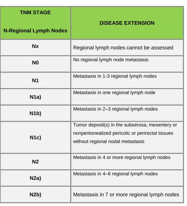
TNM STAGE
N-Regional Lymph Nodes
DISEASE EXTENSION
Nx
Regional lymph nodes cannot be assessed
N0
No regional lymph node metastasisN1
Metastasis in 1-3 regional lymph nodesN1a)
Metastasis in one regional lymph nodeN1b)
Metastasis in 2–3 regional lymph nodesN1c)
Tumor deposit(s) in the subserosa, mesentery or nonperitonealized pericolic or perirectal tissues without regional nodal metastasis
N2
Metastasis in 4 or more regional lymph nodesN2a)
Metastasis in 4–6 regional lymph nodesN2b)
Metastasis in 7 or more regional lymph nodes
Table 3. TNM classification System
34
TNM STAGE
M–Distant Metastasis
DISEASE EXTENSION
M0
No distant metastasisM1
Distant metastasisM1a)
Metastasis confined to one organ or site
(for example, liver, lung, ovary, nonregional node)
M1b)
Metastases in more than one organ/site or the
peritoneum
Table 4. TNM classification System
Data from American Joint Committee of Cancer (2009) [15].
The prognosis of patients with CRC depends on tumor invasion in the intestinal wall, the involvement of local lymph nodes and distance metastases; it is only possible to define precisely the stage of the disease in the pos-operative phase, except when there is evidence of distant metastasis [41].
2.5-CONVENTIONAL TREATMENT APPROACH
The therapeutic approach to CRC involves, in most cases surgery, chemotherapy and radiotherapy as well as new anti-angiogenic drugs. The different therapeutic options vary according to tumor staging - size, location and metastasis, as well as the physical condition of the patient.
Cancer treatment has gone through a slow process of development. It was in the 19th and early 20th centuries that major advances were made in general surgery and cancer surgery. During the final decades of the 20th century, surgeons developed greater technical expertise in minimizing the amounts of normal tissue removed during cancer operations. This progress depends not only on understanding cancer better as a disease and on better surgical instruments, but also on combining surgery with other kinds of treatments such as chemotherapy and radiation.
35
2.5.1-SURGERY
Surgery is the main treatment of CRC, which aims, regardless of the location of the tumor, the removal of the primary tumor adequate margins and conducting regional lymphadenectomy, and generally accepted that at least 12 nodes must be isolated for Correct staging tumor [46]. Approximately 92% of patients with colon cancer are subjected to surgery, as a first treatment option, mostly with curative intent [47]. For stage 0 (Tis N0 M0, T1 N0 M0), the treatment options include: a) local excision or simple polypectomy; b) segmentary resection for larger lesions not amenable to local excision. Treatment of stage I (T2 N0 M0) tumours consist in a wide surgical resection and anastomosis. For stage II (T3 N0 M0, T4 N0 M0) the standard treatment options include: a) wide surgical resection and anastomosis; b) after surgery, in high-risk patients adjuvant therapy could be considered. And for stage III (any T, N1 M0, any T, N2 M0) the treatment options are a) extensive surgical resection and anastomosis; b) after surgery the standard treatment is a doublet schedule with oxaliplatin and 5FU/folinic acid (LV) (FOLFOX4 or FLOX) [45]. Advances in surgical technique have increased the percentage of patients with potentially curable disease. These developments included the development of techniques such as block resection, total excision of the mesorecto for rectal carcinoma, laparoscopy and the technique of "no-touch" which aims at limiting the spread throughout the tumor vascular operative time. Laparoscopy is currently seen as a good alternative .The classical treatment, with the advantage that the need for shorter of hospital stay, less pos-operative pain and earlier recovery intestinal function. There is however, some controversy regarding possibility of intraperitoneal dissemination and hematogenous cells neoplastic with this surgical technique, with some studies pointing one increased risk of spreading when the laparoscopy is compared with the open surgery and other studies disagree with such fact [46].
Regarding colorectal cancer, the surgical treatment depends on the location of neoplasia. The proximal and middle rectal tumors are usually underwent low anterior resection with anastomosis primary, an ileostomy or colostomy may be necessary in the past temporary to facilitate healing, to divert the flow of fecal anastomosis. For tumors of the distal rectum amputation is indicated abdominoperitoneal with permanent colostomy and in cases where it is not possible to keep sphincter [39].
Advances in surgical techniques, has made it possible for patients with metastatic RCC, surgery may have a curative character. Patients with resectable liver metastases should undergo partial hepatectomy, thus achieving a survival at 5 years 25-30% [40].
36
2.5.2-RADIOTHERAPY
Radiotherapy is a treatment commonly used against cancer. The benefits of radiation therapy for patients with colorectal cancer include: a) killing and eliminating cancer cells and tumors; b) shrinking tumors; c) preventing cancer cells from growing and dividing. The DNA of a malignant cell is more susceptible to radiation damage than a normal cell. Radiation used before surgery to shrink a tumor can provide the best chance of successful tumor removal during surgery. Radiation administered after surgery can eliminate remaining cancer cells. Radiation therapy began with radium and with relatively low-voltage diagnostic machines. Methods and machines for delivery of radiation therapy have steadily improved. Nowadays, radiation is delivered with great precision to destroy malignant tumours and controlling damage to nearby normal tissues [15]. Radiation therapy is typically combined with chemotherapy, such as the drug, 5-FU. Adding chemotherapy to the radiation therapy improves the effectiveness of the treatment by making the tumor cells more sensitive to the radiation. This sensitivity allows the radiation to do more damage to the tumor cells. Administering the radiation and CT prior to surgery has been shown to improve the effectiveness of the surgical treatment and, also, to decrease the risk of side effects [32].
2.5.3-CHEMOTHERAPY
CT is one of the most used treatments against cancer and it’s through drugs administration. Indeed, over the years, the development and use of chemotherapy have resulted in the successful treatment of many people with cancer. The ability of CT to kill cancer cells depends on its ability to halt mitosis. Usually, chemotherapy drugs work by damaging the RNA or DNA that instructs cells on how to copy themselves in division. If cells are unable to divide, they die (apoptosis). The faster that cells divide, the more likely CT is to kill the cells, causing the tumor to shrink.
CT is called a systemic treatment because it affects the entire body.
CT drugs that affect cells only when they are dividing are called cell-cycle specific. Chemotherapy drugs that affect cells at rest are called cell-cycle nonspecific. The therapeutic modalities available for the treatment of CRC include CT and can be administered in two ways: systemic chemotherapy (intravenous or oral administration with achievement in all areas of the body) and regional chemotherapy (administration artery to a particular body area). CT may also be used at different times after surgery - adjuvant chemotherapy - with a preventive aim of recurrence and tumor prior to surgery with the aim to reduce the tumor - neoadjuvant chemotherapy [39, 45- 47].
37
In advanced cancers, CT can also be used to help reduce tumours and relieve symptoms for cancers that have spread to other organs, such as the liver.
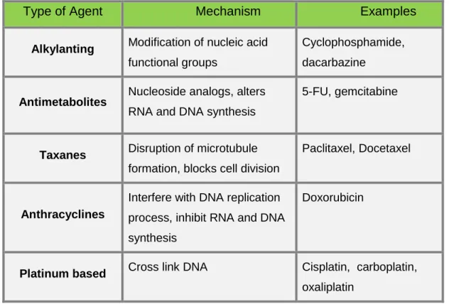
Agents and / or chemotherapy regimens more used in the treatment of CRC are: Xelox (capecitabine plus oxaliplatin), Folfox (5- fluorouracil (5-FU), leucovorin (LV), oxaliplatin) and Folfiri (5-FU, LV, irinotecan) [39 and 45- 47].
Type of Agent
Mechanism
Examples
Alkylanting Modification of nucleic acid
functional groups
Cyclophosphamide, dacarbazine
Antimetabolites Nucleoside analogs, alters
RNA and DNA synthesis
5-FU, gemcitabine
Taxanes Disruption of microtubule
formation, blocks cell division
Paclitaxel, Docetaxel
Anthracyclines
Interfere with DNA replication process, inhibit RNA and DNA synthesis
Doxorubicin
Platinum based Cross link DNA Cisplatin, carboplatin,
oxaliplatin
Table 5. Common types of chemotherapy agents and their mechanisms.
The main agent is 5- fluorouracil (5-FU), an inhibitor of Timidilato synthase [38-39]. The 5-FU is commonly administered with Leucovorin (LV), which increases the affinity of agent for the therapeutic target [39-40]. There is evidence that when compared to separate administration 5-FU, the combination 5-FU and LV increases the therapeutic response. Among patients with metastatic CRC in about 20% register to a 50% reduction in tumor size, with increased survival of 6 to 12 months [39]. The 5-FU is usually administered intravenously with the infusion better tolerated than bolus with a lower incidence of side effects such as diarrhea and neutropenia [48].
In order to improve the outcome of selected patients with metastatic CRC, monoclonal antibodies against VEGF and against epidermal growth factor receptor (EGFR) should be considered in combination with chemotherapy. Bevacizumab, an anti-VEGF
38
antibody, increases the activity of an active cytotoxic regimen, therefore, increases the survival, progression-free survival and response rate in first-line treatments.
Nowadays, several approaches are being studied to improve the activity and to reduce the side effects of chemotherapy.
The side effects of chemotherapy depend on the type and dose of drugs given and the length of time they are taken. Common side effects drugs can include:
Alopecia – although reversible and not endanger to patients’ lives, is seen as one of the most stressful secondary effects interfering with patient`s body image;
Gastrointestinal system symptoms – anorexia, nausea, vomiting, mucositis, stomatitis, diarrhoea, taste changes, smell of food aversion;
Neuropathies;
Skin colour changes;
Decreased libido;
Fatigue, tiredness.
Data from: National Cancer Institute, 2011 [74].
Radiation side effects are usually mild and reversible, and include local changes in skin, fatigue, pain related to temporarily inflammation of the nerves or muscles in radiation domain [74]. Chemotherapy because induce local and systemic immunosuppression affects adversely the training, recruitment and action of effector cells of the immune system and that can cause more side effects and precipitating a treatment failure [49].
2.6- PROGNOSIS
The stage of disease at diagnosis is a determinant factor in survival of these patients. As most recurrences occur in first four years post surgery, the survival rate at five years is a good indicator of healing [40].The analysis of survival rates at 5 years of CRC shows that when the diagnosis is made early, survival is larger, decreasing while the stage progresses. For stage I, the survival exceeds 90%, falling to 70-85% in stage II to 25-80% in stage III and less than 10% in IV [40]. On average, survival rate after the detection of metastatic disease has risen from 6 to 9 months to around 24 to 30 months [40].
Although the clinical and pathological staging be fundamental, the preoperative dosing of carcinoembryonic antigen (CEA) is also very important, since it may indicate the risk of recurrence of cancer. Unlike other tumors, primary tumor size, nodal adjusted to
39
achievement and the degree of differentiation, not directly associated with the prognosis of the patient [40].
2.7-ROLE OF NK CELLS IN CRC
NK cells are believed to play an essential role in the immune surveillance against tumors and infectious diseases.
In accordance with some investigation studies that analysed the number and activity of NK cells in patients with colon cancer, lead to the suggestion that reduced NK cell activity is associated with metastatic tumour growth in patients with colon carcinoma; and, if the decreased NK cell activity precedes the development of metastasis, it could constitute a possible a marker to identify a high risk of rapid tumour progression following curative colorectal surgery [50].
According to Halama et al [51], the early impairment of NK cells is decisive for CRC; the same is not true for other immune cells, especially for T cells which don’t have a so uniform presence during the different stages of CRC. Furthermore, they indicate a pivotal role for an escape from NK cells for CRC tumourigenesis and propose that quantification of NK cells within CRC tissue can be used as an important parameter for the detection of response therapy.
2.8-FOLLOW-UP
The main objectives of the follow-up of patients with a history of CRC are the diagnosis of metachronous polyps or new primary CRC and the detection of recurrence (liver, lung) at an early stage, in which may also be candidates for a curative treatment, like what happens with liver metastases, which may undergo surgical resection [38].
Therefore, the follow-up of these patients may improve their survival rate and includes periodic medical evaluations and conduct many additional complementary exams of diagnosis [34].
Current guidelines from the American Society of Oncology Clinical , in relation to survival rate after curative treatment of CRC, reflect a change of attitude towards the role of TC performed routinely [15].
A colonoscopy reassessment is indicated 3 years after surgical treatment, and if negative, every 5 years [52]. Flexible sigmoidoscopy is recommended every six months for 5 years for patients with rectal cancer who haven’t submitted to radiotherapy [52].
40
Although it continues to be one of the most common cancer in the last decades, deaths from CRC have been declining, in part very due to an effective screening and follow-up program as well as a more effective diagnosis.
2.9-QUALITY OF LIFE
In recent years, an increasing awareness of interpersonal and emotional repercussions of cancer and its treatments has happened, as well as its impact on the welfare of patients. Thus, the attention has been directed also to the psychosocial needs of patients, their families and health professionals and to the impact of emotional and behavioural factors at the beginning and during the course of the disease.
The diagnosis of cancer is a major source of anxiety, depression and emotional stress which affects the quality of life of patients. Approximately 20-40% of cancer patients exhibit significant levels of depression and anxiety [77].
WHO defines QOL as an individual's perception compared to their position in life, the values and cultural context and in which the individual lives and their relationship with their objectives, expectations and concerns. This is a comprehensive concept of a complex affected by physical health, psychological state of the individual mode as well as its level of independence, and how their social environment relations, as well as by personal beliefs. The concept of quality of life and so the way to deal with cancer is also unique, personal and specific to each patient.
According Psychoneuroimmunology, psychological and emotional stress induce severe alterations in several biological responses. Activation of the hypothalamus-pituitary-adrenal axis and the sympathetic nervous system appears to alter the cells of the immune system - reducing the number of NK cells and promote inflammation via multiple neuroendocrine and immune pathways.
According to an adaptation study of the satisfaction scale with social support to people with cancer illness diagnosis found that, the satisfaction with the social support relates to the all adaptation process to oncologic disease and results, specifically concerning to quality of life [53]. Based on the WHO definition of health as “a state of complete physical, mental and social welfare and not merely the absence of disease”, it becomes essential to see the patient as a whole, recognizing its needs in several aspects. The ongoing research in such an important area as cancer is undoubtedly essential. More and more is known about its causes, risk factors and development. New ways to prevent, detect and treat are also being studied always focusing on improve the quality of life of patients with cancer.
41
2.10-NOVEL APPROACH IN CANCER
A variety of modalities can be used to improve the treatment of cancer patients. A combination of those modalities of pharmacological and non pharmacological treatment of cancer is a mainstay of supportive care to those patients as presented on the current WHO guidelines.
The study and interest in learning/understanding treatment modalities called complementary or integrative medicine has grown significantly in recent years. Highlights, the importance of therapeutic relationships between doctor and patient, focus on the whole person, including all aspects of life. The central focus is the patient’s perspective, generating new strategies and multiplying treatment possibilities. According to Cassileth (2004), cancer patients usually use complementary therapies, being that about 91% of these patients used some form of complementary therapy during conventional medical treatment, in the United States [76]. Although some are expecting these therapies to have an effect on their survival time, it seems that most use these methods to improve quality of life.
One of the so called complementary therapies is the Traditional Chinese Medicine that it will discuss bellow.
42
3.
TRADITIONAL CHINESE MEDICINE – OVERVIEWTraditional Chinese Medicine (TCM) is a broad range of medicine practices sharing common concepts which have been developed in China and are based on a tradition of more 5000 years, including various forms of herbal medicine, acupuncture, massage (Tui na) exercise (Qigong) and dietary therapy.
The oldest findings known, where references to the most primary forms of care are shown, are the I Ging (The book of changes) and the Huangdi Neijing (Principles of Interne Medicine of the Yellow Emperor) [75]. It is thought that the book Huangdi Neijing was written somewhere in between the 2th century BC and the 2th century AC and establish, for the first time on the Chinese Medicine practice, some of therapeutic principles of treatment.
Many of Chinese scientific principles, since Classical China teachings and before the Yellow Emperor’s Classic of Internal Medicine, emphasized the regulatory fluctuations through circulatory functions in a simplistic manner that is similar to a sinus wave and is part of the so-called monad (Leibniz) or Taiji sign.
Over the centuries, many compilations and reassessments of the whole field of knowledge of Chinese Medicine appeared. However, it was on the 16th and 17th centuries that the preservation and dissemination of this whole field of knowledge reached its pinnacle, with publications such as Zhengjiu Dacheng (Great Compendium of Acupuncture and Moxibustion). In it was the theory behind all Chinese Medicine, such as stimulation of points and channels, which have been presented so clearly that remain current until present day.
Accordingly to Hempen, it was Soulie de Morant, at France, after 30 years of study in China that contributed, in a fundamental way, to provide the theoretical basis for the diagnosis and the introduction of Chinese Medicine in the western world [54].
Western medicine, as well as any other type of corpus medicus, presents limitations both in diagnosis as in interventions. Indeed, for a long time, Western medicine has been systematically built and developed based on scientific methodology and, as a result, on the possibility of the phenomena being measured.
Chinese Medicine emerges as another medical approach, considering the human being part of a whole, being interested primarily in the expression of life, emotions and vital body functions in order to identify possible disharmony. The basic aim is not to measure or assess a specific organic change but to evaluate the condition of a patient, reporting objective and subjective their symptoms or discomforts.
43
Currently, there is a form of modern understanding of TCM considered crucial for their integration into the Western health system and in research. The Heidelberg model fits in this context.
3.1-HEIDELBERG MODEL OF CHINESE MEDICINE
TCM is understood as “a system of sensations and clinical signs and findings designed to define the regulatory state of the body [55].
The Heidelberg Model of TCM developed by Prof. Johannes Greten was supported by the pioneering works of the medical-sinologist Prof. Manfred Porket (1974), thus adopting a Latin terminology, and also sustained by the Leibniz’ analysis of the I Ging (“The Book of Changes”).
The Heidelberg model is a scientific model that allows a rational access to Chinese medicine and it is based on symptoms that constitute the evidence of disease and pathological changes.
It is supported by a system that describes functional abnormalities through its signs and symptoms. Thus, it is understood that those signs and symptoms, as well as the feeling that the patient describes, are the result of dysfunctions in the body, particularly in the neurovegetative system.
This model of Heidelberg provides an overview of some TCM concepts by translating them as states of neurovegetative functions, being crucial to the integration of TCM in the Middle West, particularly to what regards the health system.
On the following picture it is possible to make a western physiological description of such neurovegetative functions.
44
Fig 6. Western physiological description of a vegetative sinus waveData from Greten 2010 [56].
The sinus wave symbolizes a sinus curve of regulation, a circular movement, as the classic circle of Yin/Yang, were are described the regulatory model of the phases (Wood, Fire, Metal and Water).
The upper part of the figure contains the Chinese way of description; the functional figures symbolize the clinical appearance of the mechanisms in terms of visible signs, the lower part contains the wertern description, as transmitters and neural concert of the vegetative system [56].
For instance, according to Greten (2010), stress is believed to be accompanied by a strong uprising sympathetic action, analogous to Wood [55]. At the same time, according to western medicine, vagal impulses act to counter-balance this sympathetic action and effect. In Chinese terms this is as imbalance of a vegetative functional tendency. The functional tendency, called Wood, is more or less analogous to stress and sympathetic action, adrenaline increase, muscle tension, and others. In other words, the description by groups of clinical signs in the upper part of the figure, describes the same function the western medicine describes as a transmitter and neuronal concert of vegetative system.
As a scientific medical diagnosis, besides the interpretation of symptoms, amounts to a second level of abstraction, which identifies causes and agents that can induced those changes or dysfunctions [55].

![Figure 2: The biological functions of NK cells Data from Robert D. Schereiber (2001) [8]](https://thumb-eu.123doks.com/thumbv2/123dok_br/15911872.1092771/21.892.248.731.625.1000/figure-biological-functions-nk-cells-data-robert-schereiber.webp)
![Fig 4- Colon rectal cancer overview Data from Medical Research Council [70].](https://thumb-eu.123doks.com/thumbv2/123dok_br/15911872.1092771/25.892.311.656.404.680/colon-rectal-cancer-overview-data-medical-research-council.webp)
![Table 1. Symptoms associated with CRC and respectively frequency Extrated from Camppell MS.(1992) [28]](https://thumb-eu.123doks.com/thumbv2/123dok_br/15911872.1092771/28.892.127.767.96.521/table-symptoms-associated-crc-respectively-frequency-extrated-camppell.webp)
![Fig 5 . Colonoscopy – Endoscopic views and biopsy technique Data from Johns Hopkins Medicine [32]](https://thumb-eu.123doks.com/thumbv2/123dok_br/15911872.1092771/29.892.202.772.747.1009/colonoscopy-endoscopic-views-biopsy-technique-johns-hopkins-medicine.webp)

![Fig 6. Western physiological description of a vegetative sinus wave Data from Greten 2010 [56]](https://thumb-eu.123doks.com/thumbv2/123dok_br/15911872.1092771/44.892.231.743.102.560/fig-western-physiological-description-vegetative-sinus-data-greten.webp)
![Fig 7. Four components of the functional diagnosis in TCM Data from Greten (2010) [55]](https://thumb-eu.123doks.com/thumbv2/123dok_br/15911872.1092771/46.892.185.780.114.553/fig-components-functional-diagnosis-tcm-data-greten.webp)