Ana Rita Frazão Macedo
Mestre em Microbiologia Clínica
Tuberculosis: new era for diagnosis and
surveillance using whole-genome
sequencing-based approaches
Dissertação para obtenção do Grau de Doutor em Biologia
Orientador: Doutora Isabel Portugal, Professora Auxiliar,
iMed.ULisboa - Instituto de Investigação do Medicamento
Faculdade de Farmácia, Universidade de Lisboa
Co-orientador: Doutor João Paulo dos Santos Gomes,
Investigador Auxiliar com Habilitação, Instituto Nacional de
Saúde Doutor Ricardo Jorge
Co-orientador: Doutor Jaime Mota, Professor Auxiliar,
Faculdade de Ciências e Tecnologia, Universidade NOVA de
Lisboa
Júri:
Presidente: Prof. Doutor Pedro Miguel Ribeiro Viana Baptista
Arguentes: Prof. Doutora Raquel Sá Leão
Prof. Doutora Laura Maria Brum da Cruz Martins
Vogais: João Paulo dos Santos Gomes
João Ruben Lucas Perdigão
julho, 2019
iii
Ana Rita Frazão Macedo
Mestre em Microbiologia ClínicaTuberculosis: new era for diagnosis and surveillance using
whole-genome sequencing-based approaches
Copyright © Rita Macedo
A Faculdade de Ciências e Tecnologia e a Universidade Nova de Lisboa têm o direito, perpétuo e sem limites geográficos, de arquivar e publicar esta dissertação através de exemplares impressos reproduzidos em papel ou de forma digital, ou por qualquer outro meio conhecido ou que venha a ser inventado, e de a divulgar através de repositórios científicos e de admitir a sua cópia e distribuição com objetivos educacionais ou de investigação, não comerciais, desde que seja dado crédito ao autor e editor.
As secções desta dissertação já publicadas por editores para os quais foram transferidos direitos de cópia pelos autores, encontram-se devidamente identificadas ao longo da dissertação e são reproduzidas sob permissão dos editores originais e sujeitas às restrições de cópia impostas pelos mesmos.
v
Acknowledgments
Ao Doutor João Paulo Gomes. Penso que já lhe disse tudo e não há nada que possa escrever que vá acrescentar algo ao que ele já sabe. Agradeço a amizade, o carinho, a PACIÊNCIA (!!), o incentivo e motivação, principalmente nas alturas em que desesperava e achava que não iria ser capaz. Obrigada João, sabes que não teria conseguido sem ti.
À Doutora Isabel Portugal. Que me tem acompanhado em todas estas viagens de pré e pós-graduação. O percurso tem sido longo e cheio de “caminhos desviados”, mas temo-nos sempre encontrado nas alturas certas.
Ao Doutor Jaime Mota por ter aceite o papel de meu orientador e pela simpatia e disponibilidade demonstradas ao longo deste caminho.
Aos restantes membros da Comissão de Acompanhamento de Tese (CAT), Doutora Isabel Couto, pela discussão científica e por todas as sugestões concedidas.
À Doutora Raquel Duarte que sempre me disse o que precisava de ouvir em todos os momentos. Pela amizade, pela disponibilidade e toda a contribuição e dedicação a este trabalho.
À minha equipa no laboratório, Inês João, Irene Rodrigues, Sónia Silva e Cristina Matos. Todas sabem que nunca teria conseguido sem elas; a disponibilidade que me deram ao assumir todas as tarefas extra e todos os incentivos que não me deixaram desistir. Obrigada é muito pouco. Prometo chocolates todas as semanas!
À Inês João. Minha “guru” e motivadora em todas as horas. Não sei como vou “sobreviver” sem ti.
Às minhas companheiras de almoço, e não só, que me aturaram sempre, principalmente nos meus piores momentos. Um agradecimento especial à Joana e Leonor, vocês sabem e não preciso escrevê-lo aqui. Por todos os “after works” e deixarem a vossa casa para estarem na minha.
À Andrea. Por tudo.
vi
Aos colegas da bioinformática que foram incansáveis e que tanto se cansaram em tentar satisfazer todos os meus caprichos e dúvidas (algumas ainda ficaram, mas a aluna não é das melhores...). Joana, Alexandra, Miguel e Vítor tem sido um prazer e uma oportunidade poder trabalhar convosco (e dar-vos trabalho!).
A toda a equipa da Unidade de Tecnologia e Inovação do INSA. Em especial, ao seu responsável, Doutor Luís Vieira, pelo seu empenho na implementação e otimização das metodologias de Sequenciação de Nova Geração, as quais foram essenciais para a execução deste trabalho.
Ao coordenador, Doutor Jorge Machado, e restantes responsáveis do DDI por me terem possibilitado o alcance deste objetivo.
À Faculdade de Ciências e Tecnologia da Universidade NOVA de Lisboa (FCT/UNL), em particular à Professora Isabel Sá Nogueira, Coordenadora do Programa Doutoral de Biologia.
A todos os meus amigos (alguns já aqui mencionados). Que sempre me apoiaram e me deram a força quando dela precisava.
À minha família. Mãe, Pai, Irmã, Obrigada por tudo. Embora algumas vezes com demonstração de alguma ingratidão (da minha parte, claro), só sou hoje quem sou graças a vocês. Desculpem o stress e os desabafos maldispostos durante o percurso. Prometo finalizar todas estas etapas e deixar de ser uma preocupação.
Para os meus filhos. Para saberem que mesmo nas piores circunstâncias, basta um bocadinho de força e alguém que acredite em nós. Desculpem a irritação, o cansaço e, às vezes, a falta de disponibilidade. Isto é tudo para vocês.
vii
Resumo
Desde 1993 que a OMS declarou a Tuberculose (TB) como uma emergência de saúde pública global. É, atualmente, responsável por quase 2 milhões de mortes por ano, sendo a nona principal causa de morte em todo o mundo. O principal obstáculo para o controlo efetivo da TB é a resistência aos antibacilares, havendo assim a necessidade de implementação de novas tecnologias de diagnóstico rápido que possam traduzir-se no início precoce do tratamento e bloqueio das cadeias de transmissão.
Considerando os constrangimentos para o isolamento e tempo de crescimento das estirpes de
M. tuberculosis, o principal objetivo desta dissertação consistiu em avaliar o potencial do uso de
metodologias baseadas na Sequenciação Total do Genoma (WGS) para o diagnóstico de rotina e vigilância epidemiológica. Procedeu-se à avaliação de várias plataformas bioinformáticas para previsão in silico dos perfis de resistência aos antibacilares, bem como ao desenvolvimento de “pipelines” bioinformáticas para vigilância epidemiológica. Estas abordagens revelaram uma elevada sensibilidade quando comparadas com as metodologias tradicionais, tendo sido já implementadas na rotina laboratorial do Laboratório Nacional de Referência (LNR). Adicionalmente, demonstrámos a possibilidade de usar essas mesmas metodologias diretamente em amostras clínicas, diminuindo o tempo de resposta para cinco a oito dias. Além disso, e de acordo com as novas recomendações para o tratamento da TB, iniciámos estudos para identificação de mutações associadas à resistência aos fármacos recentemente adotados, de forma a enriquecer as bases de dados e, consequentemente, a performance do diagnóstico genotípico.
Concluindo, acreditamos ter contribuído para a validação das metodologias baseadas em WGS como ferramentas para ultrapassar as dificuldades do diagnóstico e vigilância fenotípica da TB em particular, na sua capacidade de fornecer informações muito mais rápidas sobre previsão de resistência e transmissão. Por último, este trabalho esteve na base da transição tecnológica iniciada no LNR para a vigilância da tuberculose.
Palavras-chave
Tuberculose, Multirresistência, Whole-genome sequencing, Vigilância, Mutações associadas a resistência, Epidemiologia
ix
Abstract
Tuberculosis (TB) has been declared as a global public health emergency by the WHO since 1993. It still accounts for almost 2 million deaths each year, making it the ninth leading cause of death worldwide. The major obstacle for an effective TB control is antimicrobial resistance, thus, to be successful, new strategies must be addressed, for instance, the implementation of new rapid TB diagnostic technologies that could translate into early treatment initiation and blocking of transmission chains.
Considering the major constraints regarding the isolation and time of growth of M. tuberculosis strains, the main goal of this PhD dissertation was to acknowledge the potential of the use of WGS-based methodologies for routine diagnostic and epidemiological surveillance. We evaluated several software for in silico prediction of antibiotic resistance and developed bioinformatics pipelines for surveillance purposes, in particular for the identification of transmission chains. As they revealed high sensitivity, these approaches are already implemented in the routine of the Portuguese National Reference Laboratory (NRL). We also recognised the possibility to use these same approaches directly to samples collected from TB patients, lowering the time-to-results, for a complete drug resistance pattern and phylogeny analysis, for five to eight days. The validation of this methodology is ongoing and will be implemented in a near future. Additionally, and according to the new recommendations for TB treatment, we have initiated studies to identify new mutations associated with resistance to the recently adopted drugs, in order to enrich the available databases and improve the performance of the genotypic diagnostics pipelines.
This PhD dissertation highlights WGS-based methodologies as powerful tools to surpass the difficulties of phenotypic TB diagnosis and surveillance and to provide a much more rapid information regarding resistance prediction and eventual transmission chains. It also supported the technological transition performed at the NRL for TB surveillance.
Keywords
Tuberculosis, Multidrug resistance, Whole-genome sequencing, Surveillance, Resistance-associated Mutations, Epidemiology
xi
Table of contents
Acknowledgments v Resumo vii Abstract ix Table of contents xiFigure Index xiii
Table index xv
List of Abbreviations xvii
Notes of the author: thesis organization, format and outline xix
Chapter I - General Introduction 1
1. General Introduction 3
1.1 The genus Mycobacteria 3
1.1.1 Taxonomy 4
1.2 The Mycobacterium tuberculosis complex 5
1.3 History of Tuberculosis 7
1.4 Tuberculosis: pathogenesis, clinical features and diagnosis 7
1.4.1 Pathogenesis 7
1.4.2 Clinical features 9
1.4.3 Diagnosis 10
1.4.4 Drug susceptibility testing 11
1.5 Tuberculosis treatment 12
1.6 Resistance to antituberculosis drugs 13
1.7 Epidemiology of tuberculosis 17
1.8 Molecular typing of M. tuberculosis strains 18
1.9 Whole genome sequencing 18
1.9.1. Illumina Sequencing technology 18
1.9.2. Application of whole genome sequencing (WGS) to M. tuberculosis 19
1.10 Aims and general research plan 21
Chapter II - Genetic prediction of antibiotic resistance 23
2. Dissecting whole-genome sequencing-based online tools for predicting resistance in
Mycobacterium tuberculosis: can we use them for clinical decision guidance?
25
2.1 Introduction 26
2.2 Materials and Methods 28
2.2.1 Samples 28
2.2.2 Whole genome sequencing (WGS) 28
2.2.3 In silico prediction of drug-resistance using online tools 28
2.2.4 Confirmation of genotypic drug prediction 29
2.2.5 Data availability 30 2.3 Results 30 2.3.1 Genotypic/Phenotypic correlation 30 2.3.2 Analysis of discrepancies 34 2.4 Discussion 39 2.5 Conclusion 41
Chapter III - Epidemiology of multidrug resistant tuberculosis in Portugal 43
3. Trends of MDR-TB clustering in Portugal 45
Chapter IV- Development of genomic-based surveillance methodologies 49 4. Evaluation of a gene-by-gene approach for prospective whole-genome sequencing-based
surveillance of multidrug resistant Mycobacterium tuberculosis 51
4.1 Introduction 52
4.2 Material and methods 54
xii
4.2.2 Genome de novo assembly 55
4.2.3 Gene-by-gene analysis 55
4.2.4 Core-Single Nucleotide Variant (SNV)-based analysis 56
4.2.5 Data availability 57 4.3 Results 57 4.3.1 MIRU-VNTR analysis 57 4.3.2 Gene-by-gene analysis 58 4.3.3 Core-SNP-based analysis 62 4.4 Discussion 64
Chapter V - Whole-genome-sequencing of M. tuberculosis directly from clinical samples 67
5. Whole-genome-sequencing of Mycobacterium tuberculosis directly from clinical samples 69
5.1 Introduction 69
5.2 Materials and Methods 71
5.2.1 Samples 71
5.2.2 Phenotypic resistance profiles 71
5.2.3 DNA extraction 71
5.2.4 Generation of standard curves for real-time quantitative PCR (qPCR) 72
5.2.5 qPCR for quantification of MTB vs human cells 73
5.2.6 DNA capture directly from clinical samples 73
5.2.7 SureSelectXT HS target enrichment: library preparation, hybridization, and whole
genome sequencing 74
5.2.8 WGS analysis 75
5.3 Results 75
5.4 Discussion / Perspectives 80
Chapter VI - Identification of new mutations associated with decreased susceptibility to
anti-TB drugs 83
6. Identification of new mutations associated with decreased susceptibility to anti-TB drugs 85
6.1 Introduction 85
6.2 Materials and Methods 86
6.2.1. Bacterial strain and culture conditions 86
6.2.2 DNA extraction and quantification 87
6.2.3 WGS and genome assemblies 87
6.3 Preliminary results and future perspectives 88
Chapter VII - Final overview, concluding remarks and future directions 89
7. Final overview, concluding remarks and future directions 91
References
95
Supplementary material
xiii
Figure Index
Figure 1.1. Phylogenetic relationships of the different MTBC lineages 6
Figure 1.2. Phases of human tuberculosis 9
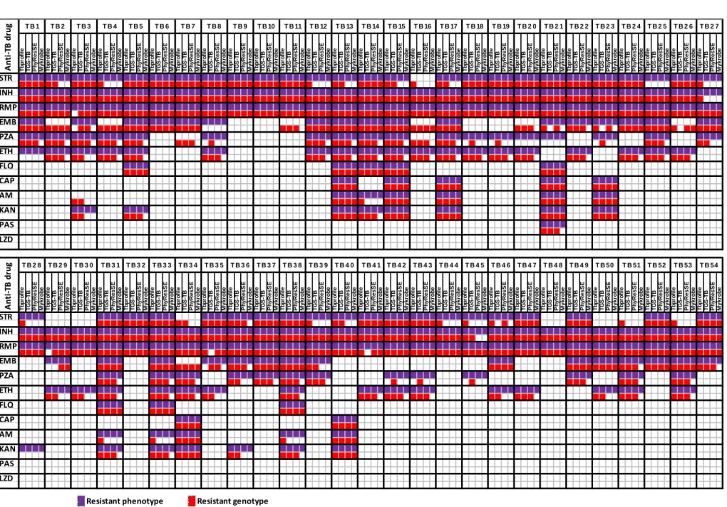
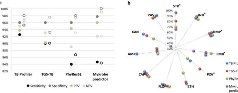
Figure 2.1. Overview of the agreement between phenotypes and genotypes predicted by
the four platforms under evaluation 31
Figure 2.2. Performance values of the bioinformatics platforms for predicting antibiotic
resistance 32
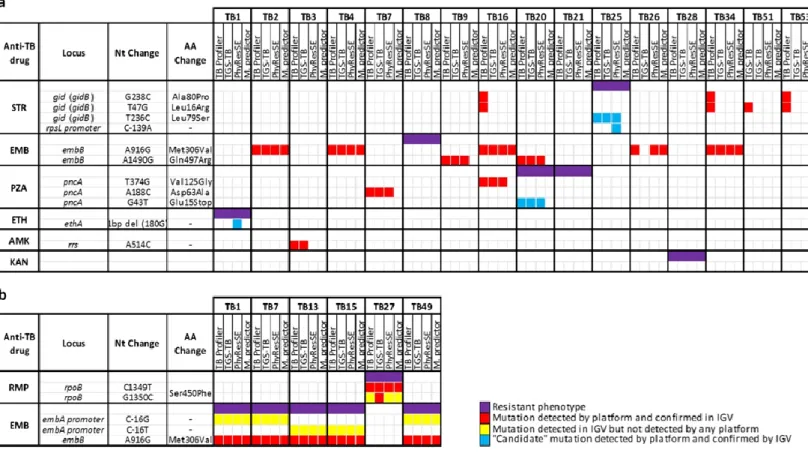
Figure 2.3. Detailed analysis of discrepancies between phenotypes and genotypes 34
Figure 2.4. Evaluation of the WGS performance for the 12 loci associated with anti-TB
resistance 36
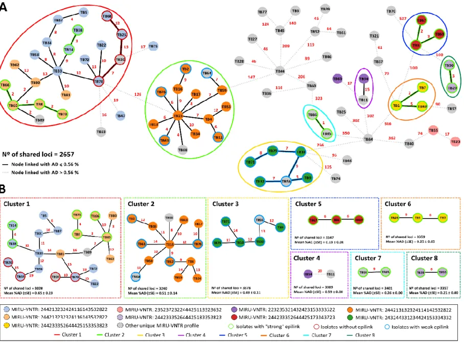
Figure 4.1. Phylogeny of 80 M/XDR-TB strains based on a dynamic gene-by-gene
approach using an extended schema (3646 loci) 59
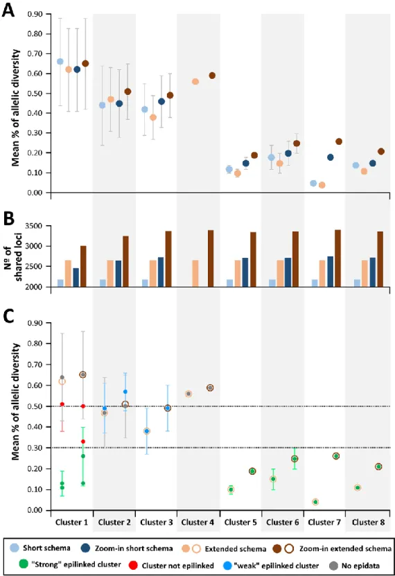
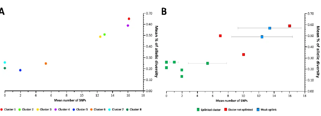
Figure 4.2. Allelic diversity with potential and confirmed clusters 60
Figure 4.3. Genetic diversity within clusters evaluated by the extended gene-by-gene and
the core-SNV approach 63
Figure 5.1. Diagnostics workflows and time-to-results 70
Figure 5.2. Schematic protocol of the M. tuberculosis DNA enrichment SureSelectXT HS
target enrichment prior to WGS 74
Figure 5.3. Results of the number of copies of human and M. tuberculosis after DNA
extraction protocol with (B) or without (A) the human-DNA depletion step 76
Figure 5.4. Percentage of reads mapping against the M. tuberculosis (Genbank
#AL123456) and human (assembly #GCA_000001405.27) reference genomes 77
Figure 5.5. Depth of coverage of the genomes sequenced directly from sputum samples 78
Figure 5.6. MST of all MTBC strains used for surveillance purposes highlighting (marked
as red dots) the phylogenetic position of the genomes that were captured directly from clinical samples.
79
Supplementary Figure 4.1. Performance of the in silico determination of MIRU-VNTR
profiles using MIRU-profiler software 127
Supplementary Figure 4.2. Phylogeny of 80 M/XDR-TB strains based on a dynamic
gene-by-gene approach using a short schema 128
xv
Table index
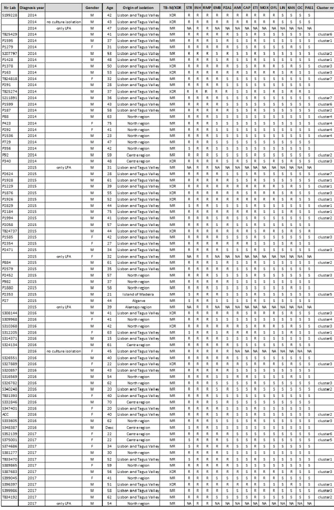
Table 3.1 Microbiological and demographic characteristics of the patients enrolled in the
study 46
Supplementary Table 4.1. Sample dataset characterization 130
xvii
List of Abbreviations
AD - allele differences AFB - acid-fast bacilli AG - arabinogalactan AMK - amikacin BDQ - bedaquiline CAP - capreomycin
CFU - colony forming units CICLO - cicloserine
CIP - ciprofloxacin DLM - delamanid
DST - drug susceptibility testing
ECDC - European Center for Disease Control and Prevention EMB - ethambutol
ETH - ethionamide FQ - fluoroquinolone GAT - gatifloxacin
GHD - General Health Directorate HIV - Human-Immunodeficiency Virus IGV - Integrative Genomics Viewer INH - isoniazid
KAN - kanamycin
LAM - lipoarabinomannan LTV - Lisbon and Tagus Valey LSP - large sequence polymorphism LVX - levofloxacin
LNZ - linezolid
MDR - multidrug resistant
MIC - Minimum Inhibitory Concentration
MIRU - Mycobacterial Interspersed Repetitive Units MTBC - M. tuberculosis complex
MST - Minimum Spanning Tree MXF - moxifloxacin
xviii
NAAT - Nucleic Acid Amplification Tests
NCCLS - National Committee for Clinical Laboratory Standards NIH - National Institute of Health
NRL - National Reference Laboratory NPV - Negative Predicted Value NTM - Non-Tuberculous Mycobacteria NTP - National TB program
OFX - ofloxacin
PAS - para-aminosalicylic acid POA - pyrazinoic acid
PPV - Positive Predicted Value PZA - pyrazinamide
Pzase - pyrazinamidase
RFLP - Restriction Fragment Length Polymorphism RIF - rifampicin
RRDR - RIF Resistance Determining Region RMP - rifampicin
SLIDs - Second-Line Injectable Drugs SNP - Single Nucleotide Polymorphism SNV - Single Nucleotide Variant SRA - Sequence Read Archive STR - streptomycin
TB - tuberculosis
VNTR - Variable Number of Tandem Repeat WGS - Whole-Genome Sequencing
WHO - World Health Organization XDR - extensively-drug resistant ZN - Ziehl-Neelsen
xix
Notes of the author: thesis organization, format and outline
This PhD dissertation is composed of seven chapters, including an Introduction, several research studies (either published or ongoing), and a final discussion. Its core is based on three manuscripts (listed below) that are presented as individual chapters and two additional chapters that include a proof of concept of methodologies to be soon implemented and submitted to a journal and an undergoing study with the major breakthroughs at the moment. The manuscripts have already been published in peer reviewed international journals, and the corresponding chapters essentially represent what was published. The chapters were organized so that they follow a rational order taking into account the objectives delineated for this PhD work. Each manuscript-based chapter is preceded by a title page describing the reference of the publication, the specific contributions of the author of the present PhD thesis, and, when applicable, the alterations that were performed regarding what is published (referred as "minor changes"). In brief, each chapter includes the following contents:
Chapter I. This chapter consists of a general introduction that intends to provide the reader with the state of the art in the subjects addressed in this doctoral dissertation around Tuberculosis, the first cause of death from an infectious disease. It includes a global overview of the major aspects of M. tuberculosis taxonomy, biology, molecular epidemiology and impact on human health, followed by insights into the genetic diversity and some already established genotype/phenotype associations. It ends with the description of the main objectives of this PhD project, which includes the specific research questions that drove the investigations carried out on behalf of each chapter.
Chapter II. Genetic prediction of antibiotic resistance
This chapter corresponds to the following published manuscript: “Rita Macedo, Alexandra Nunes, Isabel Portugal, Sílvia Duarte, Luís Vieira, João Paulo Gomes. Dissecting whole-genome sequencing-based online tools for predicting resistance in Mycobacterium tuberculosis: can we use them for clinical decision guidance? 2018. Tuberculosis. 110: 44–51”. It evaluates the use of several bioinformatics pipelines for in silico prediction of antibiotic resistance in M. tuberculosis aiming at overcoming the time-consuming laboratory procedure underlying the antibiotic susceptibility tests. The ultimate goal of this study was to implement one of the evaluated bioinformatics-based approaches in the routine practice of the National Reference Tuberculosis Laboratory at INSA.
xx
Chapter III. Epidemiology of multidrug resistant tuberculosis in Portugal
This chapter corresponds to the following manuscript: “Rita Macedo, Raquel Duarte. Trends of MDR-TB clustering in Portugal. 2019. ERJ Open Research 5: 00151-2018”. In general, this study focus on understanding the dynamics of MDR-TB emergence and transmission, to establish the rate of recent transmissions against newly developed resistant strains, to pinpoint the emergence of new cases in the population and to identify associated risk factors.
Chapter IV. Development of genomic-based surveillance methodologies
This chapter corresponds to the following manuscript: “Rita Macedo, Miguel Pinto, Vítor Borges, Alexandra Nunes, Olena Oliveira, Isabel Portugal, Raquel Duarte, João Paulo Gomes. Evaluation of a gene-by-gene approach for prospective whole-genome sequencing-based surveillance of multidrug resistant Mycobacterium tuberculosis. 2019. Tuberculosis 115: 81-88”. This study ultimately aimed to implement a whole-genome-sequencing-based approach for surveillance purposes at the National Reference Tuberculosis Laboratory at INSA, following the recommendations of the international health authorities.
Chapter V. Whole-genome-sequencing of Mycobacterium tuberculosis directly from clinical samples
This chapter corresponds to an ongoing study enrolling the PhD student and the team of the Bioinformatics Unit of the Department of Infectious Diseases at INSA. It aims at developing and optimizing a laboratory procedure to capture M. tuberculosis genomes directly from the clinical samples without the need for culture propagation of the infecting strains. If this approach turns out to be a feasible method, it could be applied upstream of the use of the bioinformatics pipeline to predict antibiotic resistance (described in Chapter II). Using these two approaches in a tandem fashion would tremendously decrease (i.e., in several weeks) the time necessary to determine the suitable antibiotic therapy.
Chapter VI. Disclosing the genetic basis of antibiotic resistance
This chapter corresponds to a long-term ongoing study enrolling the same teams as the previous study. By using in vitro selective pressure scenarios (i.e., antibiotic pressure using sub-MIC) during M. tuberculosis propagation, this study intends to identify the mutations responsible for the emergence of clones with decreased susceptibility to a specific antibiotic, and thus enrich
xxi
the available genetic databases. Ultimately, it may contribute to the accuracy improvement of the software platforms used for in silico prediction of antibiotic resistance (described in chapter II).
Chapter VII. This chapter provides a global overview of the subjects addressed throughout the chapters, highlighting the main results and conclusions achieved in this PhD dissertation. Considering that each chapter focusing a research study contains its own “Discussion”, only the most relevant results are discussed in this final chapter in order to avoid redundancy. New research questions raised with this work that can be addressed in the future follow-up of these investigations are also presented.
Considering the dissimilar layouts and in-text reference styles adopted by different journals where the manuscripts were published, all chapters were formatted in a unique style, with all references being cited by sequential numbers (in parentheses) and listed in the "References" section according to the order in which they appear in the text. In this regard, a single section of "References" is presented.
Similarly, all the supplemental material is presented at the final of this PhD thesis, enumerated accordingly with the chapter they concern to.
1
Chapter I
3
1. General Introduction
1.1 The genus Mycobacteria
Lehmann and Neumann first introduced the genus Mycobacterium to the scientific community in 1896 (1). The subsequent history of the genus has been profoundly influenced by the fact that only very few of the almost 200 currently recognized species (http://www.bacterio.net/mycobacterium.html) have been a cause of human disease, above all,
M. tuberculosis. Thus, studies of microbial physiology, structure, genetics and diagnostic tools
have mainly focused on M. tuberculosis.
Mycobacterium is the only genus in the family of the Mycobacteriaceae, as it is defined in
Bergey’s Manual of Systematic Bacteriology (2), but it is considered to be closely related to other mycolic acid-containing genera: Caseobacter, Corynebacterium, Nocardia and Rhodococcus (3). All mycobacteria are aerobic (though some species are able to grow under a reduced oxygen atmosphere), nonspore-forming, nonmotile, slightly curved or straight rods (0.2–0.6 × 1.0–10 μm). Many species form whitish or creamcolored colonies, but especially among the rapid growers, there are also many bright yellow or orange species containing carotenoid pigments (4). In some cases, the pigments are only formed in response to light (photochromogenic species), but most pigmented species also form these pigments in the dark (scotochromogenic species). The classification of Runyon separates the genus Mycobacterium into four groups (photochromogens, scotochromogens, nonphotochromogens, and rapid growers) and was introduced in the late 1950s as a systematic base for the description of mycobacteria (5). This division, based on pigmentation and growth rate, is still of use to the clinical mycobacteriologist and the separation of the genus into two major groups on the basis of the growth rate of the individual species forms the basis of the mycobacterial taxonomy. The most prominent feature of mycobacteria that is uniformly present and distinctive of the genus is the lipid-rich cell envelope (1). Indeed, it is the complex cell envelope of mycobacteria that confers these bacteria the property of ‘acidfastness’ (i.e. resistance to decolourization when stained with carbolfuchsin and decolorized with dilute hydrochloric acid). Uniformly, they do not stain well with Gram stain and should be considered gramneutral. Mycobacteria possess a cell wall polysaccharide that resembles that of gram-positive bacteria; however, the mycobacterial peptidoglycan contains lipids in place of proteins and polysaccharides (1). Furthermore, the mycobacterial envelope contains a plasma membrane that is quite similar in structure and function to the plasma membrane of other bacteria, except for the presence of lipoarabinomannan (LAM), lipomannan and phosphatidylinositol mannosides. As a whole, the cell wall component of the envelope
4
confers size, shape, protection against osmotic pressure and probably protects the plasma membrane from deleterious molecules present in the environment of the cell. In summary, the peptidoglycan confers cell shape while the next layer of the envelope, arabinogalactan esterified to the mycolic acids, provides a hydrophobic permeability barrier. Other important fatty acids are waxes, phospholipids and mycoserosic and phthienoic acids, and tuberculostearic acid (10-R-methyl-octadecanoic), a unique cell component within the Actinomycetales, including the mycobacteria (1).
In 1947, Middlebrook first described growth of tubercle bacilli in the shape of serpentine cords (‘cording’). For many years, cording was correlated with virulence and considered a distinctive feature of M. tuberculosis. However, it is now known that several mycobacterial species display cording and the correlation with virulence, if any, is unclear (1).
1.1.1 Taxonomy
The number of Mycobacterium species has increased from about 40 in 1980 (6) to over 180 in 2018 (http://www.bacterio.net/mycobacterium.html). The description of novel species is paralleled by the development of molecular methods and by the increased recognition that slow growing mycobacteria are clinically important and fast-growing mycobacteria are ecologically important. By the end of 1983, there were 52 described species, only six new species were added between 1984 and 1991, about four new species per year between 1992 and 2003 and most of the non-tuberculous mycobacteria (NTM) species were identified in recent years.
Currently the genus is broadly divided into “slow growers” and “rapid growers”. Rapid growers are those species that under optimal solid culture conditions grow visible colonies within seven days. The slow growers exceed this time, in some cases, in several additional weeks. The most notable members of the slow growers belong to the M. tuberculosis complex (MTBC), which cause tuberculosis (TB) in both humans and animals. Another slow-grower is M. ulcerans, which is the cause of the Buruli Ulcer, a neglected tropical disease with its highest incidence in sub-Saharan Africa (7). Also of note is M. avium subsp. paratuberculosis, which causes Johnes disease in cattle and has long been suspected (but not yet proven) to be a contributor to Crohn’s disease in humans (8). M. leprae causes leprosy, a disabling disease which is still endemic in isolated pockets of the world (9). All of the known rapid growing Mycobacteria are primarily environmental, with some having the ability to become opportunistic pathogens. The most
5
virulent and clinically relevant of these is M. abscessus, which can cause both wound and respiratory infections (10).
1.2 The Mycobacterium tuberculosis complex
The species M. tuberculosis belongs to the “M. tuberculosis complex” (MTBC), which is a group of closely related species that can cause TB disease in animals. Although currently defined as different species, they fall short of the minimum standard to be considered true species (i.e., more than 5% nucleotide divergence). Nevertheless, there are clear phenotypic and epidemiological differences between the members of the complex: M. tuberculosis is strictly a human pathogen; M. bovis and M. caprae can infect a wide range of animals, but of primary concern is its burden in cattle; M. africanum is mostly found in humans but it seems to be restricted geographically to West Africa (11); M. canettii is the most divergent species, differing from M. tuberculosis by at least 2% at the nucleotide level. It has unusual smooth colony morphology and a lower virulence (12). More recently, additional species of the MTBC were identified. One of those has emerged in banded mongooses (Mungos mungo) in Botswana and was named mongoose bacillus, or M. mungi sp. nov. This pathogen causes high mortality rates among banded mongooses that live in close association with humans because these animals live in human-made structures and scavenge human waste, including feces (13). Another new species is M. suricattae, isolated from free-living meerkats (Suricata suricatta) from the Kalahari Desert, South Africa, and was first reported in 2002 (14). Finally, Oryx bacilli, M. oryx, have been isolated from members of the Bovidae family, i.e., oryxes, gazelles, deer, antelope, and waterbucks, although their exact host range remains unsettled (15). However, for these novel subspecies, no cases of human disease have yet been reported to date.
The completion of the first M. tuberculosis reference genome (16) provided the opportunity to detect large sequence polymorphisms (LSPs). These LSPs were used as markers to reflect the deep evolutionary relationships between the members of the complex. Remarkably, they provided evidence that refuted the commonly proposed idea that human TB evolved from a bovine progenitor, as M. bovis was found to have diverged more recently than the other human strains (17).
Our knowledge of the MTBC was increased by sequence-based analyses of genes (18), and, more recently, by whole-genome sequencing analysis (19). This revealed the presence of seven human lineages, and one animal lineage, which includes M. bovis (Figure 1.1). M. africanum is split into two distinct lineages, West African 1 and 2. The other lineages are comprised of geographically
6
structured M. tuberculosis strains; Lineage 4, the Euro-American lineage, is the most widespread and commonly isolated (20) and Lineage 2, the East-Asian lineage, is split into Beijing and non-Beijing strains. The non-Beijing clone is of particular concern as it is typically highly drug resistant and it is supposed to have spread from East-Asia into Eastern-Europe (21,22).
In addition, these studies indicated that the MTBC had a highly clonal population structure (18) and that there was an absence of inter-genomic recombination occurring within the complex. MTBC is devoid of horizontal gene transfer, thereby exhibiting a closed genome, coupled with a low mutation rate (23). Consequently, is recognized as monomorphic bacteria, but, still, a successful pathogen that has subsisted as such since the dawn of humankind (19,24). M. canettii is an exception, as there is some evidence of recombination both within this species and with other members (12). The absence of recombination in the rest of the complex is currently unexplained, but could possibly be due to a loss of the required molecular mechanisms or a lack of opportunity due to its facultative intracellular lifestyle.
Figure 1.1. Phylogenetic relationships of the different MTBC lineages, based on SNPs of 261 mycobacterial
genomes, adapted from the work from (25) - L1 correspond to East-African-Indian (EAI) strains, L2 to Beijing strains, L3 to Dehli/Central Asian strains (Dehli/CAS), L4 to Euro-American strains (T, Haarlem, LAM, S, X), L5 to M. africanum 1 and L6 to M. africanum 2 strains. L7 strains are restricted to Ethiopia and the Horn of Africa region (26).
7
1.3 History of Tuberculosis
Tuberculosis is considered an ancient disease, and evidence for TB-like disease, confirmed by both morphological and molecular methods, has been found in skeletons dating to the Neolithic era, approximately 9,000 years ago, in the Eastern Mediterranean (27). However, some estimates place the origin of the disease much earlier – 70,000 years old – when humans first started emerging from Africa (19).
TB is thought to have killed more people than any other microbial disease throughout history (28). Its significant impact on human society is reflected by the multiple designations that were attributed to this disease throughout the centuries. Hippocrates first described it as “consumption” (or Phthisis in Greek), probably relating to the “wasting away” and weight loss experienced by the patients (29). The term “White Plague” was used during the epidemics that spread throughout Europe during the 17th and 18th centuries (30), and presumably referred to the pale complexion given by the disease. TB incidence is thought to have reached its peak in the 19th century when it is estimated that a quarter of Europeans have died from the disease (29). It is against this catastrophic scenario and prognosis that Robert Koch made his famous presentation to the Physiological society of Berlin in 1882, where he demonstrated that the tubercle was the causative agent of TB. Not only was this one of the first pathogenic bacteria to be described, but he also established the “Koch’s postulates”, which set the standard of infectious diseases’ etiology, still of relevance nowadays (28).
With the advent of antibiotics and improved public health measures, many in the western world have considered TB a disease of the past. Incidence declined gradually during the early and mid-19th century almost until the present day, although the exact reasons for this phenomenon remain unclear (28). Despite this, a third of the population is thought to be infected, and today, TB remains a disease of poverty in high and low/middle income countries, with the global burden mostly centralized in Africa, Asia and South America, where the majority of the infected people can be found (31,32).
1.4 Tuberculosis: pathogenesis, clinical features and diagnosis
1.4.1 Pathogenesis
Tuberculosis can develop through progression of recently acquired infection (primary disease), reactivation of latent infection, or exogenous reinfection (33). In immunocompetent individuals,
8
about 90% of those with TB infection never develop the disease; approximately 3-10% will develop the disease in the first 1-2 years after infection (34) and another 5% during their lifetime. The risk depends on the age of acquiring infection, being lowest in the age range of 5 to 9 years (35). Exogenous reinfection is thought to be uncommon in immunocompetent people, but life-style-related factors and chronic diseases, such as active or passive smoking (36,37), nutritional status (38) and diabetes mellitus (39) may significantly affect the risk of reactivation. In the setting of Human-Immunodeficiency Virus, HIV-1, infection, the risk of progressing rapidly to disease, once infected with M. tuberculosis, the risk of reactivation, and the risk of exogenous reinfection are all increased compared to seronegative persons (40–42).
The “life cycle of TB” starts with the inhalation of infectious droplets that reach the alveoli (Figure 1.2). They are phagocytised by the alveolar macrophages and, at this point, the immune system either manages to confine the mycobacteria, leading to a latent asymptomatic infection and the formation of granulomas, which happens for the majority of the cases, or failure can lead to an active infection (43,44). In order to control the infection, the macrophages induce production of proteolytic enzymes and cytokines that attract T lymphocytes to the site. This initial control phase can last between two to 12 weeks (45). If this is successful, a granuloma will eventually be formed, which is a nodular type lesion formed of T lymphocytes and macrophages intended to confine the mycobacteria. This primary pulmonary granuloma and associated draining lymph nodes are known as the “Ghon complex” and can be detected radiologically (46). This environment is characterised by low oxygen and pH, in which the mycobacteria are able to survive in a dormant state. The lesion can then undergo calcification and fibrosis in order to keep the infection confined. Approximately 90% of those infected with M. tuberculosis maintain the infection in this dormant state for the rest of their lives (47). Of the remaining 10%, the granuloma fails to contain the bacilli allowing them to spread to a bronchus or nearby blood vessel (45). This allows the infection to spread throughout the respiratory system where progressive lung damage occurs through the formation of cavities. In some cases it spreads to other organs such as the lymphatic system, bones and meninges and a reactivated TB can affect almost any anatomical site (48). However, only pulmonary TB is transmissible and ensures the evolutionary success and adaptation of the bacteria.
9
Figure 1.2. Phases of human tuberculosis. After inhalation of the bacteria, there is a blood-borne stage
where the immune system attempts to control the infection. In 5-10% of individuals, this will lead to active or cavitary tuberculosis, which can allow M. tuberculosis ongoing transmission through aerosols production. Adapted from (44).
1.4.2 Clinical features
Most TB cases occur as pulmonary disease with only about 17% occurring at an extrapulmonary site. However, about 70% of HIV-1 infected patients will have evidence of extrapulmonary disease or mycobacteremia and these co-infected patients are more likely to present atypically, potentially delaying TB diagnosis (1).
The classical symptoms of early and progressive active TB disease can be unspecific but the most common are fevers, night sweats, fatigue, weight loss and a chronic cough (45,49). Additionally, the disease reveals localized symptoms according to the form it takes. If untreated, the estimated fatality rate of smear positive and negative pulmonary TB is about 70% and 20%, respectively (50). Some extrapulmonary manifestations, such as milliary TB or meningitis are universally fatal in the absence of treatment (49,51). Patients with pulmonary disease, of whom, those with cavities that function as open reservoirs of large numbers of bacilli, are the most infectious and more prone to transmit the disease (52). This happens when droplets are coughed
10
up from the bronchus, aerosolised, and remain airborne for minutes to hours allowing spread to other persons. These droplet nuclei are tiny ranging from 2–5 μm in diameter and containing as few as 1–3 cells (53). The timing of the development of active TB can vary greatly from weeks to decades after infection, is most often caused by a compromised immune system, and can later become latent, and then be reactivated multiple times throughout life.
1.4.3 Diagnosis
Although TB shares many clinical and radiological features with other respiratory diseases, symptoms remain an important tool to facilitate passive case-finding (54). Radiology (X-ray) is generally used in a first evaluation for TB diagnosis. In patients with progressive primary or postprimary TB, computed tomography scanning is often performed, in addition to chest radiography. Magnetic resonance imaging may also be used to evaluate complications of thoracic disease, such as the extent of thoracic wall involvement, but is of limited value in the evaluation of patients with pulmonary TB. These chest abnormalities are merely suggestive and can be used to rule out the disease but not for confirmative diagnostic purposes (55,56). Patients suspected of having pulmonary TB should have at least two sputum specimens examined for microscopic evidence of acid-fast bacilli (AFB) (57). Fluorescence microscopy is faster to visualize and shows a higher sensitivity when compared to conventional Ziehl-Neelsen (ZN) smear microscopy (58). However, it has lower specificity than ZN smear microscopy for diagnosis, thereby suggesting a need for appropriate training, quality management, monitoring of performance and confirmation testing with ZN staining (59). In addition, specimens should be cultured in order to identify the specific mycobacterial species and for drug susceptibility testing. Mycobacteria of the MTBC are slow growers, dividing every 15 to 20 hours (60), and thus it can take over 3 weeks to see visible growth on standard culture media (61). This has significant clinical implications because therapy must be initiated before TB diagnosis is confirmed. In addition, drug susceptibility testing (DST) results are rarely available at the initiation of treatment. On the other hand, clinically significant disease can be present even in the absence of a positive culture, due to poor specimen collection, contamination and/or low bacillary count (54).
Nucleic acid amplification tests (NAAT) allow the rapid identification of MTBC (61). Several NAAT are commercially available for the laboratory-based diagnosis of TB and some of them also allow the detection of resistance to rifampicin and some other first- and second-line drugs (54). Using culture as the reference procedure, most of them reveal similar high sensitivity and specificity
11
for the detection of M. tuberculosis among sputum smear-positive patients (62,63). However, unlike culture isolation, detection by NAAT does not necessarily imply viability of the detected bacteria. Nevertheless, the WHO recommends that Xpert MTB/RIF should be used rather than conventional microscopy, culture and DST as the initial diagnostic test in adults and children suspected of having MDR-TB or HIV-associated TB (64).
1.4.4 Drug susceptibility testing
Drug susceptibility testing (DST) of M. tuberculosis should be performed on an initial isolate from all patients with TB (61). If the patient’s culture remains positive after 3 months of therapy, a new DST should be performed. Testing is done using a standard methodology such as the recommended by the National Committee for Clinical Laboratory Standards (NCCLS) (65). Drug resistance can be detected by a variety of in vitro methods that are usually contingent on demonstrating growth of the organism in the presence of a "critical" concentration of an antituberculosis drug. The two most commonly used qualitative methods are the proportion method and the BACTEC method. The agar proportion method has been proposed as the reference method for all antituberculosis drugs except pyrazinamide for which BACTEC is the reference method (65). With the proportion method, plates of drug-free agar and agar containing critical concentrations of antituberculosis drugs are inoculated with the isolate. For most drugs, resistance is determined by comparing the number of colony forming units (CFU) on drug-containing versus drug-free media, with clinically significant resistance being defined as greater than 1% growth on drug-containing media relative to drug-free media (65). Rapid broth-based methods (e.g., BACTEC, MIGIT, etc.) are recommended for initial susceptibility testing of first-line agents. The BACTEC method allows a more rapidly determination of minimal inhibitory concentrations (MICs), as the growth is facilitated by the addition of enhancing growth supplements.
The short turnover time of genotypic methods offers an attractive potential for rapid detection and characterization of drug resistance. However, genome sequencing of M. tuberculosis isolates revealed a large number of new genes, intergenic regions and nonsynonymous single nucleotide polymorphisms (SNP) showing consistent associations with drug resistance. This indicates that the genetic basis of drug resistance is more complex than previously anticipated (66) and that the available commercially and routinely used NAAT do not target all the necessary genes.
12
1.5 Tuberculosis treatment
Although TB is an ancient disease, effective drugs were not available for centuries. The pre-antibiotic therapy initially consisted of isolation of the patients in sanatoria to reduce the transmission to healthy contacts, with rest, adequate nutrition, and sunlight exposure (67–69). The first evidence of a potential anti-TB drug was made in 1940, when a dapsone-derivative compound, known as promin, was administered to a sample of infected guinea pigs. However, that compound was never subjected to human clinical trials (70–72). In 1944, Schatz and Waksman stated that streptomycin (STR), a natural substance isolated from Streptomyces
griseus, had bactericidal activity and thus could be prescribed for TB treatment. However, only
few years later, the first resistant cases arose, compromising the use of a streptomycin-based monotherapy (73). Four years later, a new synthetic drug, called para-aminosalicylic acid (PAS), was presented as an alternative drug. Following the poor results of the monotherapy, in 1952, the first regimen based on the combination of STR, PAS, and isoniazid (INH) was proposed (71,72,74).
In 1954, pyrazinamide (PZA) was discovered and ethambutol (EMB) and rifampicin (RIF) were introduced in 1961 and 1963, respectively. At this time, the duration of therapies could last two years. In 1970, trials on RIF-including regimens showed good results with a therapy of 9 months, and in 1974, the inclusion of RIF and PZA at lower dosages demonstrated the efficacy of a 6-month treatment (71,72,75,76).
The choice of the antituberculosis drugs in the different phases is not random, but it is based on the epidemiology and on the specificity of action of the drugs, which are molecules with two different mechanisms of action—bactericidal and sterilizing effect (77). The first group is crucial in the intensive phase and allows a relevant reduction of the bacterial load; the indirect consequence of this activity is the reduction of the probability of selecting drug-resistant strains. The most important drugs prescribed for that aim are INH, PZA, RIF, and STR. The sterilizing activity is performed mostly in the continuation phase because it is oriented to kill mycobacteria in a dormancy state. Antituberculosis drugs of this group are PZA and RIF (77). These general principles are accepted worldwide and the WHO defined standardized regimens (78). As such, new cases of TB in patients that were never exposed to drugs have to be treated for 6 months. The intensive phase lasts two months and should be administered a combined regimen that includes EMB, INH, PZA, and RIF. The four-months continuation phase only includes INH and RIF (77,78). Previously treated cases require a different management and a rapid and conventional DST is required before the initiation of therapy, to ensure the most appropriate regimen to choose (77).
13
It is obvious that multidrug resistant cases (MDR, i.e., in vitro resistant to at least isoniazid and rifampicin) could represent a challenge because of the poorest therapeutic options. The so-called second- and third-line antituberculosis drugs are less efficacious, more toxic, and more expensive than the first-line drugs. However, and because of the lesser effectiveness, to obtain a clinical and a microbiological cure it is mandatory to treat individuals with MDR-TB for longer periods. The WHO suggests the prescription of at least four active drugs during the intensive phase and should include PZA, one of the injectable second-line drugs (amikacin - AMK, capreomycin - CAP, or kanamycin - KAN), a new-generation fluoroquinolone (FQ), ethionamide - ETH (or prothionamide), and cycloserine (or PAS) (78,79). The duration of the first phase of the treatment should depend on the culture conversion, but it should last at least eight months, whereas the duration of the second phase should be longer than 20 months (77–79).
With the emergence of more resistant cases, first described in 2007 in patients from Africa, new therapeutic options have been proposed (80). Extensively-drug resistant (XDR) TB (defined as MDR-TB with resistance to a FQ plus a second-line injectable drug) is a considerable threat, resulting in extremely poor treatment outcomes. In a recent study of XDR-TB in South Africa, 46% of patients died after a two-year follow-up (81); the same outcome would be expected without treatment at all. As such, there is a desperately need for new or improved drugs to prevent further resistance (eventually leading to strains resistant to all drugs) (82). For this reason, several drugs approved for infectious diseases other than TB were screened and showed
in vitro and in vivo antimycobacterial activity; among them, imipenem-cilastatin, linezolid, and
meropenem-clavulanate have had a relevant role in individuals with drug-resistant TB in the last few years. The new molecules recently approved or in the last clinical trial phases are bedaquiline (a new diarylquinoline, previously called TMC 207), delamanid (previously called OPC-67683), sutezolid (PNU 100480), and PA-824 (79,83).
1.6 Resistance to antituberculosis drugs
In M. tuberculosis, drug resistance occurs through chromosomal mutations that confer resistance to individual antituberculosis drugs. These mutations occur spontaneously and at predictable rates (84). For example, mutations conferring resistance to INH and RIF occur with an estimated frequency of approximately 3 X 10⁻⁸ and 2 X 10⁻¹⁰ mutations per bacterium per generation, respectively (84). All populations of M. tuberculosis will therefore have a certain number of naturally occurring drug-resistant mutants and this probability will be influenced by
14
the size of the bacterial population and the replication rate. The probability that simultaneous resistance to INH and RIF will develop in nature is then extremely small, as it is the mathematical product of each of the separate probabilities (85). The process of development of resistance in
M. tuberculosis is basically through the selection of de novo mutations, either SNPs or indels, at loci usually termed as resistance associated genes. There are basically four mechanisms
responsible for this: i) drug target modification, as a result of non-synonymous mutations; ii) unsuccessful prodrug activation, due to mutations that prevent the prodrug of reaching its active form; iii) target overexpression, usually from mutations at the promoter region that controls the expression of the drug target; and, iv) overexpression of drug modifying enzymes, rendering the drug inactive (86).
STR interferes with protein synthesis by inhibiting genetic translation (87). Minimum Inhibitory Concentration (MIC) range between 1.0-2.0 mg/L (1.0 mg/L for M. tuberculosis H37Rv) and this drug has a moderate bactericidal activity against susceptible isolates (88). Comparing with the two other aminoglycosides used in TB treatment, KAN and AMK, STR is the least toxic (89). Resistance is usually due to mutations in rpsL and are associated with high-level resistance, in particular, the K43R mutation (90). Another mechanism of STR resistance occurs through rrs gene mutations and usually yields a lower resistance level (90,91). gidB mutations have also been detected in clinical isolates, although its role in resistance is not yet fully understood. They appear in resistant and susceptible isolates (92,93), suggesting they could be phylogeny-related (93). This is the case for the endemic MDR/XDR-TB Q1 clade in Portugal that was defined based on the A80P mutation on gidB, which is simultaneously associated with an intermediate-level resistance to STR (94,95).
INH is a synthetic prodrug that requires activation by the bacterial catalase peroxidase encoded by the katG gene and enters the cell by passive diffusion (96,97). Its efficacy is in part due to its low MIC: 0.02 mg/L for M. tuberculosis H37Rv and 0.02-0.05 mg/L in susceptible clinical isolates (88). INH has a bactericidal activity against rapidly growing mycobacteria and is bacteriostatic against slow-growers, although bactericidal activity is also observed in M. tuberculosis (98). Resistance usually develops as a consequence of katG mutations that decrease the ability of the catalase-peroxidase to convert INH to its active form. The most common mutation is a serine to threonine substitution at codon 315 (S315T), found in up to 93% of INH resistant isolates and is associated to high-level resistance (99–101). Another important mechanism of INH resistance, is the acquisition of mutations in the promoter region of the mabA(fabG1)-inhA operon (102– 104). These mutations usually lead to INH low-level resistance, and an unusual high-prevalence
15
of inhA promoter mutations (up to 91%) have been detected in Lisbon, Portugal, and in strains from M. africanum West-Africa 1 lineage (105,106).
RIF is a semi-synthetic drug derived from rifamycin (107) and binds to the β-subunit of the DNA-dependent RNA polymerase (encoded by rpoB) (108), physically blocking transcription (109). It has a bactericidal activity (110) against metabolically active bacteria, but also possesses some sterilizing activity against latent bacilli (111). RIF MIC ranges between 0.2-0.4 mg/L for susceptible clinical isolates (0.4 mg/L for M. tuberculosis H37Rv) (88) and the acquisition of resistance is usually the result of aminoacid substitutions in an 81-bp region of rpoB, named RIF resistance determining region (RRDR) (112,113). Besides aminoacid substitutions, deletions or insertions in rpoB have been reported in some studies (114). The most common substitutions occur in codons 450 (prevalence of 31.0-76.9% in RIF-resistant isolates), 445 (7.7-43.0%) and 435 (3.4-28.6%), according to M. tuberculosis RpoB numbering (92,112,113,115–119). RIF resistance level depends on the mutation, e.g., S450L and H445D result in high-level resistance whereas D435V mostly results in an intermediate-level resistance (91,120,121). It is of most importance to note that resistance to RIF rarely emerges before resistance to other drugs, especially INH, and, for this reason, is considered a marker for prediction of MDR-TB cases (122).
EMB is an antimycobacterial drug synthesized from ethylenediamine and targets the cell wall biosynthesis by inhibiting the arabinosylation of the cell wall arabinogalactan (AG) and lipoarabinomannan (LAM) (123). It has a bacteriostatic activity against metabolically active bacilli (124,125) and MIC range of 0.5-2 mg/L for susceptible isolates (0.5 mg/L for M.
tuberculosis H37Rv) (88). EMB resistance is mainly associated with mutations in embB (126) and
the most common ones occur in codon 306, usually involving the substitution of a methionine by a valine, leucine or isoleucine (103,127–129). Although strains bearing embB306 mutations have been associated with a higher level of resistance (130), the molecular basis of EMB resistance is not fully determined, as mutations at this site have been described to appear in both resistant and sensitive strains (91,103,129).
PZA is a nicotinamide synthetic prodrug that enters the cell by passive diffusion where it is converted by the bacterial pyrazinamidase (PZase)/nicotinamidase into pyrazinoic acid (POA) in an acidic pH environment (131). However, there is recent evidence showing that PZA can also act on neutral pH conditions (132). Concerning resistance in clinical isolates, the main mechanism supporting PZA resistance is the acquisition of mutations in pncA, which have been identified by numerous other authors in about 72.0-99.9% of the PZA resistant isolates studied (92,131,133,134).
16
The second-line injectable drugs (SLIDs) for TB treatment are KAN, AMK and CAP. Although KAN and AMK are, such as STR, aminoglycosides and CAP is a macrocyclic peptide, these drugs share the same action mechanism and cross-resistance between the three has been well documented (135,136). KAN, AMK and CAP have shown in vitro bactericidal activities but CAP has also a bactericidal effect against latent M. tuberculosis bacilli (137). These three drugs act through the inhibition of the genetic translation due to ribosomal binding. Mutations in the 16S rRNA-encoding rrs gene can mediate cross-resistance between CAP, KAN and AMK, being the most common mutation the A1401G, present in 49.3-88.6% of the resistant isolates (91,103,136). On the other hand, the C1402T mutation, mediates resistance to KAN, CAP, but not to AMK whereas the G1484T mutation also mediates resistance to the three drugs (KAN, AMK and CAP) (136,138). KAN resistance has also been linked with eis overexpression, which is responsible only for low-level KAN resistance (139). CAP resistance can also be mediated by tlyA mutations (140). Nevertheless, tlyA mutations in CAP resistant isolates are rare and it is more likely that CAP resistance develops because of KAN/AMK cross-resistance (140).
FQs are quinolones fluorinated at the central ring system. These are broad-spectrum antibacterial drugs that target the bacterial DNA gyrase and topoisomerase IV, therefore inhibiting DNA replication (141). FQ resistance has been reported to be increasing as a result of previous exposure to FQ prior to TB diagnosis and treatment as it is commonly used for treatment to other bacterial infections (79). Gatifloxacin (GAT) and moxifloxacin (MXF) appear to induce FQ resistant mutants at a lower rate than ciprofloxacin (CIP) and levofloxacin (LVX) (142). Furthermore GAT and MXF are more effective (lower MIC) than CIP and ofloxacin (OFX) and can be used to treat FQ low-level resistant isolates (143). Von Groll et al has also observed almost complete cross-resistance between OFX, MXF and GAT (144). The molecular basis of FQ resistance has been associated with gyrA and gyrB mutations (145). The most common mutations associated with FQ resistance occur in gyrA in codons 94 and 90, in 46.2-71.9% and 4.0-43.0% isolates, respectively (103,143,144,146). Mutations occurring in gyrB have also been described, although at a lesser frequency and some with questionable role in FQ resistance (142,146).
The molecular mechanisms responsible for the resistance to the remaining second and third-line anti-TB drugs, e.g. third-linezolide, PAS and cycloserine, are still poorer investigated and, as such, the correlation between genotypic and phenotypic DST has low sensitivity.
17
1.7 Epidemiology of tuberculosis
The WHO declared TB as a global public health emergency in 1993 and, at the present, almost three decades after, TB still accounts for almost 2 million deaths each year, making it the ninth leading cause of death worldwide (147,148).
The major obstacle for an effective TB control is, undoubtedly, the antimicrobial resistance (149). The new WHO’s End TB framework is aiming towards TB elimination by 2035 but, to be successful, this new strategy must effectively address increasingly different challenges (148). One of them regards the use and the development of new anti-TB drugs and efficient vaccines. The other refers to the implementation of TB diagnostic technologies that could translate into proper care, early treatment initiation and blocking of the transmission chains.
In 2017, 10 million people fell ill with TB, and 1.6 million died from the disease (including 0.3 million among people with HIV) (31). WHO estimates that there were 558 000 new RIF-resistant cases, of which 82% were also MDR-TB. According to this, about 500 000 new MDR-TB cases have occurred in 2017 (31), which, accordingly to the latest estimates of 2014, will likely result in the death of approximately 78 400 of these patients (about 16%). XDR-TB associated mortality and treatment success are even lower than those for MDR-TB: 28% and 30%, respectively (148). Across the WHO European region alone, 15 363 (17.7%) new MDR-TB cases were reported in 2017 (150). However, only approximately 40% of all RIF-resistant and MDR-TB cases are currently being detected in Europe, due to the lack of universal DST coverage or rapid testing, and, as such, the increase of primary MDR-TB transmission is being potentiated (150). Although it is estimated that 54 million lives were saved through TB diagnosis and treatment between 2000 and 2017, the detection rate is still very far from the WHO established target of 85%, making the deployment of adequate molecular testing an urgent matter and the knowledge of the resistance targets a pressing need (31).
In Portugal, TB incidence has been steadily decreasing in the last years, with an average of about 5% per year and the proportion of patients with MDR-TB remained stationary with 1% of the cases (151). In 2017, the report of the Portuguese National TB program (NTP) reported 1607 new cases of pulmonary TB with 12 MDR-TB cases (151).
Considering that M/XDR-TB are more difficult and expensive to treat compared to drug sensitive TB, survival rates are poorer, and knowing that primary transmission is the major cause for this epidemic, controlling these cases is the key for successful TB control programs and for achieving the targets of the “End-TB Strategy” (148).
18
1.8 Molecular typing of M. tuberculosis strains
The development of molecular techniques to differentiate strains within a species has been useful both as a public health and as a research tool. Over the last decades, several techniques have been developed taking advantage of the most variable loci in the M. tuberculosis genome. The first typing method developed was based on restriction fragment length polymorphism (RFLP) analysis using the insertion sequence element IS6110 as a probe (152). Another commonly used technique, spoligotyping, targets specific repeat sequences found in multiple copies at a single locus in the M. tuberculosis genome (the direct repeat locus) using a DNA probe (153). Variable number of tandem repeat (VNTR) typing is the most recently developed method, based on the presence and number of mycobacterial interspersed repeat loci (MIRU) and is currently recognised as the gold standard (154–156). The three methods vary in their reliability, resolution and time-on-hands, but there is no clear winner with different laboratories across the world preferring either one method or employing all of them at once.
Despite the undeniable usefulness of these genotyping techniques, it is equally undeniable that they have their limitations. By design, these loci are at the extremes of variation so are unrepresentative of the genome as a whole. As such, they can only provide us with a basic idea of relatedness, and are difficult to resolve with temporal information. Their major issue for public health applications is that they can also lack discriminatory power as isolates with identical DNA fingerprints may not always be epidemiologically linked (157).
1.9 Whole genome sequencing
1.9.1. Illumina Sequencing technology
In the last decade there have been major advances in the so called “next generation” sequencing technologies in order to allow sequencing to become more high throughput and affordable. There several technologies currently available, but the Illumina platforms currently dominate the high-throughput sequencing market, and have been used for all the sequencing analysis carried out for this thesis, so will be discussed in more detail.
19
Illumina sequencing is similar to the Sanger method in that it is based on a sequencing-by-synthesis approach, where a polymerase is used to sequencing-by-synthesise a complementary strand to the single stranded target DNA with terminator nucleotides used to halt the synthesis. However, the Illumina technology utilises reversible terminators so that the chain termination process is not permanent and synthesis can continue after each base is detected. Fluorescently tagged nucleotides are used to determine which base is being incorporated as the synthesis proceeds one base at a time. In order to achieve this, “libraries” of the target DNA need to be prepared. First, the genomic DNA is fragmented with an aim to produce lots of overlapping fragments within a specific size range (for bacterial genomes this is most often 250-500bp). Adaptors are attached to the fragments, which serve four functions: ligation to the flowcell, as primers for PCR amplification, sequencing primer-binding sites and as index tags to allow multiplexing of multiple libraries in a single run. After adaptor ligation, a PCR step is then typically used to enrich for DNA fragments with the adaptors in the correct orientation. The DNA is then denatured to produce single strands, which are then ligated to a flowcell, where each fragment is amplified to form clusters of clonal DNA, which will increase the intensity of the fluorescent signal. The sequencing reaction is carried out with modified versions of the four nucleotides (dATP, dGTP, dCTP, dTTP) with a different fluorescent dye and blocking group. When a base is incorporated as complementary to the template strand, the fluorescent dye is photographed and then removed. This allows the sequence of the millions of DNA fragments to be determined at once, one base at a time (158).
1.9.2. Application of whole genome sequencing (WGS) to M. tuberculosis
It took 13 years and 3.8 billion dollars (159) for the completion of the Human Genome Project in 2003 (160). Since then, advances in sequencing technology have made it possible to sequence an entire human genome in a few days and costing a few thousand dollars. As impressive as this is, bacterial genomes are megabases long as opposed to gigabases and WGS is starting to become a clinical reality for infection diagnosis. First, its higher resolution compared with the traditional genotyping technologies allows academic researchers to better understand bacterial population structure, mutation rates and evolutionary processes. Using genome wide SNP as the basis for these kinds of studies means we can start to understand temporal parameters, as these units of variation are likely to be more clock-like associated than those studied using traditional genotyping techniques. The second major advantage of WGS is that it can provide information on variants other than SNP. Both mapping and de novo assembly approaches can