4(35): 5604-5611, 2014 SCIENCEDOMAIN international www.sciencedomain.org
Comparative Study of the Prevalence of
Occlusal Anomalies in Down Syndrome
Children and Their Siblings
Viviana Macho
1*, David Andrade
1, Cristina Areias
1, Ana Coelho
1and Paulo Melo
11
Faculty of Dentistry, University of Oporto, Portugal.
Authors’ contributions
This work was carried out in collaboration between all authors. Author VM designed the study, wrote the protocol, and wrote the first draft of the manuscript. Author AC performed the statistical analysis. Authors CA and AC managed the literature searches. Authors DA and PM managed the analyses of the study. All authors read and approved the final manuscript.
Received 13th July 2014 Accepted 27th July 2014 Published 12th August 2014
ABSTRACT
Aims: The aim of this study was to characterize occlusal anomalies in a Portuguese population with Down Syndrome (DS) and to compare its distribution with that of their siblings.
Study Design: A sibling-matched, population-based and cross-sectional survey was performed.
Methodology: This study involved 132 children with DS and 84 of their siblings, aged 2 to 26 years. Data were gathered through the use of a complete questionnaire and clinical observation. Data analysis was performed by using SPSS® v.20.0 and any p-value <.05 was considered significant.
Results: Seventy six percent of the DS children and 72% of their siblings showed class I of Angle. There were no statistically significant differences between both groups regarding class I (P=.60). Only 4% of the DS children presented class II versus 22% of the siblings (P=.001). Twenty percent of the DS children and 6% of the siblings presented class III (P=.008). The DS group presented occlusal anomalies more
anterior crossbite (P=.001). Deep overbite was only found in the siblings group (P=.001). Conclusions: The results of this study suggest that children with Down syndrome have more occlusal anomalies than general population.
Keywords: Down syndrome; malocclusion; occlusal anomalies; Trisomy 21.
1. INTRODUCTION
Down syndrome (DS) is characterized by generalized hypotonia, neurological changes, great risk of respiratory problems and infections, dental anomalies and orofacial dysmorphology [1-5].
Underdevelopment of the midface bones is common, causing a shortened palate in the anteroposterior dimension [6-8].
The most frequent occlusal anomalies stem from variations in vertical and transversal
occlusions (anterior open bite, posterior crossbite and reductions in the maxillary arch) [9-11]. These anomalies cause problems related to oral functioning (chewing, swallowing
and speaking) [12-14].
Children with DS are known to have a high prevalence of occlusal anomalies, with serious implications in their growth and this should be one of the main concerns within the need for treatment of these patients.
The objective of this study was to characterize occlusal anomalies in a Portuguese population with DS and to compare its distribution with that of their siblings.
2. METHODOLOGY
A sibling-matched, population-based, cross-sectional survey design was done at the Department of Dentistry of Oporto and Lisbon’s Universities (Portugal). The ethical committee of Oporto University approved this study. Consent was obtained according to the Declaration of Helsinki of 2002.
The recruitment of children with Down syndrome was done by mailing detailed study information to the main organizations working with this population in Portugal. Two centers were set up, one in the south, Lisbon, and the other one in the north, Oporto. Inclusion criteria implemented for the DS group were: (1) cytogenetic diagnosis of trisomy 21, (2) adequate cooperation, and (3) signed informed consent by legal guardians. Study inclusion criteria for the control group were: (1) be sibling of children with DS, (2) adequate cooperation, and (3) signed informed consent by legal guardians. The control groups were sibling-matched based on closeness in age. DS children without siblings were also included. The population included 138 children with Down syndrome and 86 of their siblings, aged between 2 and 26 years. Children were compared in different subgroups: [2,6], [6,12] and [13,26]. Since 8 children (6 with DS and 2 siblings) of the group [2,6] didn’t allow a correct evaluation of their oral cavity they were excluded.
Data were gathered through the use of a complete questionnaire and clinical observation. Clinical examination of every participant was carried out by a certified dentist (one examiner) aided by two dental assistants. In order to perform dental observation the dentist used an intra-mirror, a probe, compressed air and a millimetric rule. No radiographic examination was undertaken.
Clinical examination recorded unilateral or bilateral posterior crossbite, anterior crossbite, anterior open bite and deep overbite for all patients. Molar relationship was recorded for patients with erupted permanent first molars using Angle’s classification of Class I, Class II or Class III [15]. The World Health Organization criteria were used to assess malocclusions [16].
Variables were analyzed by groups (DS versus siblings) and by age category within groups. The age categories are proxies for primary dentition, mixed dentition and permanent dentition.
To conduct a descriptive analysis of the sample appropriate summary statistics were used. Chi-square and Fisher Exact Tests were applied and a p-value of .05 was considered to be statistically significant. The analysis was performed using the statistical analysis program SPSS® (Statistical Package for Social Sciences), version 20.0.
3. RESULTS
The sample included 216 children (132 with DS, 84 siblings) aging between 2 and 26 years (Table 1).
Table 1. Sample characteristics
Total (n=216) DS (n=132) Siblings (n=84) P n (%) n (%) n (%) Gender Male 118 (55) 70 (53) 48 (57) 0,55* Female 98 (45) 62 (47) 36 (43) Age (recoded) [2,6] 37 (17) 25 (19) 12 (14) 0,44* [6,12] 97 (45) 61 (46) 36 (43) [13,26] 82 (38) 46 (35) 36 (43)
DS–Down syndrome, S–Siblings; *Independent chi-square test
Table 2. Evaluation of angle’s class in down syndrome children and their siblings
Class Total(n=149) DS(n=78) Siblings(n=71) P
n (%) n (%) n (%)
Class I 110 (74) 59 (76) 51 (72) .60*
Class II 19 (13) 3 (4) 16 (22) .001*
Class III 20 (13) 16 (20) 4 (6) .008*
DS–Down syndrome, S–Siblings; *Independent chi-square test
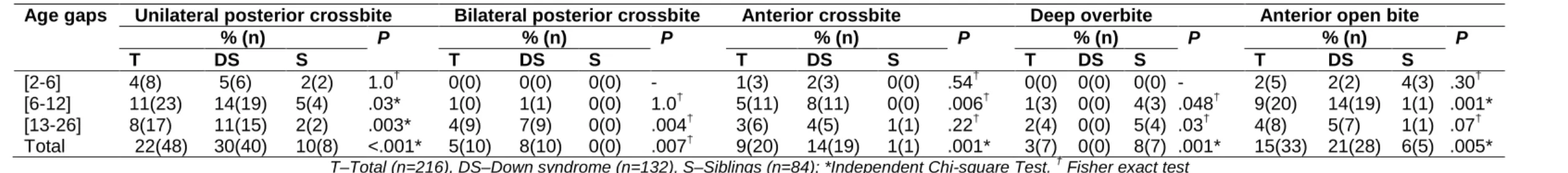
Table 3. Evaluation of the presence of occlusal anomalies
Age gaps Unilateral posterior crossbite Bilateral posterior crossbite Anterior crossbite Deep overbite Anterior open bite
% (n) P % (n) P % (n) P % (n) P % (n) P T DS S T DS S T DS S T DS S T DS S [2-6] 4(8) 5(6) 2(2) 1.0† 0(0) 0(0) 0(0) - 1(3) 2(3) 0(0) .54† 0(0) 0(0) 0(0) - 2(5) 2(2) 4(3) .30† [6-12] 11(23) 14(19) 5(4) .03* 1(0) 1(1) 0(0) 1.0† 5(11) 8(11) 0(0) .006† 1(3) 0(0) 4(3) .048† 9(20) 14(19) 1(1) .001* [13-26] 8(17) 11(15) 2(2) .003* 4(9) 7(9) 0(0) .004† 3(6) 4(5) 1(1) .22† 2(4) 0(0) 5(4) .03† 4(8) 5(7) 1(1) .07† Total 22(48) 30(40) 10(8) <.001* 5(10) 8(10) 0(0) .007† 9(20) 14(19) 1(1) .001* 3(7) 0(0) 8(7) .001* 15(33) 21(28) 6(5) .005*
Fifty five percent of the sample was male, with no significant statistical differences in the gender element in both groups (P=.55).
As the permanent first molar erupts normally at the age of six, the sample considered for the evaluation of Angle’s class was of 149 individuals (only children who already presented the permanent first molar erupted were included) (Table 2 above).
Seventy six percent of the DS individuals and 72% of their siblings showed class I and no statistically significant differences between both groups were found (P=.60). Only 4% of the DS children presented class II versus 22% of the siblings (P=.001). Twenty percent of the DS children and 6% of the siblings presented class III (P=.008).
During children’s clinical examination the presence of occlusal anomalies was also observed (Table 3 above).
Posterior crossbite was found to be more frequent in DS children, in both unilateral (P<.001) and bilateral (P=.007) cases. Fourteen percent of the DS group showed anterior crossbite versus 1% of the siblings (P=.001). Anterior open bite was also more frequent in DS children: 21% against 6% of the siblings group (P=.005).
Only 8% of the siblings showed deep overbite and none of the DS children presented it (P=.001).
4. DISCUSSION
The sample used in this study presented a good distribution with respect to age and sex parameters.
Oredugba [17] found 51% of class I and 47% of class III among 43 individuals with DS and only 5% of class III in the control group (P<.05). In this study most of the cases were class I in both groups. In respect to class II, differences were found between both groups, with a higher prevalence of it in the siblings. When comparing class III prevalence, the DS group showed a higher value (P=.008). These results are similar to Soares et al. [18], who have concluded that class III is more frequent in DS individuals. This is a consequence of the midface hypoplasia [17]. Anatomically, the facial mid-third is underdeveloped but the mandible follows normal development (pseudo-progeny). This midface dysplasia also contributes to the narrow maxilla [19].
A 38% prevalence of posterior crossbite (unilateral and bilateral) was found in the DS group: 30% presented it unilaterally and 8% bilaterally, the last being found only in the DS group, with a highly significant difference when compared to their siblings, supporting the data saying that maxilla hypoplasia is usually found in these patients. Oliveira et al. [9] found a 31% prevalence of posterior crossbite and Soares et al. [18] identified posterior crossbite in 39% of their sample. These results are similar to the ones found in this study.
Though in this study only 14% of the DS children were diagnosed with anterior crossbite, Oliveira et al. [9] found a 33% prevalence of it and Soares et al. [18] found 26%. However, when comparing the DS group with their siblings a significant difference between them was found. Anterior crossbite is primarily attributed to the anteroposterior deficiency of the
arch towards the front of the maxilla. Anterior crossbite is not due to the increased growing of the mandible but due to the maxilla underdevelopment (pseudo-progeny) [1].
An anterior open bite prevalence of 21% was found in the DS group. These results are similar to the ones found by Oliveira et al. [9], 21%, and Carlstedt et al. [20], 20%. Oral and facial musculature, in particular tongue and lips, are hypotonic. Tongue gives the impression of being abnormally large on account of muscle weakness and of an anterior and low position in the mouth (relative macroglossia) [18]. Tongue thrust and posture might hamper eruption enough to cause anterior open bite and to influence the shape of the dental arch and the position of the teeth [9].
Deep overbite was not found in DS children, despite of an 8% prevalence of deep overbite in the siblings group. Oliveira et al. [9] also found a low prevalence of deep overbite (5%) in DS children. Normally these children have a high palate, hypertrophy of the tonsils, hypotonia and nasal obstruction, which lead to mouth breathing and therefore tend to exhibit an anterior open bite instead of a deep overbite.
In this study the prevalence of occlusal anomalies found in mixed dentition [6,12] was higher in the DS group than in their siblings. To improve the oral health of people with DS, health programs must incorporate intervention methods to prevent and treat malocclusions as early as possible. Therefore, there’s a need to do a complete radiographic examination to identify hipodontia and other anomalies; occupational therapy to strengthen orofacial musculature; early mixed dentition orthodontic examination to screen for habits; and airway assessment, including consideration of tonsillectomy, palate expansion and tongue crib appliances [19,21]. Patients with DS should not be excluded from the general population with regard to dental care. From the ethical perspective, dentists must accept the responsibility and commitment to contribute with their knowledge to improve the quality of life of these people [9]. Though children with DS can be excellent orthodontic patients, orthodontic prognosis may be poor because of their learning disability, parafunctional habits and severe periodontal disease [17]. This investigation reinforces the need for further studies on this issue, including studies of the functional and psychosocial impacts of malocclusions in DS patients.
Limitations of this study include the absence of panoramic and lateral cephalometric radiographs, lack of a habit history and absence of information about sleep symptoms. Even though the study included children from all over the country, the majority of them belonged to the surrounding areas of the two observation locations, Oporto and Lisbon. As such, this might also be considered a limitation to the present study, since different observation locations would have allowed a wider representative group.
5. CONCLUSION
Children with Down syndrome present posterior crossbite, anterior crossbite and anterior open bite more frequently than their siblings.
Children with Down syndrome have great risk of developing occlusal anomalies, with serious implications in their growth and this should be a concern when treating these patients.
CONSENT
All authors declare that written informed consent was obtained from the patient (or other approved parties) for publication of this research paper.
ETHICAL APPROVAL
All authors hereby declare that all experiments have been examined and approved by the appropriate ethics committee and have therefore been performed in accordance with the ethical standards laid down in the 1964 Declaration of Helsinki.
COMPETING INTERESTS
Authors have declared that no competing interests exist.
REFERENCES
1. Desai SS. Down syndrome: A review of the literature. Oral Surg Oral Med Oral Pathol Oral Radiol Endo. 1997;84:279-85.
2. Abanto J, Ciamponi AL, Francischini E, Murakami C, Rezende NP, Gallottini M. Medical problems and oral care of patients with Down syndrome: A literature review. Spec Care Dentist. 2011;31:197-203.
3. Hennequin M, Faulks D, Veyrune JL, Bourdiol P. Significance of oral health in persons with Down syndrome: A literature review. Dev Med Child Neurol. 1999;41:275-83. 4. Macho V, Seabra M, Pinto A, Soares D, Andrade D. Oral and craniofacial
characteristics in Trisomy 21 ActaPediatr Port. 2008;39:190-4.
5. Mouradian WE. The face of a child: Children's oral health and dental education. J Dent Educ. 2001;65:821-31.
6. Bauer D, Evans CA, Begole EA, Salzmann L. Severity of occlusal disharmonies in down syndrome. Int J Dent. 2012;2012:872367. doi: 10.1155/2012/872367. Epub 2012.
7. Shore S, Lightfood T, Ansell P. Oral disease in children with Down syndrome: Causes and prevention. Community Pract. 2010;83:18-21.
8. Korbmacher H, Limbrock J, Kahl-Nieke B. Orofacial development in children with Down's syndrome 12 years after early intervention with a stimulating plate. J OrofacOrthop. 2004;65:60-73.
9. Oliveira AC, Paiva SM, Campos MR, Czeresnia D. Factors associated with malocclusions in children and adolescents with Down syndrome. Am J Orthod Dentofacial Orthop. 2008;133:489.e1-8.
10. Alio J, Lorenzo J, Iglesias MC, Manso FJ, Ramirez EM. Longitudinal maxillary growth in Down syndrome patients. Angle Orthod. 2011;81:253-9.
11. Moraes ME, Moraes LC, Dotto GN, Dotto PP, Santos LR. Dental anomalies in patients with Down syndrome. Braz Dent J. 2007;18:346-50.
12. Winter K, Baccaglini L, Tomar S. A review of malocclusion among individuals with mental and physical disabilities. Spec Care Dentist. 2008;28:19-26.
13. Oliveira A, Pordeus I, Torres C, Martins M, Paiva S. Feeding and nonnutritive sucking habits and prevalence of open bite and crossbite in children/adolescents with Down syndrome. Angle Orthod. 2010;80:748-53.
14. Azuma S, Kohzuki M, Saeki S, Tajima M, Igarashi K, Sugawara J. Beneficial effects of orthodontic treatment on quality of life in patients with malocclusion. Tohoku J Exp Med. 2008;214:39-50.
15. Guideline on management of the developing dentition and occlusion in pediatric dentistry. Pediatr Dent. 2008-2009;30:184-95.
16. World Health Organization. Oral health surveys: Basic methods. Geneva; 1997. 17. Oredugba FA. Oral health condition and treatment needs of a group of Nigerian
individuals with Down syndrome. Downs Syndr Res Pract. 2007;12:72-6.
18. Soares, KA, Mendes RF, Prado Júnior RR, Rosa LC, Costa KC. Prevalence of malocclusion in patients with Down syndrome in the city of Teresina-PI. RGO. 2007;57:187-91.
19. Faulks D, Collado V, Mazille MN, Veyrune JL, Hennequin M. Masticatory dysfunction in persons with Down's syndrome. Part 1: Aetiology and incidence. J Oral Rehabil. 2008;35:854-62.
20. Carlstedt K, Henningsson G, Dahllöf G. A four-year longitudinal study of palatal plate therapy in children with Down syndrome: effects on oral motor function, articulation and communication preferences. Acta Odontol Scand. 2003;61:39-46.
21. Backman B, Grever-Sjolander AC, Holm AK, Johansson I. Children with down Syndrome: Oral development and morphology after use of palatal plates between 6 and 18 months of age. Int J Paediatr Dent. 2003;13:327-35.
© 2014 Macho et al.; This is an Open Access article distributed under the terms of the Creative Commons Attribution License (http://creativecommons.org/licenses/by/3.0), which permits unrestricted use, distribution, and reproduction in any medium, provided the original work is properly cited.
Peer-review history:
The peer review history for this paper can be accessed here: