Universidade de Lisboa
Faculdade de Farmácia
Pancreatic cancer. New screening
biomarkers and detection methods
Mariana Amaral Colaço de Andrade Gaspar
Mestrado Integrado em Ciências Farmacêuticas
Universidade de Lisboa
Faculdade de Farmácia
Pancreatic cancer. New screening
biomarkers and detection methods
Mariana Amaral Colaço de Andrade Gaspar
Monografia de Mestrado Integrado em Ciências Farmacêuticas
apresentada à Universidade de Lisboa através da Faculdade de
Farmácia
Orientadora: Professora Doutora Ana Cristina Ferreira da
Conceição Ribeiro, Professora auxiliar FFUL
Resumo
O cancro do pâncreas posiciona-se em quarto lugar como o de maior letalidade, com mais de 90% dos doentes diagnosticados nos estadios III e IV. A taxa de sobrevivência de cinco anos é de apenas 8,5%, o que pode ser explicado pela ausência de sintomas específicos e pela falta de uma estratégia de diagnóstico padronizada, de sensibilidade e precocidade acrescida. Pelo facto de o tumor primário necessitar de pelo menos cinco anos para obter capacidade metastática, o diagnóstico early-stage (como forma de melhorar a qualidade de vida e sobrevivência dos pacientes com cancro pancreático) é possível. Urge assim, uma atualização baseada na especificidade e sensibilidade, na atualização de biomarcadores, facilmente implementados na estratégia laboratorial e, economicamente concretizável.
4
Abstract
Pancreatic cancer ranks fourth as the most lethal with more than 90% of patients being diagnosed in stages III and IV. The five-year survival rate is only 8.5%, which can be explained by the absence of specific symptoms and by the lack of a standardized guideline for an early-stage diagnostic strategy with high sensitivity. Due to the fact that the primary tumor requires at least five years to obtain metastatic capacity, early-stage diagnosis (as a way to improve the life and survival chance of pancreatic cancer patients) is possible. However, there is an urge for an update in specificity and sensitivity and an update in biomarkers easily implemented in laboratory strategy and economically feasible.
Agradecimentos
A realização desta tese (e todo o mestrado integrado) não teria sido possível sem o apoio incondicional da minha família, orientadora e amigos.
À professora Doutora Ana Cristina Ribeiro, agradeço por toda a sua disponibilidade, dedicação, tempo, energia e por tudo o que me ensinou. Obrigada por todo o seu apoio e orientação durante estes meses. Não teria sido possível sem a professora. À minha família, obrigada pela paciência, pelo apoio e pela compreensão, por me terem proporcionado esta possibilidade, por saber que posso sempre contar convosco.
6
Table of Contents
1 INTRODUCTION ... 1 1.1 Pancreatic Cancer ... 2 1.2 Epidemiology ... 5 1.3 Etiology ... 81.3.1 Non-modifiable risk factors ... 8
1.3.2 Modifiable risk factors ... 10
1.4 Current Diagnosis Methods ... 13
1.4.1 Multidetector computed tomography (MDCT) ... 14
1.4.2 Magnetic Resonance Imaging (MRI) ... 14
1.4.3 Endoscopic Ultrasonography with fine needle aspiration (EUS) ... 15
1.4.4 Positron Emission tomography (PET) ... 15
1.4.5 Biopsy ... 15 1.4.6 Blood test ... 15 1.5 Therapeutics ... 16 1.5.1 Surgery ... 16 1.5.2 Chemotherapy ... 16 1.5.3 Radiation Therapy ... 16 2 PANCREATIC CARCINOGENESIS ... 16 2.1 Pathology ... 16 2.2 Genetics ... 17
3 CURRENT STATE OF THE ART IN DIAGNOSTICS ... 18
3.1 Novelties in Pancreatic Biomarkers ... 18
3.1.1 Traditional tumor biomarkers ... 19
3.1.1.1 Carbohydrate Antigen 19-9 (CA19-9) ... 19
3.1.1.2 Other conventional biomarkers ... 23
3.1.2 Metabolites ... 23
3.1.3.1 Somatic mutations ... 25
3.1.3.2 Epigenetic modulations ... 26
3.1.4 Autoantibodies ... 28
3.1.5 Cell-free noncoding RNA (ncRNA) ... 29
3.1.5.1 microRNA (miRNA) ... 30
3.1.5.2 Long non-coding RNA (lncRNA) ... 32
3.1.6 Inflammatory factors and growth factors ... 34
3.1.7 Circulating tumor cells (CTCs) ... 36
3.1.8 Exosomes ... 36
3.1.8.1 Proteins ... 37
3.1.8.2 DNA with somatic mutations ... 37
3.1.8.3 miRNAs ... 38
3.1.9 Soluble stroma-related Biomarkers ... 38
3.2 Glycosylation in Cancer ... 40
3.2.1 Glycomic aberration vs. biomarkers ... 41
3.3 Lectins as a tool ... 47
3.3.1 Biomedical Application ... 48
3.3.2 Immobilized-Lectin Affinity Chromatography ... 48
3.3.3 Enzyme-linked Lectin Assay ... 49
3.3.4 Lectin Histochemistry ... 50
3.3.5 Lectin Blotting ... 51
3.3.6 Lectin Array ... 52
4 CONCLUSIONS ... 53
5 CHALLENGES AND FUTURE DIRECTIONS ... 56
BIBLIOGRAPHY ... 57
ANNEXES ... 96
1 Introduction
Cancer is one of the biggest public health problems worldwide, as it is the major cause of mortality and morbidity (Mordente et al., 2015). The lifetime risk of developing cancer is of almost 40% (National Cancer Institute, 2018).
Cancer is a term defining a wide variety of pathologies that can occur in almost any part of the body. Its common properties include local invasion and distant spread. It develops when there is an accumulation of genetic damage in cells over time, due to the failing of the normal corrective processes (figure 1). The cells acquire abnormal qualities, namely rapid cell division, immortality and invasion of surrounding tissue (World Cancer Research Fund/American Institute for Cancer Research, 2018).
Figure 1. Hallmarks of cancer
(adapted from Hanahan et al., 2011, with permission from Elsevier)
There are many tumor causing factors. Its composition, evolution, diagnosis and treatment also vary according to its subtype (La Thangue et al. 2011).
2
very promising, non-invasive and quick way for early diagnosis, staging and monitoring disease progression and therapeutic response.
1.1 Pancreatic Cancer
The pancreas is a gland localized behind the stomach, containing two different parts: exocrine and endocrine. While the exocrine pancreas is responsible for the production of digestive enzymes, the endocrine pancreas produces hormones, like insulin and glucagon, being therefore included in the metabolism of glucose (World Cancer Research Fund, 2018).
Pancreatic cancer is not a single disease but a large number of neoplasms being divided into the two main pancreas functions: exocrine and endocrine.
The majority of the pancreatic cancers develop in the exocrine pancreas (about 95%) with most of them being localized in the pancreas head (World Cancer Report, 2014), namely adenocarcinoma (Vincent et al., 2011, Ferlay et al., 2012, Hidalgo et al., 2013). The most aggressive pancreatic neoplasm is the ductal adenocarcinoma (Hruban et al., 2007). Nevertheless, the early-detection of ductal adenocarcinoma improves the overall survival (Matsuno et al., 2004). Concerning the endocrine tumors, they make up for less than 5% of cases (Vincent et al., 2011, Ferlay et al., 2012, Hidalgo et al., 2013) and have a better five-year survival rate (65%) (Hruban et al., 2007). The most common benign pancreatic neoplasm is the serous cystadenoma (Tseng et al., 2005). Almost all patients with this type of tumor are cured (World Cancer Report, 2014). Pancreatic cancer is the fourth cancer with more related deaths (Siegel et. al 2018). Because of the diagnosis difficulties, it is normally not diagnosed in time, having an extremely high mortality and poor prognosis (Bunger et al., 2011). According to Brierly et al. (2017), more than 90% of patients are diagnosed only at stages III and IV. According to the National Cancer Institute (2018), the number of deaths caused by pancreatic cancer has recently outweighed the ones caused by breast cancer (table 1).
Table 1. Comparison of breast and pancreatic cancer
(adapted from National Cancer Institute, 2018)
Due to the fact that there is a lack of standardized international guideline (Zhang et al., 2018a) regarding its diagnostic strategy, even for high-risk populations (Bunger et al., 2011), nor specific symptoms (World Cancer Research Fund, 2018), pancreatic cancer is normally diagnosed in metastatic stage (Stathis et al., 2010). Metastasis is the most prevailing motive of death in cancer patients (Leary et al., 2010). It consists in the dissemination and growth of the neoplastic cells away from the primary tumor (Yachida et al., 2010).
After diagnosis, very few individuals survive five years, namely 8,5% (National Cancer Institute, 2018). These poor rates have not been improving in the last 40 years (figure 2) (Zhang et al., 2018a).
Figure 2. Five-year survival rate
(adapted from National Cancer Institute, 2018)
Almost 80% of the diagnosed patients have spread cancer, either in regional or metastatic phase (Costello et al., 2012), having no opportunity to go through a curative treatment, as the surgical resection (which is the only curative option) is only possible in early stages (Yachida et al., 2010). According to the National Cancer Institute (2018), only 9,7% of diagnosed patients with pancreatic cancer are at local stage (figure 3). This majority of patients do not have symptoms or radiologically visible manifestations either as the disease progresses to the metastatic phase (Ghaneh et al., 2007, Hidalgo et al., 2010) making the early detection, with the current available methods, still very difficult. Nevertheless, the survival rates improve, if the pancreatic cancer is diagnosed,
4
and treated, at an early stage, confined to the primary site (Herreros-Villanueva, et al. 2016).
Figure 3. Percent of cases by stage of diagnosis
(adapted from National Cancer Institute, 2018)
In addition to that, cancer screening programs for other cancers, like breast, colorectal, cervical and lung cancer, have proven to reduce its mortality rates (Hakamaet et al., 1986, Lynge et al., 1989, Mandel et al., 1999, Sigurdsson et al., 1999, Tabár et al., 2000, Nyströmet et al., 2002, Atkin et al., 2010, Aberle et al., 2011, Schoenet et al., 2012, Hur et al., 2016), due to the fact that screening programs enable and increase the likelihood of an early-stage diagnosis. In early-stage, the tumor is less aggressive and still not metastatic making the chances of undergoing through curative measures majorly higher.
According to Yachida et al. (2010) and Poruk et al. (2013), in pancreatic cancer there are required at least five years, for the primary tumor to acquire the metastatic ability. During these five years the early diagnosis and prevention of deaths from pancreatic cancer could be fulfilled. Studies of Lennon et al. (2014), show that from the first mutation in the pancreas to the development of mutation sites, there could be a window of opportunity of detection of up to two decades. This is why, one of the most promising ways to improve the prognosis of pancreatic cancer is to develop effective early-detection strategies (Zhou et al., 2017).
Nevertheless, current methods are not sufficient to improve the early-diagnosis (Rulyak et al., 2003). The current diagnostic methods include, Computed Tomography (CT), Magnetic Resonance Imaging (MRI) and invasive endoscopic techniques
(Bunger et al., 2011), which are not efficient nor comfortable for the patient. Especially in comparison over plasmatic biomarkers, for which you only need a blood sample (which remain the most promising approaches to improving long-term survival (Llop et al., 2018).
1.2 Epidemiology
According to the World Health Organization (2019) “Epidemiology is the study of the distribution and determinants of health-related states or events (including disease), and the application of this study to the control of diseases and other health problems.” It is therefore a basic science of public health that deals with incidence, distribution and possible control of diseases, among others.
Pancreatic cancer is a disease occurring almost three times more frequently in developed countries (figure 4), as is the case of Europe, Asia and Northern and South America (Ferlay et al., 2010).
Figure 4. Estimated age-standardized incidence rates worldwide in 2018, for pancreas cancer
(adapted from Word Health Organization, IARC, 2018)
Incidence and mortality rates have been increasing in Europe (Bosetti et al., 2013), and North America (Kohler et al., 2015).
Seeing specifically where most of the new cases are estimated to have occurred in 2018 (figure 5), Asia stands out, making 46,7% of all new cases worldwide. Europe comes in second place (before North America) with 28.9% (Globocan, 2018).
Estimated age-standardized incidence rates (World) in 2018, pancreas, both sexes, all ages
< 1.5 1.5–2.8 2.8–4.4 4.4–7.2 ≥ 7.2 No data Not applicable ASR (World) per 100 000
All rights reserved. The designations employed and the presentation of the material in this publication do not imply the expression of any opinion whatsoever on the part of the World Health Organization / International Agency for Research on Cancer concerning the legal status of any country, territory, city or area or of its authorities, or concerning the delimitation of its frontiers or boundaries. Dotted and dashed lines on maps represent approximate borderlines for which there may not yet be full agreement.
Data source: GLOBOCAN 2018 Graph production: IARC (http://gco.iarc.fr/today)
World Health Organization © International Agency for Research on Cancer 2018
6
Figure 5. Estimated number of new cases of pancreatic cancer in 2018
(adapted from Globocan, 2018, http://gco.iarc.fr)
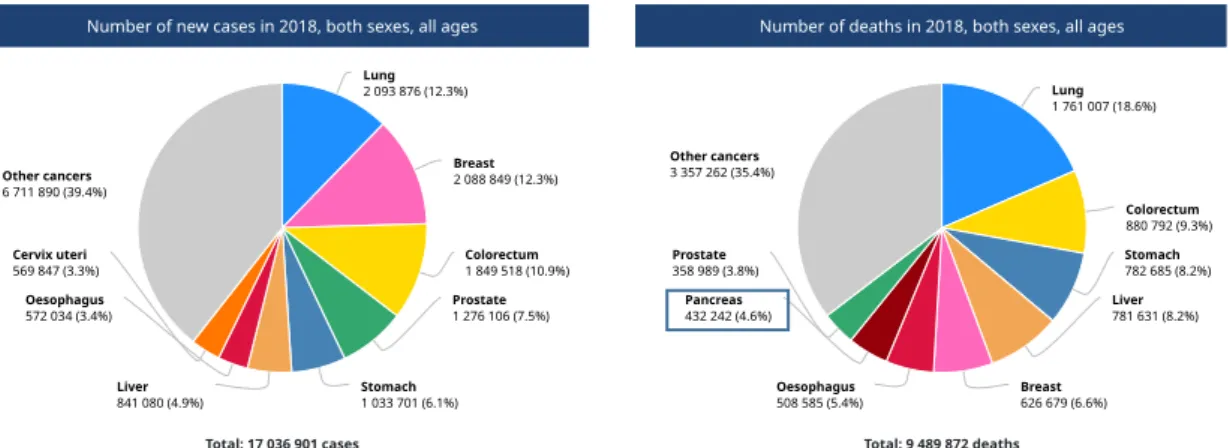
In 2018, despite not being one of the cancers with more new cases, pancreatic cancer counts to one of the eight with more deaths related to it, namely 4,6% of all cancer-related deaths (figure 6).
Figure 6. Number of new cases and deaths of cancer worldwide
(adapted from Globocan, 2018, http://gco.iarc.fr)
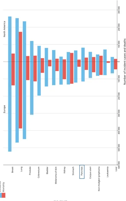
Comparing the number of incident cases and deaths in Europe and North America for Pancreatic Cancer (figure 7), the estimated age-standardized incidence is similar in both continents in 2018 (7.7 in Europe to 7.6 in North America) (Globocan, 2018).
Estimated number of new cases in 2018, pancreas, both sexes, all ages
Asia 214 499 (46.7%) Europe 132 559 (28.9%) North America 56 002 (12.2%)
Latin America and the Caribbean
35 270 (7.7%) Africa 16 059 (3.5%) Oceania 4 529 (0.99%) Total : 458 918
Data source: Globocan 2018 Graph production: Global Cancer Observatory (http://gco.iarc.fr)
All cancers excl. non-melanoma skin cancer
Source: Globocan 2018Number of new cases in 2018, both sexes, all ages
Total: 17 036 901 cases Lung 2 093 876 (12.3%) Breast 2 088 849 (12.3%) Colorectum 1 849 518 (10.9%) Prostate 1 276 106 (7.5%) Stomach 1 033 701 (6.1%) Liver 841 080 (4.9%) Oesophagus 572 034 (3.4%) Cervix uteri 569 847 (3.3%) Other cancers 6 711 890 (39.4%)
Number of deaths in 2018, both sexes, all ages
Total: 9 489 872 deaths Lung 1 761 007 (18.6%) Colorectum 880 792 (9.3%) Stomach 782 685 (8.2%) Liver 781 631 (8.2%) Breast 626 679 (6.6%) Oesophagus 508 585 (5.4%) Pancreas 432 242 (4.6%) Prostate 358 989 (3.8%) Other cancers 3 357 262 (35.4%)
Cancer incidence and mortality statistics worldwide and by region
Incidence Mortality
Both sexes Males Females Both sexes Males Females
New cases Cum. risk
0-74 (%) New cases Cum. risk 0-74 (%) New cases Cum. risk 0-74 (%) Deaths Cum. risk 0-74 (%) Deaths Cum. risk 0-74 (%) Deaths Cum. risk 0-74 (%) Eastern Africa 324 872 13.20 126 435 11.32 198 437 14.91 227 315 10.03 92 885 8.69 134 430 11.23 Middle Africa 94 004 10.69 40 484 10.42 53 520 11.08 67 819 8.17 30 243 7.83 37 576 8.57 Northern Africa 279 108 14.10 132 309 14.49 146 799 13.80 176 562 9.30 95 609 10.53 80 953 8.14 Southern Africa 108 899 19.05 47 442 21.07 61 457 17.97 61 004 11.23 29 712 13.39 31 292 9.77 Western Africa 224 163 11.19 87 225 9.84 136 938 12.53 150 430 8.09 62 105 7.14 88 325 9.02 Caribbean 106 577 19.48 54 918 21.55 51 659 17.62 62 416 10.36 33 960 11.60 28 456 9.25 Central America 245 529 14.20 109 886 13.93 135 643 14.49 117 613 6.80 56 800 6.76 60 813 6.84 South America 992 055 19.81 480 644 21.46 511 411 18.55 485 630 9.45 250 873 10.71 234 757 8.40 North America 1 896 063 29.18 970 083 31.02 925 980 27.59 692 970 9.59 363 883 10.73 329 087 8.55 Eastern Asia 5 587 812 21.45 3 090 552 24.14 2 497 260 18.83 3 444 684 12.86 2 129 585 16.32 1 315 099 9.33 South-Eastern Asia 975 838 15.11 470 912 16.11 504 926 14.37 625 763 10.06 342 407 11.85 283 356 8.50 South-Central Asia 1 719 178 10.15 848 206 10.44 870 972 9.92 1 159 624 7.17 614 633 7.79 544 991 6.59 Western Asia 390 616 17.17 204 447 19.51 186 169 15.31 219 719 10.11 128 940 12.67 90 779 7.77
Central and Eastern Europe 1 202 972 24.44 595 190 28.87 607 782 21.52 692 484 13.78 381 727 18.74 310 757 10.13
Western Europe 1 212 725 29.37 658 666 32.66 554 059 26.30 545 750 10.97 305 922 13.25 239 828 8.81
Southern Europe 872 216 26.79 479 229 30.78 392 987 23.20 419 269 10.57 244 944 13.49 174 325 7.90
Northern Europe 623 404 29.07 326 588 31.26 296 816 27.07 272 206 10.29 145 393 11.54 126 813 9.13
Australia and New Zealand 163 754 30.51 87 701 32.97 76 053 28.10 58 263 9.30 32 678 10.60 25 585 8.04
Melanesia 14 594 19.02 6 435 19.63 8 159 18.84 8 881 12.49 4 154 13.22 4 727 12.02 Polynesia 1 539 23.68 805 26.56 734 21.05 838 13.08 472 15.52 366 10.77 Micronesia 983 18.93 528 21.29 455 16.48 632 11.99 370 14.56 262 9.44 Low HDI 656 909 11.55 262 657 10.27 394 252 12.79 456 679 8.64 192 111 7.72 264 568 9.52 Medium HDI 2 790 875 11.79 1 344 186 12.13 1 446 689 11.54 1 847 037 8.15 975 365 9.00 871 672 7.37 High HDI 6 405 942 19.73 3 417 482 21.96 2 988 460 17.67 3 999 220 12.32 2 413 616 15.41 1 585 604 9.31 Very high HDI 7 174 973 27.15 3 790 116 30.18 3 384 857 24.59 3 182 854 10.49 1 763 971 12.76 1 418 883 8.45 World 17 036 901 19.42 8 818 685 21.38 8 218 216 17.71 9 489 872 10.58 5 347 295 12.65 4 142 577 8.66
Incidence, both sexes
Asia (50.9%) Europe (23%) North America (11.1%) LAC* (7.9%) Africa (6.1%) Oceania (1.1%)
Mortality, both sexes
Asia (57.4%) Europe (20.3%) North America (7.3%) Africa (7.2%) LAC* (7%) Oceania (0.72%)
5-year prevalence, both sexes
Asia (44.4%) Europe (27.2%) North America (14.2%) LAC* (8.1%) Africa (4.9%) Oceania (1.4%) Population Number Asia 8 673 444 Europe 3 911 317 North America 1 896 063 *Latin America and the Carribean 1 344 161 Africa 1 031 046 Oceania 180 870 Total 17 036 901 Population Number Asia 5 449 790 Europe 1 929 709 North America 692 970 Africa 683 130 *Latin America and the Carribean 665 659 Oceania 68 614 Total 9 489 872 Population Number Asia 17 147 410 Europe 10 504 195 North America 5 467 820 *Latin America and the Carribean 3 116 179 Africa 1 874 428 Oceania 523 388 Total 38 633 420
Most pancreatic cancer patients are diagnosed at stage III or IV (79%), with only 21% having an early-diagnosis (stage I and II) (National Cancer Registration and Analysis Service, 2016, ISD Scotland, 2016).
Figure 7. Estimated number of new cases and deaths in 2018, comparing Europe and America (Globocan, 2018, http://gco.iarc.fr)
E
st
im
a
te
d
n
u
m
b
e
r
o
f
n
e
w
ca
se
s
in
2
0
1
8
,
b
o
th
se
xe
s,
a
g
e
s
0
-7
4
Eu ro pe 10 0 00 0 20 0 00 0 30 0 00 0 40 0 00 0 50 0 00 0 60 0 00 0 0 10 0 00 0 20 0 00 0 30 0 00 0 40 0 00 0 50 0 00 0 60 0 00 0 N or th A m er ic a Br ea st Lu ng Pr os ta te Co lo re ct um Bl ad de r M el an om a of s ki n Ki dn ey St om ac h Pa nc re as Co rp us u te ri N on -H od gk in ly m ph om a Le uk ae m ia Li ve r Th yr oi d Can cer si te N um be r of in ci de nt c as es a nd d ea th s In ci de nc e M or ta lit y D at a so ur ce : G lo bo ca n 20 18 G ra ph p ro du ct io n: G lo ba l C an ce r O bs er va to ry ( ht tp :/ /g co .ia rc .fr )8
1.3 Etiology
Tumorigenesis is the progress of the cancer cells into a clinically significant disease, taking years. It is a complex process which can have innumerous causes and may involve lifestyle, namely diet and physical activity, environmental factors and host factors, like inheritance or epigenetic changes (figure 8). As for the host factors they can influence the probability of cancer development, with the accumulation of genetic damage over time. The interaction of these three types of factors over time is critical to the development (or not) of a malignancy (Annex 1).
Figure 8. Factor affecting cancer process
(adapted from World Cancer Research Fund, 2018)
1.3.1 Non-modifiable risk factors
Pancreatic cancer, like all cancers and many other diseases, has a higher risk of occurring more frequently with age (World Cancer Research Fund, 2018). The risk increases especially after 65 years, with most diagnoses being made between the ages of 60 and 80 (Ferlay et al., 2012).
In addition to that, it also varies with race, as the black population has an increased risk by 1.5.
Regarding pancreatic cancer in specific, it has the highest incidence of venous
(Nakchbandi et al., 2008). Patients with pancreatic cancer and venous thromboembolism also have a higher risk of poor prognosis and recurrence (Kondo et al., 2018) (table 2).
According to Klein et al. (2018), individuals with non-0 blood group also have an increased risk of pancreatic cancer.
Positive family history and some inherited genetic disorders also enhance the risk
of pancreatic cancer, as are the Lynch syndrome, Peutz-Jeghers syndrome, familial atypical multiple mole melanoma syndrome, among others (Greer et al., 2007). Mutations in specific genes also increase de risk of developing this type of cancer (gene PRSS1, K-ras, p16, p53, BRCA2, ATM) (Slebos et al., 2000).
An established risk factor is also the history of previous pancreatic diseases, as the case of pancreatitis. Although a correlation of higher risk can be made, most patients with pancreatitis do not develop cancer (Zheng et al., 2013).
Considering that the adult obtained height, has as cause the number of cell divisions during fetal life and childhood, health and nutrition in childhood and age of sexual maturity, which changes hormonal microenvironment producing alterations in the level of growth factors (higher circulating levels of IGF-1 (Gunnell et al., 2001, Bray et al., 2006). Tall people have undergone more cell divisions, so there can be a higher potential for error during DNA replication, which can result in cancer (Le Roith et al., 2001) (table 2).
10 Table 2. Summary non-modifiable risk factors
1.3.2 Modifiable risk factors
As already known, cigarette smoking (or chewing) is one of the biggest causes of cancer, especially lung cancer. Tobacco use is proven to be the leading cause of pancreatic cancer too (Secretan et al., 2009). It is estimated that 20% of pancreatic cancers are also caused by tobacco (IARC, 2012). The risk is increased by 74% for current smokers and 20% for former ones. Former smokers have an equal risk as non-smokers after 20 years (Lowenfels et al., 2006). According to Parkin et al. (2010), this risk may even be bigger. It is definitely the leading identified cause of pancreatic cancer. Tobacco and its smoke contain more than 7000 chemical compounds, many identified as carcinogens (World Cancer Report, 2014). These carcinogens contribute through many pathways to the development of the neoplasm: DNA binding, mutations, inflammation, oxidative stress and epigenetic changes.
Another cause of a major part of the pancreatic cancers is body fatness (especially abdominal) and physical inactivity. It also increases the risk of almost all gastrointestinal cancers. With an increase of body fatness, there can also occur
insulin resistance and diabetes (Hursting et al., 2003), influencing levels of many
other circulating hormones. This is why new-onset diabetes can be an early sign of pancreatic cancer (World Cancer Report, 2014). Due to the fact that insulin resistance is increasing in obesity, the pancreas compensates with and increased insulin
Risk factor Referance
Age (> 65 years) Ferlay et al., 2012, World Cancer Research
Fund/American Institute for Cancer Research, 2018
Black population World Cancer Research Fund/American Institute
for Cancer Research, 2018
Venous thromboembolism Kondo et al. (2018)
Non-0 blood group Klein et al. (2018)
Positive family history, some inherited genetic disorders
Slebos et al., 2000, Greer et al., 2007
Previous pancreatic diseases Zheng et al., 2013
Bigger attained adult
obtained height
Le Roith et al., 2001, World Cancer Research Fund/American Institute for Cancer Research, 2018
production. This hyperinsulinemia (Calle et al., 2004), is an environment propitious to carcinogenesis, discouraging apoptosis (World Cancer Research Fund, 2018). Due to the fact that adipocytes (fat cells) produce pro-inflammatory factors (Calle et al., 2004), most obese individuals have a chronic inflammation (Rexrode et al., 2003), which in turn can also promote carcinogenesis (Loffreda et al., 1998).
By contributing to weigh control and influencing the overall metabolic state (World Cancer Research Fund, 2018), regular physical activity has proven to reduce the risks, as the reduction of sweetened food/ beverages and the consumption of vegetables, fruits and whole grains. Physical activity has also proven immunomodulatory effects (improves innate and acquired immunity) and promoting in this way a diminished risk of carcinogenesis (McTiernan et al., 2008, Fridenreich et al., 2010). Exercise also decreases oxidative stress improving in this way DNA repair mechanisms (Fridenreich et al., 2010).
Alcohol, if consumed in abuse (more than three drinks a day) (World Cancer Research
Fund, 2018), has a dose-response association (Baan et al., 2009). Especially, but not only, ethanol is genotoxic contributing to carcinogenesis (World Cancer Report, 2014), being classified as a group 1 carcinogen (Duell et al., 2012) firstly in 1988, by the IARC. It is thought that its metabolites, like acetaldehyde might have a higher carcinogenic potential (World Cancer Research Fund, 2018).
The vast majority of the pancreatic juice samples studied by Maekawa et al. (2018) had bacterial DNA, namely Enterococcus faecalis, suggesting that it may be involved in the progression from pancreatitis to cancer. In 2018, about 16% of the total cancers were due to infections with viruses, bacteria and microparasites, as Helicobacter pylori, hepatitis B and C viruses (World Cancer Report, 2014). Through compromising the immune system, infection with HIV could also increase the risk of virus-related viruses.
Exposure to all types of radiation, like ionizing, ultraviolet and electromagnet one, can increase the risk of malignancies (World Cancer Report, 2014). This is both true for natural (like the sun) and man-made sources.
General air pollution from vehicles, households and a wide range of industries and drinking-water contamination may have carcinogens, as is the case of aromatic hydrocarbons (HAP), benzenes, asbestos, nitrates and nitrites and arsenic (World Cancer Report, 2014).
Plastic is produced using Bisphenol A. This is the reason why more than 90% of USA, Europe and Asia are exposed and probably contaminated by it through food or liquids (Vandenberg et al., 2010). In the 1980s it was established that Bisphenol A was not a
12
50 μg/kg/day (www.epa.gov/iris/subst/0356.htm). However, nowadays, with more than a hundred studies published since then, it is proven that this substance has an influence in the endocrine system (Keri et al., 2007), causing changes in the cancer cells at concentrations within the limits of exposure (Vandenberg et al., 2009). This evidence is however contested (Hengstler et al., 2011).
Pharmacological drugs, including the ones used as antineoplastic agents in cancer
therapy could also potentially induce cancer development (as prevent it) (World Cancer Report, 2014). Due to its unspecific genotoxicity, antineoplastic drugs could be responsible for inducing second cancers in supposed cured patients (table 3).
Concluding, many cancers can be prevented, by not engaging in those modifiable risk factors. The recommendations by the World Cancer Research Fund can be seen figure 9.
Figure 9. Cancer Prevention Recommendations
(adapted from World Cancer Research Fund, 2018)
Diet, Nutrition, Physical Activity and Cancer: a Global Perspective 82
Figure 8: Our Cancer Prevention Recommendations as an overarching ‘package’
While following each individual Recommendation offers cancer protection benefit, most benefit is gained by treating all ten Recommendations as an integrated pattern of behaviours relating to diet, physical activity and other factors that can be considered as a single overarching ‘package’ or way of life.
Table 3. Modifiable risk factors
1.4 Current Diagnosis Methods
Currently, the only way to diagnose asymptomatic pancreatic cancer is using imaging techniques (Winter et al., 2006, Poruk et al., 2013). Imaging the pancreas is also used for cancer staging, monitoring the response to treatment and detection of metastatic lesions.
Among others, the techniques used are computed tomography (CT), magnetic resonance imaging (MRI), positron emission tomography (PET), and endoscopic ultrasonography (EUS) (Morana et al., 2010, Appel et al., 2012, Fusaroli et al., 2012, Raman et al., 2012, Conrad et al., 2013) (figure 10).
Risk factor Referance
Tobacco use Secretan et al., 2009
Body fatness, physical
inactivity
Hursting et al., 2003
Insulin resistance and
diabetes
Hursting et al., 2003
Alcohol World Cancer Research Fund/American Institute
for Cancer Research, 2018
Infections with viruses,
bacteria and microparasites
Maekawa et al. (2018), World Cancer Report, 2014
All types of radiation World Cancer Report, 2014
Air pollution World Cancer Report, 2014
Bisphenol A Vandenberg et al., 2009
14
Figure 10. Algorithm for evaluation of a patient with suspicion of pancreatic cancer
(adapted from Zhang et al., 2018a)
1.4.1 Multidetector computed tomography (MDCT)
For patients with suspicion of pancreatic adenocarcinoma MDCT is the most available and best-validated diagnostic method (Zhang et al., 2018a). In addition to that it also the cheapest method, safe and non-invasive (Ahn et al., 2009). It takes an image in which the disease and the rest of the pancreatic tissue can be well distinguished, as well as examination of distant disease (Brennan et al., 2007). For these reasons, it is normally the first method of diagnosis being used (National Comprehensive Cancer Network, 2017). Nevertheless, it is a nephrotoxic method which exposes the patient to radiation (Al-Hawary et al., 2014).
1.4.2 Magnetic Resonance Imaging (MRI)
MRI has a superior imaging, allowing theoretically a diagnosis in earlier stage, which has proven to be not true in the practical case (with the same sensitivity as CT scanning) (Treadwell et al., 2016). It is radiation free, but expensive and not possible if metal implants are present. For these reasons it normally is not the first choice (National Comprehensive Cancer Network, 2017).
background pancreatic parenchyma with wide anatomic coverage, and thus allowing comprehensive examination of local and distant disease in one single section[8].
Numerous international guidelines endorse the use of CT as the initial modality in diagnosis of suspected pancreatic cancer[9,10]. In particular, MDCT is best
performed according to a dedicated pancreas protocol[10].
Despite some inter-institutional variability, the standard MDCT pancreas protocol is a helical type scan that takes interval images of 0.5 to 1 sub-millimetres, with two phases: pancreatic parenchymal phase at 40 to 50 seconds and portal venous phase at 65 to 70 seconds. The majority of modern scanners are 128 and 256
slice scanners. It includes the administration of both intravenous high iodine concentrated contrast, injected at a rate of 3 to 5 mL per second and ingestion of neutral oral contrast. The pancreatic phase is described as the intermediate between the arterial and hepatic phase where maximal enhancement of the pancreas is achieved to see the contrast between tumour and pancreatic parenchyma, as well as visualization of the peri-pancreatic arteries and veins[11]. The image is usually
reconstructed in the following ways: (1) axial views at 2 to 5 mm thickness; (2) coronal and sagittal views with multi-planar reformats at 2 to 3 mm thickness; and (3) vascular evaluations with maximum intensity projections
2049 May 21, 2018|Volume 24|Issue 19|
WJG|www.wjgnet.com
Diagnostic modalities Advantages Limitations
MDCT Most commonly available
Best validated Cheapest
Nephrotoxicity Radiation exposure
MRI Superior imaging
Depiction of local pancreatic disease Iodine-free and no radiation
Expensive Less available
Contraindicated with some metal implants
EUS +/- FNA Safe and less invasive
High sensitivity Able to detect small lesions Able to take histological sample
Less available in some countries Operator dependent Inability to detect distant metastasis
PET/CT Metastatic disease detection
Clarification of equivocal CT findings Monitoring recurrence and response to adjuvant therapy
Expensive Less available Radiation and contrast exposure
Table 1 Benefits and limitations of pancreatic cancer diagnostic modalities
CT: Computed tomography; MDCT: Multi-detector computed tomography; MRI: Magnetic resonance imaging; EUS: Endoscopic ultrasound; FNA: Fine needle aspiration; PET: Positron emission tomography.
Clinical suspicion of pancreatic cancer CT or MRI with pancreas protocol MDT review1 Mass in pancreas on imaging No mass in pancreas on imaging
Metastatic disease No metastatic disease Metastatic disease No metastatic disease
Biopsy confirmation of metastatic site EUS + FNA Biopsy confirmation of metastatic site If ongoing clinical suspicion, consider EUS +/- FNA to confirm absence of pancreatic cancer
Figure 1 Algorithm for the evaluation of a patient that has clinical suspicion of pancreatic cancer. 1Multi-disciplinary review should involve a panel including
gastroenterologist, surgeon, medical and or radiation oncologist, diagnostic imaging and pathologist. CT: Computed tomography; MDCT: Multi-detector computed tomography; MRI: Magnetic resonance imaging; EUS: Endoscopic ultrasound; FNA: Fine needle aspiration.
1.4.3 Endoscopic Ultrasonography with fine needle aspiration (EUS)
EUS consists on an upper gastrointestinal endoscopic examination with an echoendoscope positioned in the stomach near the pancreas under sedation (Zhang et al., 2018a). It Is a safe, less invasive diagnosis method, having high sensitivity and detecting small lesions (Puli et al., 2013). It also allows the collection of a sample for biopsy (by needle aspiration) (Hewitt et al., 2012).
However, it is not always available, is operator dependent and it does not allow the detection of distant metastasis (Chen et al., 2012).
1.4.4 Positron Emission tomography (PET)
PET consists on enhancing the metabolism of glucose in cancer cells. These overexpress glucose transporter 1 and can accumulate glucose like this (Llop et al., 2018). For detection, Fluorine 18-fluorodeoxyglucose (FDG) is used. FDG is a glucose analogue, used as a radiotracer. FDG-PET has yet a low sensitivity and specificity, resulting often in false-negatives (due to hyperglycemia) and false-positives (as FDG levels increase in inflammations for example) (Matsumoto et al., 2013). PET has been showing to have better, more sensitive results for monitoring response to treatment in comparison to CT and for detecting recurrence (Sperti et al., 2010, Kinupe et al., 2017). Due to its wide anatomic coverage it is easier for metastatic detection in the entire body (Dibble et al. 2012, Lee et al., 2014).
1.4.5 Biopsy
Confirmation procedure, consisting on removing a small sample of tissue to be examined under microscope. This can occur by fine-needle aspiration (inserting a needle into the pancreas) or during EUS.
1.4.6 Blood test
The best, most invasive, easier and quicker way to diagnose is by a simple blood test, looking for biomarkers. Biomarkers can be used with different goals, namely early detection, therapeutic monitoring, follow-up after surgery and to guide treatment decisions (Llop et al., 2018).
For pancreatic cancer the only used is CA19-9, with no diagnostic goal though. It is currently FDA-approved for monitoring the disease.
16
1.5 Therapeutics
Currently the only curative option is surgery, which can only be performed in early-stage pancreatic cancer (Costello et al., 2012).
Unlike in most cancers, cytotoxic agents are still first-line treatment for pancreatic cancer (Shi et al., 2018). Nevertheless, there are not many pharmacological agents that can be used and therefore do not vary much with the type of pancreatic cancer. According to Shi et al. (2018), the levels of CA19-9 should be monitored while choosing the therapeutic for patients with advanced pancreatic cancer.
1.5.1 Surgery
The only option that actually heals the patient. It can consist in removing only the part of the pancreas where the tumor is located or the entire pancreas. A person without pancreas can live normally needing insulin and enzyme replacement.
1.5.2 Chemotherapy
Consists in the use of toxic pharmacological substances injected or taken orally isolated or in combination with radiation therapy (Shi et al., 2018). In combination it is normally used in cases of wide spread pancreatic cancers or in cases where the tumor has first to be shrunken so that surgery is possible. It is also possible after surgery to prevent recurrence of the tumor.
1.5.3 Radiation Therapy
Cancer cells are destroyed with the use of highly energetic radiation, such as X-rays and protons. As chemotherapy, it can be used when surgery is not an option, to reduce the tumor before surgery or after surgery. Often in combination with chemotherapy.
2 Pancreatic Carcinogenesis
2.1 Pathology
Tumor mass consists of pancreatic stellate cells, immune cells, lymphatic and vascular endothelial cells, pathologically increased nerves and extracellular matrix (Duan et al., 2017).
The infiltrating ductal adenocarcinoma deduces an intense desmoplastic stromal reaction (Li et al., 2012, Rahib et al., 2014), creating a very complex tumor
microenvironment (Jiang et al., 2018). This complexity explains the lethality of this cancer, promoting its development (O’Neil et al., 2012, Duan et al., 2017). Due to its complexity, biopsies may mislead to false negative results (Olive et al., 2009).
Desmoplasia is the growth of fibrous or connective tissue. Although it may occur in benign circumstances like in scar tissue after surgery, it normally is associated with malignant neoplasms (causing dense fibrosis around it and invading healthy tissues like this) (Jiang et al., 2018).
Pancreatic stellate cells are thought to be the most responsible for the desmoplasia (Masamune et al., 2015). They occur normally in the pancreas, but they can be activated when in contact with pancreatic cells, synthesizing biologically active molecules (Pothula et al., 2016). They can also inhibit apoptosis and promote stem cells phenotypes of pancreatic cancer cells (Erkan et al., 2008), being therefore responsible for the resistance of pancreatic cancer to chemotherapy, distant metastasis and a poor prognosis (Hwang et al., 2008).
2.2 Genetics
Around 10% pancreatic cancer is of familial origin, meaning that some inherited mutated genes increase the risk of its development (Shi et al., 2009).
Exosome sequencing of ductal adenocarcinoma showed 16 significantly mutated genes (World Cancer Report, 2014). Including one oncogene (KRAS), three tumor suppressor genes (CDKN2A, TP53, SMAD4), MLL3, ATM, TGFBR2, ARID1A, and SF3B1 (Jones et al., 2008).
Biankin et al. (2012) also discovered novel mutated genes: genes involved in chromatin modification (EPC1 and ARID2) and DNA damage repair, and other mechanisms (ZIM2, MAP2K4, NALCN, SLC16A4, and MAGEA6).
Studying the mutated genes, does not only help in monitoring high-risk populations, but also in the development of personalized therapy, like the poly (ADP-ribose) polymerase inhibitors or mitomycin C for cancers with BRCA2 or PALB2 mutations (World Cancer Report, 2014).
18
3 Current State of the Art in Diagnostics
3.1 Novelties in Pancreatic Biomarkers
There is no current reliable early-state diagnostic biomarker for pancreatic cancer (Zhang et al., 2018b).
Due to the fact that most patients are only diagnosed with advanced pancreatic cancer, the samples to be studied for biomarkers come from them, making the identification of early-stage biomarkers more difficult (Resovi et al., 2018). This is why the use of in vivo models is fundamental.
Biomarkers are the most promising early-diagnosis method, due to its low cost, convenience, quickness and minimal invasiveness (Llop et al., 2018). However, it has been showed recently that no single biomarker could be reliable enough for cancer diagnosis, making a combination of biomarkers and incorporation of other clinical factors (as imaging techniques) a better approach (Capello et al., 2017, Chang et al., 2017). Figure 11 illustrates the potential early-stage pancreatic cancer biomarkers currently being studied.
Figure 11. Overview of circulating biomarkers for early detection of pancreatic cancer
(adapted from Zhang et al., 2018b)
Circulating biomarkers of pancreatic cancer
333 Am J Cancer Res 2018;8(3):332-353
ed by a large amount of extracellular matrix, creating a hurdle for pathologic biopsy [10]. Effective biomarkers that can be obtained in a less invasive manner have become a research focus. The ideal biomarkers should be easily detected with satisfactory sensitivity and spe- cificity and should distinguish PC from other benign pancreatic lesions. In the context of early detection, the identification of preneo-plastic conditions, such as pancreatic intraepi-thelial neoplasia (PanIN), intraductal papillary mucinous neoplasms (IPMNs) and mucinous cystic neoplasms (MCNs), is of great impor-tance [11].
Blood is easily accessible and relatively stable, making serum an ideal specimen in which to discover biomarkers. However, biomarkers se- creted into serum are extremely dilute and pro- bably obscured by other more-abundant serum proteins [12]. Technological advances in the last decade have provided more opportunities to discover circulating biomarkers based on “omics” analyses, including methods focused on proteins, nucleic acids, circulating tumor cells (CTCs), and exosomes. Numerous proteins
of low abundance can be analyzed by mass spectrometry-based approaches and proteo- mic technologies. Next-generation sequencing techniques provide deeper insight into soma- tic mutations and epigenetics analysis of the genome and broaden the characterization of circulating tumor DNA (ctDNA) and cell-free RNA. With the development of cell tracking techniques and flow cytometry, it is now possi-ble to capture and analyze CTCs and exosomes.
Thus, in this review, we summarize recent prog-ress in the early detection of PC using va- rious types of circulating biomarkers (Figure 1). CA19-9, other carbohydrate antigens, and carcinoembryonic antigen
CA19-9, also called sialyl Lewis a, is the only biomarker approved by the US FDA for monitor-ing the progression and therapeutic response of PC; it has also been widely used in the dia- gnosis of PC for a long time [13, 14]. However, the reported sensitivity (ranging from 69% to 98%) and specificity (ranging from 46% to 98%) of CA19-9 are moderate for PC screen- ing [15-18].
Figure 1. Overview of major circulating biomarkers for early detection of pancreatic cancer. Various biomarkers can be detected in plasma or serum from PC patients.
Soluble stroma-related Biomarkers
3.1.1 Traditional tumor biomarkers
3.1.1.1 Carbohydrate Antigen 19-9 (CA19-9)
CA19-9 is a modified Lewis blood group carbohydrate antigen (Young et al., 2018), called siayil Lewis A (SLea). It is embedded on cell surface molecules, namely
glycoproteins, gangliosides and mucins (Goh et al., 2017, Young et al., 2018), and is expressed in pancreatic and hepatobiliary diseases (Zhang et al., 2018a). It is the only FDA approved biomarker for pancreatic cancer (Locker et al., 2006), widely used with ELISA (Yue et al., 2011) but with many limitations (Wong et al., 2008).
It is used for monitoring the response of pancreatic cancer to treatment (Wong et al., 2008, Yue et al., 2011), as a prognostic biomarker (national cancer observatory) and for monitoring recurrence after surgical resection (Ballehaninna et al., 2012) having a lack of sensitivity and specificity for early-diagnosis (Huang et al., 2014). Their values to diagnosing pancreatic cancer vary according to different authors: 82 and 90% respectively (Llop et al., 2018) and according to Huang et al. (2014), it has a variable sensitivity of ~85% and specificity of ~85%. It has been shown that CA19-9 is upregulated up to two years before the diagnosis of pancreatic cancer, showing its good potential (O’Brien et al., 2015).
According to a study of Haab et al. (2015), most studies performed with CA19-9 do not use the same standards, varying from institution to institution. Feng et al. (2013), performed a large cohort, using the same rigorous conditions in every institution and key control groups. The goal was to understand if the different results obtained until that point, had divergent results due to the use of different standards. For this study they compared two antibody-based assay kits. As a global result, they reached similar AUC of 0.77 (area under the curve, it measures the accuracy with an AUC of 1 representing a perfect test) and comparable to the ones obtained in previous studies. However, these results varied a lot between patients, showing here a potential to be farther studied (Young et al., 2018).
As a prognostic biomarker it performs better in symptomatic patients (Bellehaninna et al., 2012, Huang et al., 2014). Nevertheless, it is not reliable when used alone (Singh et al., 2011).
According to Llop et al. (2018), it is not expressed in 10-20% of the Lewis antigen-negative population, due to a genetic deficiency of focusyltransferase activity (Capello
20
et al., 2017). In general, about 5-10% of the whole population does no express it (Herreros-Villanueva et al., 2016, Root et al., 2018).
In addition to that, it can lead to false-negatives, as only 65% of patients with pancreatic cancer have a high expression of CA19-9 (Gold et al., 2013).
It can also be falsely positive when other pathologies are present, like biliary infection, inflammation or obstruction, chronic pancreatitis and obstructive jaundice (Marrelli et al., 2009), hepatic and pancreatic cysts (Llop et al., 2018), other benign gastrointestinal conditions (Locker et al., 2006) or other cancers, like colorectal and breast (Llop et al., 2018). It has also racial and sex expression variations, being highest in Caucasians (Resovi et al., 2018).
In order to overcome these limitations and improve efficiency a combination of CA19-9 and other biomarkers has good potential (Faca et al., 2008, Chang et al., 200CA19-9, Brand et al., 2011 Gold et al., 2013, Lennon et al., 2014).
Many of these combinations with CA19-9 are being studied. The combination with
albumin and IGF (Insulin-like growth factor 1) (Goh et al., 2017), can distinguish
between pancreatic cancer and chronic pancreatitis with a high sensibility (93,6%) and high specificity (95%) (Park et al., 2012, Ferri et al., 2016).
Makawita et al. (2013), studied regenerating islet-derived 1 beta (REG1B), syncollin (SYCN), anterior gradient homolog 2 protein (AGR2), and lysyl oxidase-like 2 (LOXL2). The combination between CA19-9, SYNC and REG1B had the best results, with an AUC of 0.9.
In combination with ICAM-1 (Intercellular Adhesion Molecule 1) and OPG (Osteoprotegerin), Brand et al. (2011), reported a sensitivity for diagnosis from 88% and a specificity of 94%. It is a highly selective combination that had negative results from almost all breast, lung and colorectal cancers.
In combination with cathepsin D and MMP-7 (matrix metalloproteinase-7) it even has a higher AUC (Park et al., 2012).
Zeh et al. (2005) reported a combination of CA19-9 and serum cytokines (HGF,
MCP-1, IP-10, Eotaxin) with very good results: sensitivity 85.7% and specificity of 92.3% to
distinguish PC from healthy controls and sensitivity of 98%, specificity of 96.4% to distinguish PC from chronic pancreatitis. CA19-9 in combination with albumin,
C-reactive protein and interleukin 28 demonstrated sensitivity was 99.39% for
all-stages, 96.10% for early-stage and 98.80% for advanced-stage pancreatic cancer at 90% specificity when discriminating between cancer and healthy individuals.
Cohen et al. (2017), studied the combination of four plasma proteins (CA19-9, CEA
(Carcinoembryonic antigen), HGF (Hepatocyte growth factor), OPN (osteopontin)) with ctDNA testing for KRAS mutations. Although it outperformed
CA19-9 alone, it had a sensitivity of only 64%, making an optimization of the assays crucial to see if it could be an option to test furtherly.
Capello et al. (2017) researched already studied protein candidates to come with the best combination, namely TIMP1 (TIMP metallopeptidase inhibitor 1), LRG1
(Leucine-rich alpha-2-glycoprotein 1) and CA19-9, which outperformed CA19-9.
Combining CA19-9 with MUC5AC (Mucin 5AC), which in literature seems to have very good biomarker properties (is over-expressed in pre-cancerous lesions), Kaur et al. (2017), found it to have a better performance in ELISA.
The same was proven by Kim et al. (2017) for the combination thrombo-spondin-2
(THBS2) and CA19-9.
Sefrioui et al. (2017) studied a panel consisting in CA19-9, ctDNA and CTC analysis. In comparison with EUS-FNA alone it increased significantly the sensitivity and specificity.
According to Llop et al. (2018), combining CA19-9 with miR-16 and miR196-a it improves its performance to an AUC from 0,98.
22
Table 4. Summary of CA19-9 combinations under study as early-stage diagnostic pancreatic cancer biomarkers
Biomarker Panel (concentration) AUC, sensitivity, specificity Referance Carbohydrate Antigen 19-9 (CA 19-9) AUC: 0.77; Sensitivity: ~85%; Specificity: ~85% Feng et al. (2013) Huang et al, 2014 CA 19-9, albumin and IGF Sensitivity: 93,6%; Specificity: 95% Goh et al., 2017 Park et al., 2012 Ferri et al., 2016
CA 19-9, SYNC, REG1B AUC: 0.9; Makawita et al., 2013
CA 19-9, ICAM-1, OPG Sensitivity: 78%;
Specificity: 94% Brand et al., 2011 CA 19-9, cathepsin D, MMP-7 AUC: 0.9 Sensitivity: 88%; Specificity: 80% Park et al., 2012 CA 19-9, HGF, MCP-1, IP-10, Eotaxin Sensitivity: 85.7%; Specificity: 92.3% Zeh et al., 2005 CA 19-9, albumin, C-reactive protein and interleukin
Sensitivity: 99.39%; Specificity: 90%
Zeh et al., 2005
CA19-9, CEA, HGF, OPN with ctDNA testing for KRAS mutations
Sensitivity: 64%;
Specificity: 99.5%
Cohen et al., 2017
CA19-9, TIMP1, LRG1 AUC: 0.95;
Sensitivity:75%;
Specificity: 95%
Capello et al., 2017
CA19-9, MUC5AC AUC: 0.87;
Sensitivity:83%; Specificity: 83% Kaur et al., 2017 CA19-9, THBS2 Sensitivity:87%; Specificity: 98% Kim et al., 2017
CA19-9, ctDNA and CTC analysis
Sensitivity: 78%; Specificity: 91%
Sefrioui et al., 2017
CA19-9, miR-16 and miR196-a
3.1.1.2 Other conventional biomarkers
There are many other conventional biomarkers (Zhang et al., 2018b), namely CA242, CA72-4, CA125, CEA (Bunger et al., 2011). Although they have limited early-diagnostic potential (Zhang et al., 2015), they are promising for Lewis-negative population (in which CA19-9 has no utility).
An example of this high specificity seen for Lewis-negative patients are CEA and CA125 (98% and 93,8% respectively) (Luo et al., 2017).
However, this would imply first testing a possible patient regarding the state of the Lewis-group, which is not ideal
3.1.2 Metabolites
According to Hanahan et al. (2011), due to the fact that cancer cells can rewire metabolically, they can survive and proliferate even with oxygen and nutrient deficiency (called Warburg effect (Warburg et al., 1956)). In the specific case of pancreatic cancer, these cells survive in hypoxia, desmoplasia (Jiang et al., 2018) and hypovascularization.
A malignancy could therefore be diagnosticated in the presence of aberrant low-molecular weight substances (Denkert et al., 2012) deriving from this abnormal biochemical state (Vermeersch et al., 2013).
Because of the different demands of pancreatic cancer cells, especially metabolic ones, they can rewire the metabolism. This happens normally with a mutation of the oncogene KRAS (which is normally present in pancreatic cancer (Biankin et al., 2012)). These different metabolic demands are supported by the fibroblasts, which mediate metabolite exchange (Ozdemir et al., 2014, Rhim et al., 2014) and pancreatic stellate cells (Sousa et al., 2016).
Alterations in the metabolism include alterations in metabolic enzyme, accumulation of intermediates (Zhou et al., 2012), making amino acids, lipids, fatty acids and bile acids in serum potential biomarkers (Kobayashi et al., 2013, Ritchie et al., 2013, Zhang et al., 2013). One amino acid who is being studied is palmitic acid in serum. According to Di Gangi et al. (2016), it has been showed to have better results in early-diagnosis of pancreatic cancer than CA19-9 with an AUC of 1.0.
Mayers et al. (2014), also studied plasmatic amino acids, coming to the conclusion that an increase deriving from muscle catabolism could increase the risk of developing pancreatic cancer (Young et al., 2018).
24
Four other serum metabolites showed a higher sensitivity and lower false-negative rate when comparing with CEA and CA19-9, namely xylitol, 1,5-anhydro-D-glucitol, histidine, and inositol (Sakai et al., 2016).
There are extreme changes during different stages of pancreatic cancer. Metabolomics is therefore a sensitive indicator for monitoring pre-cancerous lesions and diagnosing early-stage (Yuan et al., 2016). For example, Kynurenate and methionine levels are only upregulated in pre-cancerous lesions and decreased in cancer.
Yuan et al. (2016) studied 82 metabolites in a prospective cohort study, concluding that two metabolites (isocitrate and aconitate) were associated with survival of pancreatic cancer patients. Both of these metabolites are intermediates of the tricarboxylic acid cycle. In addition to that, inherited ACO1 genotypes also influenced survival of patients.
Nowadays there have been identified already 50 differences in serum metabolites between pre-cancerous lesions and pancreatic cancer, making metabolites a good potential panel for biomarkers (LaConti et al., 2015).
Different enzymes and intermediates influence the concentration of a single metabolite, making the development of a panel with a combination of biomarkers crucial for a good diagnostic.
As for other potential biomarkers, they still have to be studied in larger populations and under standardized conditions (Zhang et al., 2018b) (table 5).
Table 5. Principal Metabolites being studied as early-stage diagnostic pancreatic cancer biomarkers
3.1.3 Cell-free DNA
By apoptosis or necrosis nucleic acids can be freed into the extracellular environment, originating cell-free DNA (cfDNA) (Zhang et al., 2018b). Cui et al. (2008), showed that patients with solid tumors normally have elevated levels of cfDNA.
Biomarker Panel AUC, sensitivity, specificity
Referance
Palmitic acid AUC: 1.0;
Sensitivity: 100%; Specificity: 100%
Di Gangi et al., 2016
Xylitol, 1,5-anhydro-D-glucitol, histidine, and inositol
Sensitivity: 84.1%; Specificity: 84.1%
Circulating tumor DNA (ctDNA) carries mutations corresponding to the somatic mutations in the primary tumor (Kinugasa et al., 2015), and it is believed that it correlates to its burden (Fleischhacker et al., 2007, Diaz et al., 2014, Newman et al., 2014), meaning that analyzing ctDNA from liquid biopsies could indicate tumor state and genetics. Bettegowda et al. (2014), showed that in stage I to III of pancreatic cancer, about 40% of patients have detectable ctDNA an about 90% for stage IV tumors.
Liquid biopsies are one of the most recent approaches in oncology, but still not suitable to replace tissue biopsies (Imamura et al., 2016, Pishvaian et al., 2016, Riva et al., 2016).
3.1.3.1 Somatic mutations
In pancreatic cancer, inactivating mutations in tumor suppressor genes, like CDKN2A, TP53, SMAD4, BRCA2 are found mostly in late states. In contrast to this, the most predominant genetic characteristics are KRAS mutations (Zhang et al., 2018b). They occur even in premalignant lesions and in high rate, being detected in serum, pancreatic juice and feces (Jones et al., 2008). However, it is not specific for pancreatic cancer as it also occurs in other diseases like chronic pancreatitis (Yanagisawa et al., 1993).
Its importance consists in controlling patients with pancreatic intraepithelial neoplasia. Almost 90% of these patients have a KRAS mutation with its rate correlating to the grade of the disease implying that a KRAS mutation is an early event during tumorigenesis (Kanda et al., 2012). There was also found a relation between KRAS mutations and clinical stage (Castells et al., 1999).
This potential biomarker still needs to be furtherly studied, as results of current studies do not always match (Mullcahy et al., 1998, Maire et al., 2002).
The detection rates vary from 35% of plasma samples of pancreatic cancer patients (Uemura et al., 2004) and 48% (Bettegowda et al., 2014) to 63% of pancreatic patients (Kinugasa et al., 2015).
Mutations in TP53 and SMAD4, in comparison with KRAS mutations, do not occur very often (Zhang et al., 2012). This is the reason why there are only a few studies on them. These mutations occur normally later, making them not suitable for early-stage diagnosis (Maitra et al., 2003) (table 6).
26
Table 6. Somatic Mutations of Cell-free DNA as early-stage diagnostic biomarkers
3.1.3.2 Epigenetic modulations
Epigenetic modifications of cfDNA (cell-free DNA), especially alterations in methylation pattern are very common in cancer, as it is the case of hypermethylation of tumor genomic DNA and hypermethylation of tumor suppressor genes (Zhang et al., 2018b). Sato et al. (2003), discovered several targets of abnormal DNA methylation in pancreatic cancer. UCHL1, NPTX2, SARP2, CLDN5, FOXE1, CDH3.
CD1D, KCNK12, CLEC11A, NDRG4, IKZF1, PKRCB and KRAS are currently under clinical study after resulting in 75% sensitivity and 95% specificity (Kisiel et al., 2015). Other methylation biomarkers associated to the disease are ppENK, cyclin D2, sparc-7, Osteonectin and TSLC1. There has not been found further validation and clinical application for them yet (Fukushima et al., 2003, Matsubayashi et al., 2003, Sato et al., 2003).
There are, in addition to that, abnormal methylation profiles in specific cfDNA regions. Although P16 and proproenkephalin promoters are hypermethylated in plasmatic DNA, their detection rates are from about 30% and 25% accordingly (Jiao et al., 2007). After analyzing 30 plasma samples, Liggett et al. (2010), combined some targets, particularly 14 gene promoters differentiating chronic pancreatitis patients from controls (sensitivity of 81,7% and specificity of 78%) and from pancreatic cancer patients (sensitivity of 91,2% and specificity of 90,8%) according to its methylation status. The panel included, CCND2 (cyclin D2), DAPK1 (death-associated protein kinase 1), ESR1 promA (estrogen receptor 1 promoter A), HMLH1 (human mutL homolog 1), MGMT (O-6-methylguanine-DNA methyltransferase), MUC2, (mucin 2, oligomeric mucus/gel-forming), MYOD1 (myogenic differentiation 1), CDKN2B (cyclin-dependent kinase inhibitor 2B), CDKN1C (cyclin-(cyclin-dependent
kinase inhibitor 1C), PGK1 (phosphoglycerate kinase 1), PGR-proximal (pro-gesterone receptor proximal promoter), RARb (retinoic acid receptor beta), RB1 (retinoblastoma 1), SYK (spleen tyrosine kinase).
Biomarker Panel AUC, sensitivity, specificity
Referance
KRAS mutation Detection rate: 35-63%
of pancreatic cancer patients Uemura et al., 2004 Kinugasa et al., 2015 TP53 and SMAD4 mutations
Lower detection rates than KRAS mutation
A combination of CCND2, SOCS1 and THBS1 has also a very good potential for early-stage diagnosis, with a sensitivity of 76% and a specificity of 59% (Melnikov et al., 2009).
Studying the methylation status of BNC1 (Basonuclin 1) and ADAMTS1 (ADAM Metallopeptidase with Thrombospondin Type 1 Motif 1) in cfDNA could also be used for early-stage detection (81% sensitivity, 85% specificity) (Yi et al., 2013).
Still, epigenetic modifications need to be furtherly studied but have a very good potential. The incidence of aberrant DNA methylation at select cpg islands higher than incidence of genetic mutations and they have fewer false-negatives. This aberrant epigenetic alteration is an early event during tumorigenesis, it leads to gain/loss of function of critical molecules in cancer cells and the DNA methylation status is stable, making it easily detected with great sensitivity, even with contamination (Zhang et al., 2018b) (table 7).
28
Table 7. Principal methylation patterns of gene promoters in cfDNA as biomarkers
3.1.4 Autoantibodies
Cancer patients normally develop an immune system dysfunction (Zhang et al., 2018b). Though the immune response is not strong enough to have clinical manifestations (Kobold et al., 2010), circulating autoantibodies could be potential biomarkers for diagnosing cancer.
In some types of cancer, including pancreatic, there are produced autoantibodies against tumor-associated antigens, ie. misfolded, overexpressed, aberrantly modified, ectopically expressed and mutated tumor proteins, (Desmetz et al., 2011, Kaur et al., 2012). These autoantibodies can indirectly reflect altered genetics and proteomics but can only be detected in low frequency (Zhang et al., 2018b). Due to tumor heterogeneity, detection is even more difficult.
Biomarker Panel
(Methylation status of gene promoters in cfDNA measured) AUC, sensitivity, specificity Referance UCHL1, NPTX2, SARP2, CLDN5, FOXE1, CDH3. CD1D, KCNK12, CLEC11A, NDRG4, IKZF1, PKRCB and KRAS Sensitivity: 75% Specificity: 95% Kisiel et al., 2015 ppENK, cyclin D2, sparc-7, Osteonectin and TSLC1
With no validation yet Fukushima et al., 2003, Ueki et al., 2002, Matsubayashi et al., 2003, Sato et al., 2003 CCND2, DAPK1, ESR1 promA, HMLH1, MGMT, MUC2, MYOD1, CDKN2B, CDKN1C, PGK1, PGR-proximal, RARb, RB1, SYK Sensitivity: 91,2% Specificity: 90,8% Liggett et al., 2010 CCND2, SOCS1, THBS1 Sensitivity: 76% Specificity: 59% Melnikov et al., 2009 BNC1, ADAMTS1 Sensitivity: 81%; Specificity: 85% Yi et al., 2013
Despite the fact that, in a study by Bracci et al. (2012), there were analyzed several autoantibodies, concluding that there are significant differences in expression levels in pancreatic cancer patients, their diagnostic value was poor with an AUC < 0.7.
Recently, the main focus on autoantibodies research has been Anti-mucin 1 antibodies (MUC1). MUC1 is a membrane associated glycoprotein that is overexpressed in some cancers, including pancreatic one and is associated with CA19-9 (Zhang et al., 2018b). In the last years, Gold et al. (2006), researched a monoclonal antibody against MUC1 with a sensitivity of 77% and a specificity of 95% for diagnosing pancreatic cancer. Other autoantibodies being researched include autoantibodies to two acidic isoforms of glycolytic enzyme enolase (ENOA 1 e 2), which seem to be more frequent in patients with normal CA19-9 levels (Tomaino et al., 2011), Ezrin (Capello et al., 2013), vimentin isoform (Hong et al., 2006), and calreticulum isoforms (Hong et al., 2004) (table 8). Neoantigen are new immunogenic protein sequences produced by malignancies. They are absent from the normal human genome and are primarily created by tumor-specific mutations in the genome. They could be potential targets for diagnoses and immunotherapy (Schumacher et al., 2015).
Table 8. Autoantibodies with potential as biomarkers
3.1.5 Cell-free noncoding RNA (ncRNA)
Genes code proteins, but only about 2% of all genes. At least 75% of them do not encode anything, being called noncoding RNA (ncRNA) (Djebali et al., 2012). ncRNA include microRNA, small interfering RNA, piwi-interacting RNA, small Cajal
body-Biomarker Panel AUC, sensitivity, specificity Referance Monoclonal antibody against MUC1 Sensitivity: 77% Specificity: 95% Gold et al., 2006 ENOA 1 e 2 Sensitivity: 62% Specificity: 97% Tomaino et al., 2011 Ezrin AUC: 0.9 Sensitivity: 93.2% Specificity: 75.5% Capello et al., 2013
Vimentin isoform With no validation yet Hong et al., 2006