First diabetic retinopathy prevalence study in
Portugal: RETINODIAB Study
—Evaluation of the
screening programme for Lisbon and Tagus
Valley region
Marco Dutra Medeiros,
1,2,3Edgar Mesquita,
4Ana Luísa Papoila,
5,6,7Victor Genro,
1João Filipe Raposo
1,8For numbered affiliations see end of article.
Correspondence to Dr Marco Dutra Medeiros, Portuguese Diabetes Association (APDP), Rua Salitre 118, Lisbon 1250-203, Portugal; marcodutramedeiros@gmail. com
Received 4 February 2015 Accepted 10 March 2015
To cite: Dutra Medeiros M, Mesquita E, Papoila AL, et al. Br J Ophthalmol Published Online First: [ please include Day Month Year] doi:10.1136/ bjophthalmol-2015-306727
ABSTRACT
Background/aims In Portugal, so far, there is no study or even accurate data on the prevalence of diabetic retinopathy (DR), based on a large representative sample and on a long-term follow-up. The objective of our study was to determine the prevalence of DR based on a national screening community-based programme. Methods A 5-year retrospective analysis of the RETINODIAB screening programme results was implemented in Lisbon and Tagus Valley area between July 2009 and October 2014. We estimated the prevalence of retinopathy for all patients with type 2 diabetes and studied the association between known risk factors and retinopathy emergence at theirfirst screening. Results Throughout this period, from a total of 103 102 DR readable screening examinations, 52 739
corresponded to patients who attended RETINODIAB screening at entry. Globally, DR was detected in 8584 patients (16.3%). Of these, 5484 patients (10.4%) had mild non-proliferative (NP) DR, 1457 patients (2.8%) had moderate NPDR and 672 (1.3%) had severe NPDR. Finally, 971 patients (1.8%) had proliferative DR requiring urgent referral to an ophthalmologist. The presence of any DR, non-referable DR or referable DR was strongly associated with increasing duration of diabetes and earlier age at diagnosis.
Conclusions The prevalence rate of DR in our study (16.3%) was slightly lower than other published international data. The RETINODIAB network proved to be an effective screening programme as it improved DR screening in Lisbon and Tagus Valley surrounding area.
INTRODUCTION
Diabetic retinopathy (DR) is the leading cause of legal blindness in the working-aged population of industrialised societies.1–3 In 2013, 382 million people had diabetes; this number is expected to rise to 592 million by 2035, according to the International Diabetes Federation (IDF).4 5
It is well established that the effectiveness of the laser treatment depends on the accuracy and timely treatment of DR among patients with diabetes mel-litus, particularly those with a high risk of DR.6 Indeed, DR represents an excellent paradigm for screening as laid out in the principles for screening of human disease described by Wilson and Jungner in 1968.7 8
In order to decrease by about 30% the new cases of blindness caused by diabetes, the declaration of St. Vincent (1989) called for the implementation of
national strategies for screening for DR in a system-atic manner.9 WHO, IDF and the Directorate-General of Health (DGS) co-organised (1997) the Fourth Meeting in Lisbon for the Implementation of the St. Vincent Declaration on Diabetes Care and Research in Europe, which was attended by dele-gates from 60 countries.10 This conference rein-forced once again the need for greater engagement from all signatory countries for the St. Vincent Declaration in order to address diabetes complica-tions, particularly DR. This international challenge was strengthened at the Liverpool meeting in 2005.11Despite all, it is only in the last decade that significant progress has been made in implementing screening programmes to detect and monitor DR.
Portugal currently has a population of 10.6 million, predominantly Caucasian, whose majority (around 8.5 million) is located on the western coast (∼80%).12 According to the National Observatory for Diabetes, nearly one million Portuguese have diabetes, the equivalent to 13% of the population between 20 and 79 years.13 Of these, about 400 000 people are undiagnosed. It is imperative that these people are identified through early diagnosis in order to signifi-cantly reduce the incidence of serious complications.
The Portuguese Diabetes Association (APDP) is the world’s oldest diabetes association and a senior member of the IDF. From the moment it was founded, early in the 20th century, APDP has been driven by a single overarching objective: to improve the quality of life of people with diabetes. Involved nationally in dia-betes advocacy and the provision of education, as well as the delivery of care, APDP has become a key player in the healthcare arena in Portugal.
Following a pilot regional DR screening pro-gramme which was launched in 2008, the Diabetic Retinopathy Screening Service for Lisbon and
Tagus Valley—RETINODIAB—was commissioned
and driven by APDP. This screening programme is supported by the Regional Health Administration of Lisbon and Tagus Valley (ARSLVT) and follows the norms of the DGS, which is a public body of the Ministry of Health. The major aim of the pro-gramme was to identify all undiagnosed sight-threatening DR in order to ensure timely onward referral to Lisbon area hospital eye services.
Herein, the authors describe the first Portuguese study regarding the prevalence of DR, as well as focus on the screening programme for DR (2009– 2014) implemented in the area of Lisbon and Tagus Valley.
METHODS
The RETINODIAB network
RETINODIAB (Study Group for Diabetic Retinopathy
Screening) is a m-health screening system carried out by APDP, which focuses on clinical aspects of DR screening. Its primary aim is to promote the advancement of knowledge on all aspects of DR through an active cooperation between ophthalmologists and other specialists such as endocrinologists, internists and neurologists. Additionally, APDP has fostered the development of important scientific studies in epidemiology and diabetology in Portugal.14 15
Lisbon and Tagus Valley area
Lisbon and Tagus Valley is one of thefive Regions of Portugal (Nomenclature of Territorial Units for Statistics (NUTS) II divi-sions). It corresponds to 13% of the Portuguese territory, it has a population of 3.7 million (34.7% of the total population) and it represents 44% of the national Gross domestic product (GDP). There are 15 primary care groups (ACES) in this area (figure 1) organised according to the five existing NUTS III (subregions: Greater Lisbon, Setúbal Peninsula, Middle Tagus and Lezíria West Coast).
APDP screening protocol—RETINODIAB
The RETINODIAB screening programme was held in several primary care health units covered by the APDP protocol. Each screening centre is equipped with a non-mydriatic camera (model CR-2, Canon, Tokyo, Japan). Two 45° non-stereoscopic retinal digital photographs per eye were taken in a scotopic environment, one centred on the posterior pole and the other on the optic disc. Despite all efforts, in several patients it was impossible to obtain an image with minimum quality. In these specific cases, orthoptists have proceeded to iatrogenic pupil dilation. The remaining possible causes for deficient acquisition of fundus were documented in the clinical report and those patients were referred to a specialist within a maximum period of 3 months. After the capture of images, they are compressed in the Digital Imaging and Communications in Medicine (DICOM) protocol and transmitted through the internet to APDP reading centre. All images were classified according to The International Clinical Diabetic Retinopathy Scale.16As clin-ically significant macular oedema is not discernible on non-stereoscopic images, maculopathy was defined as the presence of hard exudates or haemorrhages within 1 disc diameter of the fovea. Patients who had undergone panretinal laser treatment
were classified as having proliferative retinopathy. Both eyes were assessed for DR and the worse grade from the two eyes was used in the analysis. Retinal images were considered not gradable if retinas of both eyes could not be visualised properly —that is, retinal vessels were not visible within 1 disc diameter of the fovea andfine vessels were not visible across the surface of the optic disc. Where only one eye was gradable, the presence or absence of DR relied on this eye. The reader-automatically generated report displays diagnosis of DR level, diagnosis of
non-diabetic ocular disorders and recommendations for
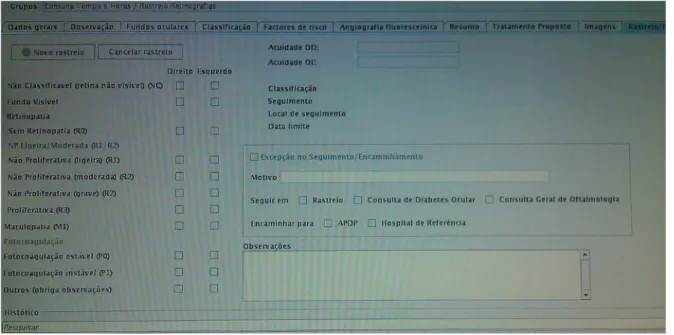
follow-up (figure 2).
APDPSoft software
The APDPSoft is a software developed since 1999, which accompanies the evolution of the services provided by the APDP. Currently, this software supports and monitors several valences, especially in terms of clinical datafile, markings man-agement, laboratory parameters, invoicing the health subsys-tems, integration of numerous diagnosis equipments as well as an effective liaison with the electronic services of the Ministry of Health. It makes the storage of clinical data, fundus photo-graphs and statistics. All stored images were downloaded by cer-tified ophthalmologists at the RETINODIAB Reading Centre comprising three readers.
Statistical methods
The features of the study participants were described using means (SD) for continuous variables and percentages for cat-egorical variables. The age at diagnosis was categorised into four groups (40–49 years, 50–59 years, 60–69 years and >70 years) for all analyses at first screening. The duration of diabetes was discretised into four categories (<5 years, 5–9 years, 10–15 years and >15 years). Adjusted ORs and corre-sponding 95% CIs were calculated. Furthermore, we defined referable diabetic retinopathy (RDR) to all patients graded as moderate non-proliferative (NP) DR, severe NPDR or prolifera-tive retinopathy DR (with or without maculopathy) or maculo-pathy with mild retinomaculo-pathy. This category relates to those who would, according to the guidelines, need referral to the hospital eye service for further clinical evaluation. Univariable and
multivariable logistic regression models were used to assess the association of the collected variables with retinopathy status, separately for each subset of diabetes (any DR; RDR; non-referable diabetic retinopathy (NRDR)). A level of significance of α=0.05 was considered. All data were analysed using the Statistical Package for the Social Sciences for Windows V.22.0 (IBM Corp. Released 2013. IBM SPSS Statistics for Windows, V.22.0. Armonk, New York: IBM Corp.).
RESULTS
Our 5-year retrospective analysis included data for all patients with type 2 diabetes, diagnosed over the age of 40 years, and who attended APDPSPbetween July 2009 and October 2014.
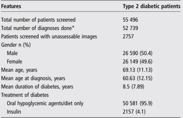
Throughout this period, from a total of 103 102 DR readable screening examinations, 52 739 corresponded to patients who attended RETINODIAB screening at entry. The baseline features of study participants are included intable 1. Patients’ mean age is 69.13 (SD=11.13) years. Women accounted for 49.6% (n=26 149) of all patients. The mean duration of diabetes was 8.5 years (SD=7.89).
Overall, not all screening examinations resulted in assessable images. In our present study, retinal photographs of at least one eye could not be graded in 2757 of the 55 496 total screening patient examinations performed at entry (4.96%). This subset of patients was not included in thefinal group used to calculate several preva-lence rates (total number of DR diagnoses performed).
The prevalence of the different categories of DR is shown in
table 2, regarding to the total number of assessable images. Globally, DR was detected in 8584 patients (16.3%). Of these, 5484 patients (10.4%) had mild NPDR, 1457 patients (2.8%) had moderate NPDR and 672 (1.3%) had severe NPDR. Finally, 971 patients (1.8%) had proliferative DR requiring urgent referral to an ophthalmologist. A total of 732 patients (1.4%) had maculopathy.
The results of the multivariable logistic regression analysis are shown intable 3.
Men had increased odds of all severities of DR compared with women. The odds of all grades of DR increased with the duration of diabetes. There was a 2.50-fold, 4.99-fold and 8.20-fold increased odds of any DR associated with a duration
of diabetes of 5–9, 10–15 and≥15 years compared with <5 years (reference subgroup) and a 2.38-fold, 4.19-fold and 5.03-fold increased odds of NRDR in the same subgroups, respectively. For RDR subset of patients, the odds increased by a factor of 2.80 with a known duration of diabetes of 5–9 years and 6.37-fold for a known duration of diabetes of 10–15 years. Finally, for patients with a duration over 15 years the odds increased 12.43-fold compared with the reference subgroup.
Additionally, the ORs of any DR, NRDR and RDR groups were significantly lower with the increasing of age at diagnosis of diabetes. For instance, the odds of any DR increased to 1.12, to 1.48 and to 2.00 in the age ranges 60–69 years, 50–59 years and > under 50 years, respectively, compared with the reference group (> 70 years).
Regarding insulin treatment, all patients with diabetes under insulin therapy had a significant increased odds for all different DR subgroups.
DISCUSSION
Until now, there have been no studies addressing the prevalence of DR in Portuguese type 2 diabetic population, which include a large sample size and a long-term follow-up.
Diabetes has a high prevalence in Portugal. The PREVADIAB Study14which was carried out by APDP found a diabetes preva-lence of 11.7%, with a significant difference between men (14.2%) and women (9.5%). While 6.6% (approximately 508 000 people) had previously been diagnosed with diabetes, 5.1% (around 393 000 persons) were undiagnosed. On the other hand, the prevalence of ‘pre-diabetes’ (impaired fasting glucose, impaired glucose tolerance or both) in the population was 23.3%. In our study, we reported a prevalence of any DR, NRDR and RDR in patients with type 2 diabetes of 16.3%, 10.4% and 5.9%, respectively.
The teleophthalmology network constitutes an efficient means to overcome the lack of ophthalmologists.17 In accord-ance with the aforementioned, Portugal may have about one million people with diabetes, of whom 700 000 diagnosed and on medical treatment and who should be consulted annually according to the criteria stated above. According to the Portuguese Ophthalmology Society (SPO), each of the 930 Portuguese ophthalmologists (2012 data) would have to observe about 753/each year, an infeasible number in terms of logistics specialty requirements. Moreover, screening centres or mobile units using non-mydriatic cameras should be allocated in areas with a high rate of poverty and a low number of ophthalmolo-gists, like the West Region of Portugal, which is covered by RETINODIAB programme. Additionally, the centralisation of the network around a central reading headquarters provides a quality control and uniformity between graders.
The epidemiological studies addressing DR prevalence in type 2 diabetes have varied worldwide, at least partly due to different ethnic populations and different sample sizes.18 19Nevertheless, the comparison of the DR prevalence rates between published studies is difficult due to the lack of uniformity regarding the different grading protocols employed. In France, Massin et al carried out several epidemiological studies addressing DR screening in Paris and the surrounding area.20They documented a prevalence of any DR around 24%. Several UK screening pro-grammes evaluated the prevalence of DR for type 2 diabetes. The Scottish programme ascertained the DR prevalence in 47 090 newly diagnosed patients with type 2 diabetes and
reported at 19.3% for any DR and 1.9% for RDR.21
Furthermore, Thomas et al22 undertook a cross-sectional ana-lysis of 86 390 patients with type 2 diabetes in Wales. They documented a prevalence of any DR and sight-threatening DR of 30.3% and 2.9%, respectively. Similarly, the presence of DR, non-sight-threatening and sight-threatening, was strongly asso-ciated with either variables: increasing duration of diabetes and earlier age at diagnosis. In Iceland, the prevalence of DR was higher in type 2 diabetes, estimated at 41.0%.23The prevalence rate of DR in our study (16.3%) was slightly lower than other published data. Indeed, all patients previously diagnosed with DR had been already previously forwarded, and only patients whose retinal status was unknown were included in this study.
Regarding logistic regression analysis, the duration of diabetes was a significant risk factor for the presence of any DR, NRDR and referable diabetic retinopahy (RDR) in subjects with type 2 diabetes. Multivariable adjusted ORs were much higher in all subgroups, in the longer time frame (over 15 years), compared with other shorter periods of time. The strong association with disease duration demonstrates the importance of early detection and enrolling to a screening programme. Moreover, a later age Table 1 Baseline features of study participants at time of first
screening event
Features Type 2 diabetic patients
Total number of patients screened 55 496 Total number of diagnoses done* 52 739 Patients screened with unassessable images 2757 Gender n (%)
Male 26 590 (50.4)
Female 26 149 (49.6)
Mean age, years 69.13 (11.13)
Mean age at diagnosis, years 60.63 (12.15) Mean duration of diabetes, years 8.5 (7.89) Treatment of diabetes
Oral hypoglycemic agents/diet only 50 581 (95.9)
Insulin 2157 (4.1)
*Total number of examinations with gradable photographs (used to calculate prevalences).
Table 2 The prevalence of diabetic retinopathy (DR) at first screening for all patients successfully screened
DR status Patients Per cent 95% CI
Total 52 739 100% No DR 44 155 83.7 Any DR 8584 16.3% (16 to 16.5) Mild NPDR 5484 10.4 (10.1 to 10.7) Moderate NPDR 1457 2.8% (2.6 to 2.9) Severe NPDR 672 1.3% (1.2 to 1.4) PDR 971 1.8% (1.7 to 2.0) Maculopathy 732 1.4% (1.3 to 1.5) Non-referable DR
Mild NPDR without maculopathy 5258 9.99% (9.9 to 10.1) Referable DR 3326 6.31% (6.2 to 6.4)
Mild NPDR with maculopathy 226 Moderate NPDR without maculopathy 1220 Moderate NPDR with maculopathy 237 Severe NPDR without maculopathy 516 Severe NPDR with maculopathy 156 PDR without maculopathy 858 PDR with maculopathy 113
of diagnosis has a protective effect regarding all grades of DR. Indeed, all patients who were diagnosed over the age of 70 years have twofold lower risk of developing any DR com-pared with all patients with diabetes diagnosed under 50 years old. This is in accordance with several studies that advise a vari-able schedule distribution of screening intervals according the individual patients’ risk. In fact, increasing the length of the screening intervals for lower risk cases would involve less screening episodes, with resulting benefits in terms of health costs.24
The main purpose of RETINODIAB implementation was to improve DR screening in Lisbon and Tagus Valley surrounding area in order to efficiently perform, within an acceptable time frame, all eye examinations according to the guidelines of the DGS.25
This study provides thefirst estimate of the prevalence of DR for subjects over the age of 40 years and not receiving ophthal-mological assistance in Portugal. In line, to the best of our knowledge, this study represents the second largest reported international community-based DR screening network. We will continue to follow these patients to better define all clinical and epidemiological data regarding this diabetic population.
Author affiliations
1Portuguese Diabetes Association APDP, Lisbon, Portugal
2Department of Ophthalmology, Central Lisbon Hospital Center, Lisbon, Portugal 3NOVA Medical School, Universidade NOVA de Lisboa, Lisbon, Portugal 4
University of Minho, Braga, Portugal
5CEAUL, Lisbon, Portugal
6Statistics and Informatics Department, NOVA Medical School, Universidade NOVA
de Lisboa, Lisbon, Portugal
7
Epidemiology and Statistics Unit, Research Centre, Central Lisbon Hospital Center, Lisbon, Portugal
8Department of Public Health/CEDOC, NOVA Medical School, Universidade NOVA de
Lisboa, Lisbon, Portugal
Acknowledgements The authors would like to thank all the people involved in the RETINODIAB network, namely computer engineers, administratives, orthoptists and graders.
Contributors All authors have contributed to the planning, conduct and reporting of the work described in the article.
Funding The APDP screening network was supported by a grant from the Portuguese Ministry of Health.
Competing interests None.
Ethics approval APDP ethics committee, as well as from Directorate-General of Health (DGS),, on behalf of the Ministry of Health.
Provenance and peer review Not commissioned; externally peer reviewed.
REFERENCES
1 Mokdad AH, Ford ES, Bowman BA, et al. Diabetes trends in the U.S.: 1990–98. Diabetes Care2000;23:1278–83.
2 MacKinnon JR, Forrester JV. Diabetic retinopathy. In: Wass JAH, Shalet SM, Eds. Oxford textbook of endocrinology and diabetes. Oxford, UK: Oxford University Press, 2002:1764–78.
3 Franck RN. Diabetic retinopathy.New Eng J Med2004;350:48–58. 4 Guariguata L, Whiting DR, Hambleton I, et al. Global estimates of diabetes
prevalence for 2013 and projections for 2035.Diabetes Res Clin Pract 2014;103:137–49.
5 International Diabetes Federation. IDF Diabetes Atlas. 6th edn. Brussels, Belgium: International Diabetes Federation, 2013. http://www.idf.org/diabetesatlas (accessed 21 Nov 2014).
6 Diabetic Retinopathy Study Research Group. Photocoagulation treatment of proliferative diabetic retinopathy: clinical application of Diabetic Retinopathy Study (DRS)findings report number 8.Ophtalmology1981;88:583–600.
7 Cuckle HS, Wald NJ, Lindenbaum RH. Maternal serum alpha-fetoprotein measurement: a screening test for Down syndrome.Lancet1984;1:926–9. 8 Wilson JMG, Jungner G. Principles and practice of screening for disease. WHO
Chronicle. Geneva: World Health Organization, 1968. 22(11): 473. Public Health Papers, #34
9 World Health Organization/International Foundation Europe. Diabetes care and research in Europe: the Saint Vincent declaration.Diabetes Med 1990;7:360.
10 Fourth Meeting for the Implementation of the St.Vincent Declaration. Diabetes Care and Research in Europe. Improvement of Diabetes Care. Lisbon, Portugal, 26 February–1 March 1997.
11 Screening for diabetic retinopathy in Europe: 15 years after the Saint Vincent Declaration. Proceedings of the Liverpool Declaration, 17–18 November 2005, Liverpool, UK. http://www.drscreening2005.org.uk (accessed 21 Nov 2014). 12 Census—Final results: Portugal—2011.” Statistics Portugal. 20 November 2012.
http://www.ine.pt (accessed 25 Nov 2014).
13 Diabetes: Factos e Números 2013− Relatório Anual do Observatório Nacional da Diabetes; Novembro/2013. https://www.dgs.pt/documentos-e-publicacoes/ diabetes-factos-e-numeros-2013.aspx (accessed 6 Dec 2014).
14 Gardete-Correia L, Boavida JM, Raposo JF, et al. First diabetes prevalence study in Portugal: PREVADIAB study.Diabet Med2010;27:879–81.
15 Pinto-Figueiredo L, Moita J, Genro V, et al. Diabetic retinopathy in a population of 1,302 insulin dependent diabetics (IDDM) diagnosed before 30 years of age. Int Ophthalmol1992;16:429–37.
Table 3 Multivariable logistic regression analysis for the association between age at diagnosis, gender, duration of diabetes and diabetes treatment with the presence of any diabetic retinopathy (DR), non-referable diabetic retinopathy (NRDR) and referable diabetic retinopathy (RDR)
Any DR OR (95% CI) NRDR OR (95% CI) RDR OR (95% CI) Age at diagnosis of diabetes
≥70 years (n=12 678) 1.00 1.00 1.00 60–69 years (n=16 443) 1.12 (1.02 to 1.21)* 1.06 (0.97 to 1.16) 1.34 (1.15 to 1.56)* 50–59 years (n=14 569) 1.48 (1.36 to 1.61)* 1.34 (1.22 to 1.47)* 1.89 (1.62 to 2.19)* <50 years (n=9049) 2.00 (1.83 to 2.19)* 1.61 (1.45 to 1.78)* 2.73 (2.34 to 3.19)* Gender Female (n=26 149) 1.00 1.00 1.00 Male (n=26 590) 1.23 (1.17 to 1.30)* 1.16 (1.10 to 1.23)* 1.27 (1.18 to 1.37)* Duration of diabetes <5 years (n=20 322) 1.00 1.00 1.00 5–9 years (n=12 998) 2.50 (2.30 to 2.72)* 2.38 (2.17 to 2.62)* 2.80 (2.38 to 3.29)* 10–15 years (n=12 103) 4.99 (4.61 to 5.40)* 4.19 (3.84 to 4.58)* 6.37 (5.50 to 7.37)* > 15 years (n=7316) 8.20 (7.52 to 8.94)* 5.03 (4.56 to 5.55)* 12.43 (10.70 to 14.45)* Insulin treatment No (n=50 581) 1.00 1.00 1.00 Yes (n=2157) 3.22 (2.93 to 3.54)* 2.10 (1.89 to 2.33)* 2.90 (2.59 to 3.25) *p<0.001.
16 Wilkinson CP, Ferris FL III, Klein RE, et al. Global Diabetic Retinopathy Project Group. Proposed international clinical diabetic retinopathy and diabetic macular edema disease severity scales.Ophthalmology2003;110:1677–82.
17 Massin P, Erginay A, bem Mehidi A, et al. Evaluation of a new non-mydriatic digital camera for detection of diabetic retinopathy.Diabet Med2003;20:635–41. 18 Williams R, Airey M, Baxter H, et al. Epidemiology of diabetic retinopathy and
macular edema: a systematic review.Eye (Lond)2004;18:963–83.
19 Sivaprasad S, Gupta B, Crosby-Nwaobi R, et al. Prevalence of diabetic retinopathy in various ethnic groups: a worldwide perspective.Surv Ophthalmol
2012;57:347–70.
20 Schulze-Döbold C, Erginay A, Robert N, et al. Ophdiat(®):five-year experience of a telemedical screening programme for diabetic retinopathy in Paris and the surrounding area.Diabetes Metab2012;38:450–7.
21 Looker HC, Nyangoma SO, Cromie D, et al. Scottish Diabetic Retinopathy Screening Collaborative; Scottish Diabetes Research Network Epidemiology Group. Diabetic retinopathy at diagnosis of type 2 diabetes in Scotland.Diabetologia2012;55:2335–42. 22 Thomas RL, Dunstan FD, Luzio SD, et al. Prevalence of diabetic retinopathy within a
national diabetic retinopathy screening service.Br J Ophthalmol2015;99:64–8. 23 Kristinsson JK, Stefansson E, Jonasson F, et al. Screening for eye disease in type 2
diabetes mellitus.Acta Ophthalmol Scand1994;72:341–6.
24 Aspelund T, Thornórisdóttir O, Olafsdottir E, et al. Individual risk assessment and information technology to optimise screening frequency for diabetic retinopathy. Diabetologia2011;54:2525–32.
25 Norma da DGS n° 006/2011 de 27/01/2011 (Diagnóstico Sistemático e Tratamento da Retinopatia Diabética. http://www.dgs.pt/ms/7/default.aspx?pl=&id=5519& acess=0 (accessed 1 Dec 2014).