UNIVERSITY OF TRÁS-OS-MONTES AND ALTO DOURO
Study of the preventive role of physical exercise
on mutagenesis
DNA damage and repair mechanisms in human lymphocytes:
effects of physical exercise training
ACADEMIC DISSERTATION OF PHILOSOPHY DOCTOR IN SPORT SCIENCES
Jorge Frederico Pinto Soares
SUPERVISORS:
Maria Paula Gonçalves da Mota
Isabel O’Neill de Mascarenhas Gaivão
Jorge Frederico Pinto Soares
Study of the preventive role of physical exercise on
mutagenesis
DNA damage and repair mechanisms in human lymphocytes:
effects of physical exercise training
UNIVERSITY OF TRÁS-OS-MONTES AND ALTO DOURO
Vila Real, Portugal, 2015
v
Soares,J.P. (2015). Study of the preventive role of physical exercise on mutagenesis. DNA damage and repair mechanisms in human lymphocytes: effects of physical exercise training. University of Trás-os-Montes and Alto Douro, Research Centre in Sport, Health and Human Development.
vii
This academic dissertation was
submitted with the purpose of obtaining a
doctoral degree in Sport Sciences according
to the provisions of Portuguese Decree-Law
107/2008 of June 25th.
ix
Funding
The present thesis was supported by Portuguese Science and Technology
Foundation (FCT) grant SFRH/BD/66438/2009 under the Human Potential Operating
Program, supported by the European Social Found (ESF).
xi
“It is the mark of an educated mind to be able to entertain a thought without accepting it.”
xiii
xv
A
CKNOWLEDGEMENTS
The elaboration of this thesis could not be possible without the help and precious contribution of many kind people.
I owe my gratitude to my supervisor Professor Maria Paula Mota for supervising this work with her continuous optimism, enthusiasm, encouragement and support. I would like to thank the vast scientific knowledge that she shared. Thank you for every single thing and for the friendship during this academic course.
I would like to thank to my supervisor Professor Isabel Gaivão for all the support, knowledge, and connections provided in and outside of UTAD. I have to thank you your friendship during this course. To all the participants who willingly “gave their all” to this study, without them and their continuous effort none of this would be possible. Thank you for everything.
To Professor Dr. Amélia Silva, Professor Dr. Maria Manuel Oliveira and Professor Dr. Francisco Peixoto, although not being supervisors of this work thank you for all the availability. Undoubtedly this work had a lot of your support.
Professor Manuela Matos of the genetics department, my kindest thank you for all the time and resources provided.
Professor Vicente once again thank you for always being there, thank you for the friendship, joy and motivation.
To Dr. João Paulo Teixeira and Dr. Susana Pinho e Silva from INSA Ricardo Jorge IP, I would like to leave my appreciation for the availability of all the equipment, microscope and software, without them all would have been more difficult and without the proper quality standard. Thank you for the hospitality.
I would like to leave an appreciation for the important contribution of Sandra Silva at the beginning of this work. After this, there will always be a kind friendship.
To all the staff and friends Sr. Escaleira, D. Lurdes, D. Ana Fraga, Dr. Helena Ferreira who put up with my haste and lack of time.
To my kind colegues Vanessa Almeida, Zirlene Santos, Ana Inês, António Cortinhas, Teresa Bento, Renata Oliveira, Guilherme Borges, Ana Pereira, Rita Bodas and Carla Afonso, thank you for everything and then some.
To all of my friends who helped in one way or another and in the end made this a simple process. To my “Nené”, thank you for being part of my life…
I cannot thank to my parents, brothers and aunt enough, because I owe them everything. Thank you…
xix
T
ABLE OF
C
ONTENTS
Acknowledgements ... xv
Table of Contents ... xix
List of Publications ... xxi
Figures Index ... xxiii
Table Index ... xxv
List of Abbreviations ... xxvii
Abstract... xxix
Resumo ... xxxi
Chapter 1 – General Introduction ... 1
Chapter 2 – Studies ... 9
Study 1: Aging and DNA damage in humans: a meta-analysis study ...11
Study 2: How can age and lifestyle variables affect DNA damage, repair capacity and endogenous biomarkers of oxidative stress? ...31
Study 3: Effects of combined physical exercise training on DNA damage and repair capacity: role of oxidative stress changes ...57
Study 4: Effects of physical exercise training in DNA damage and repair in different genetic polymorphisms of the hOGG1 (Ser326Cys)...83
Chapter 3 – General Discussion ... 101
Chapter 4 – Overall limitations ... 109
Chapter 5 – Overall Conclusions ... 113
Chapter 6 – Future Research Perspectives ... 117
xxi
L
IST OF
P
UBLICATIONS
Published works:
Soares, J. P., Cortinhas, A., Bento, T., Leitao, J. C., Collins, A. R., Gaivao, I., & Mota, M. P. (2014). Aging and DNA damage in humans: a meta-analysis study. [Research Support, Non-U.S. Gov't]. Aging (Albany NY), 6(6), 432-439.
Soares, J. P., Silva, A. M., Fonseca, S., Oliveira, M. M., Peixoto, F., Gaivao, I., & Mota, M. P. (2015). How can age and lifestyle variables affect DNA damage, repair capacity and endogenous biomarkers of oxidative stress? Exp Gerontol, 62, 45-52. doi: 10.1016/j.exger.2015.01.001
Works submitted for publication:
Soares, J. P., Silva, A. M., Oliveira, M. M., Peixoto, F., Gaivão, I., & Mota, M. P. (2015). Effects of combined physical exercise training on DNA damage and repair capacity: Role of oxidative stress changes (accepted for publication).
Soares, J. P., Silva A. I.,Silva, A. M., Almeida, V., Matos, M., Gaivão, I., Mota, M. P., (2015). Effects of physical exercise training in DNA damage and repair activity in humans with different genetic polymorphisms of the hOGG1 (Ser326Cys).
xxiii
F
IGURES
I
NDEX
STUDY 1:
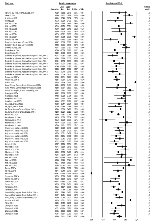
Figure 1 - Forest plot, the effect size (r) of each study (relative weight of each study in the age-related DNA damage). ...20
STUDY 2:
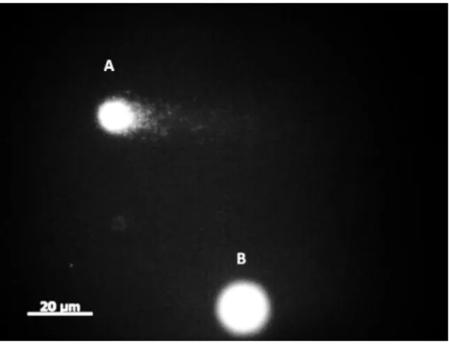
Figure 1 Fluorescent micrograph of comet assay from lymphocytes stained with ethidium bromide. (A) High DNA damage and (B) Low DNA damage. ...41
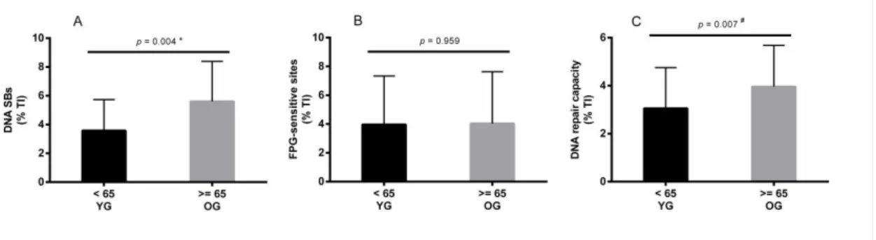
Figure 2 - DNA damage and repair in lymphocytes, measured with the comet assay. A) DNA strand breaks; B) FPG sensitive-sites; C) DNA repair capacity (OGG1). Mean values are shown with SD. Significant differences: * p values were analysed by unpaired t test (normal distribution variables); # p values were analysed by Mann-Whitney test (non-normal distribution variables). ...46
Figure 3 - Biomarkers of antioxidant capacity (TAC) and of oxidative stress (MDA). D) Total antioxidant capacity by age groups; E) Lipoperoxidation (log10 MDA) by age groups. Mean values are shown with SD. p values were analysed by unpaired t test (MDA) (normal distribution variable); p values were analysed by Mann-Whitney test (TAC) (non-normal distribution variable). ...46
STUDY 3:
Figure 1 - Fluorescent micrograph of comet assay from lymphocytes stained with ethidium bromide. (A) Low DNA damage, (B) High DNA damage. ...69
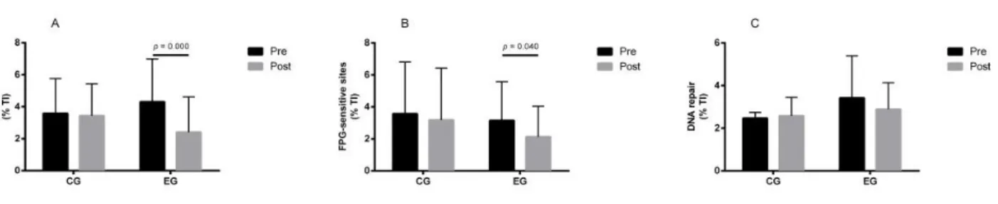
Figure 2 - DNA damage and repair in lymphocytes, measured with the comet assay in control group (CG) and in exercise group (EG), pre-training and post-training. A) DNA strand breaks; B) FPG sensitive-sites; C) DNA repair capacity (OGG1). Mean values are shown with SD. Significant differences: p values, within groups, were analysed by related variables Wilcoxon test (non-normal distribution variables). ... 73
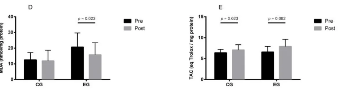
Figure 3 - Biomarkers of antioxidant capacity (TAC) and of oxidative stress (MDA) in control group (CG) and in exercise group (EG), pre-training and post-training. D) Lipoperoxidation (MDA); E) Total antioxidant capacity (TAC). Mean values are shown with SD. Significant differences: p values, within groups, were analysed by related variables Wilcoxon test (non-normal distribution variables). ...74
xxiv STUDY 4:
Figure 1 - Characterization of the OGG1 genotypes. Wild-type (lane 1) genotype Ser326Ser, mutant heterozygote (lane 2) Ser326Cys, and mutant homozygote (lane 3) Cys326Cys. The positions of the molecular weight markers (M) are indicated as 100 bp, 200 bp and 500 bp. ...93
Figure 2 - Data presented are mean + SD measured in the wild-type group (Ser/Ser genotype) and in the Mutant (Ser/Cys and Cys/Cys genotype); A) DNA strand breaks expressed in percentage of tail intensity (%TI); B) FPG sensitive-sites expressed in percentage of tail intensity (%TI); C) DNA repair activity (OGG1) expressed in percentage of tail intensity (%TI); D) Total antioxidant capacity (TAC). a (p) - statistical differences (p ≤ 0.05) within groups, analysed by Wilcoxon test. b (p) - statistical differences (p ≤ 0.05) between each genotype group (hOGG1) in pre-training and post-training evaluation, analysed by Mann-Whitney test. ...95
xxv
T
ABLE
I
NDEX
STUDY 1:
Table 1 - Effect size of the moderator variables (Sex, Tobacco, Sample/Tissue, and Technique) on age-related DNA damage. PBMC – peripheral blood mono-nuclear cells; SCSA – sperm chromatin structure assay; SCGE–CA – Single cell gel electrophoresis- Comet Assay; ELISA - enzyme-linked immunosorbent assay; HPLC – High Performance/Pressure Liquid Chromatography; MN- micronucleus; SCE – sister chromatid exchange...19
STUDY 2:
Table 1 - Characteristics of the Subjects and Lifestyle Variables by Age Group ...45 Table 2 - Correlations between Biomarkers of Damage (DNA and log10 MDA), DNA Repair Capacity, TAC and Lifestyle Variables. ...47
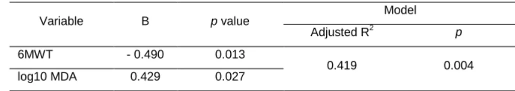
Table 3 - Multiple regression analysis for DNA strand breaks ...48
STUDY 3:
Table 1 - Anthropometric, lifestyle, DNA and oxidative stress variables of the subjects in the Control Group (CG) and Experimental Group (EG) pre and post-training ...73
Table 2 - Physical fitness assessments, pre-training and post-training of the Experimental Group. ...73
STUDY 4:
Table 1 - Mean ± standard deviation values regarding the subject’s anthropometric characteristics and age. ...94
xxvii
L
IST OF
A
BBREVIATIONS
1RM one repetition maximal strength 6MWT six minute walking test
8-oxoG 8-Oxoguanine BMI body mass index CA comet assay CG control group
CMJ counter-movement jump DNA deoxyribonucleic acid DNA SBs DNA strand breaks EG experimental group
ELISA enzyme-linked immunosorbent assay FPG formamidopyrimidine DNA glycosylase HPLC high-performance liquid chromatography IPAQ inventory physical activity questionnaire MDA malondialdehyde
MN micro nucleus test MG mutant group n number of subjects
OGG1 8-Oxoguanine DNA glycosylase PBMC peripheral blood mononuclear cell
PCR-RFLP polymerase chain reaction, restriction fragment length polymorphism PSS perceived stress scale
ROS reactive oxygen species SCE sister chromatid exchange SCSA sperm chromatin structure assay SCGE single cell gel electrophoresis assay SD standard deviation
Sets series
SPPB short physical performance battery STS sit-to-stand test
TAC total antioxidant capacity Wk week
xxviii
WTG wild-type group X2 chi-square
Physical exercise, DNA damage and repair
xxix
A
BSTRACT
Age-related DNA damage is regarded as one of the possible explanations of aging. To better understand the question of age-related DNA damage in humans and to identify possible moderator variables, a meta-analysis was conducted. A total of 76 correlations from 36 studies with 4676 participants were included. Based on our analysis, a correlation between age and DNA damage was found (r = 0.230, p = 0.000; 95% confidence interval = 0.111 - 0.342). The test for heterogeneity of variance indicates that the study´s results are significantly high (Q (75) = 1754.831, p = 0.000). Moderator variables such as smoking habits, technique used, and the tissue/sample analyzed, are shown to influence age-related DNA damage (p=0.026; p=0.000; p=0.000, respectively). Nevertheless, sex did not show any influence on this relation (p=0.114).
Oxidative stress has been regarded as one of the main causes of several damages in DNA. Thus, considering the modulator role of the lifestyle in the production of ROS and consequently in the antioxidant protection, doubts arise regarding how these variables could affect the DNA steady-state, in both damage and repair activity. Considering this and the results from the previous meta-analysis, the purpose of the 2nd study was to investigate how age and lifestyle might affect DNA damage, DNA repair capacity (8-Oxoguanine DNA glycosylase activity (OGG1)) and biomarkers of oxidative stress. Sixty-one healthy men (40 to 84 yrs) were enrolled in this study. The results showed that DNA strand breaks (DNA SBs) and DNA repair capacity were greater in the older group (≥ 65 yrs) compared to the younger group (< 65 yrs) (p<0.05). FPG-sensitive sites, total antioxidant capacity and lipid peroxidation (MDA) were not statistically different between groups. The correlation test showed that DNA damage variables were not correlated with any lifestyle variable excepting DNA SBs which was correlated with aerobic capacity (6 min walking test (6MWT)). DNA SBs and DNA repair were positively correlated with age. The multiple regression analysis revealed that aerobic capacity (6MWT) and lipid peroxidation (MDA) were the predictors for the variation of DNA SBs (41.9%). In conclusion these results suggest that DNA SBs damage increases with age but not FPG-sensitive sites. Moreover, base excision repair capacity increases with age without the increase of oxidative damage to DNA. The most predictable variables of DNA SBs were aerobic capacity and MDA.
Considering the results from the previous study that suggested a possible role of physical exercise training on DNA damage, the purpose of the 3rd study was to identify the effects of 16 weeks of combined physical exercise in DNA damage and DNA repair activity,
Physical exercise, DNA damage and repair
xxx
considering physical fitness improvement. Besides, we aimed to investigate the role of oxidative stress in those changes. Fifty-seven healthy men (40 to 74 yrs) were enrolled in this study. The sample was divided into two groups: the experimental group (EG), composed of 31 individuals, submitted to 16 weeks of combined physical exercise training; and the control group (CG) composed of 26 individuals who did not undergo any specifically orientated physical activity. An improvement of overall physical performance was observed in the EG, after the physical exercise training. A significant decrease in DNA SBs and FPG-sensitive sites was found after the physical exercise training, with no significant changes in OGG1 activity. An increase was observed in antioxidant activity, and a decrease was found in MDA levels after physical exercise training. These results suggest that physical exercise training induces protective effects against DNA damage (DNA SBs and FPG-sensitive sites) possibly related to the increase in antioxidant capacity.
A question emerged from the previous study, as to how physical exercise training affect the individuals with different genetic polymorphisms of the hOGG1 (Ser326Cys) which encode OGG1 repair enzyme. The main purpose of this pilot study was to investigate the possible influence of genetic polymorphisms of the hOGG1 (Ser326Cys) gene on DNA damage and repair activity by OGG1, in response to 16 weeks of combined physical exercise training. Thirty two healthy Caucasian men (40 - 74 years old), were submitted to a training of 16 weeks of combined physical exercise. The subjects with Ser/Ser genotype were considered as wild-type group (WTG), Ser/Cys and Cys/Cys genotype were analyzed together as mutant group (MG). The results from the 4th study showed no differences between DNA damage (both SBs and FPG-sensitive sites) and repair activity (OGG1) between genotype groups (in the pre-training condition). Regarding post-training, the results revealed a decrease in DNA SBs in both groups, a decrease in FPG-sensitive sites and an increase in TAC in the WTG but no changes in MG. No significant changes in OGG1 activity were observed in either genotype groups with physical exercise training. This preliminary study suggests the possibility of different responses in DNA damage to the physical exercise training, considering the hOGG1 Ser326Cys polymorphism.
The overall findings of this thesis showed evidences of the positive effects of physical exercise training in physical fitness, in DNA protection to damage, and in related changes in oxidative stress variables.
Keywords: physical exercise training; DNA damage; DNA repair; aging; oxidative stress; physical fitness.
Physical exercise, DNA damage and repair
xxxi
R
ESUMO
O aumento de danos de DNA com a idade tem sido considerado como uma das possíveis explicações do envelhecimento. Para um melhor entendimento desta questão, ao nível da literatura disponível e para identificar possíveis variáveis moderadoras, foi realizada uma meta-análise em estudos com humanos. Foram incluídos um total de 76 correlações, a partir de 36 estudos com 4676 participantes. Com base na análise dos estudos, foi encontrada uma correlação entre a idade e danos de DNA (r = 0,230, p = 0,000; IC95% = 0,111-,342). O teste de heterogeneidade da variância indica resultados significativamente elevados (Q (75) = 1754,831, p = 0,000). As variáveis consideradas moderadoras na relação idade e danos de DNA foram os hábitos tabágicos, técnica usada e o tecido/amostra analisada (p = 0,026; p = 0,000; p = 0,000, respectivamente). A variável sexo não mostrou qualquer influência sobre a correlação entre a idade e os danos de DNA (p = 0,114).
Considerando o papel modulador do estilo de vida na produção de ROS e, consequentemente, na protecção antioxidante, surgem dúvidas sobre como é que os danos de DNA e a sua capacidade de reparação podem ser afectados por estas variáveis. Tendo em conta o anteriormente exposto e os resultados da meta-análise, o objectivo do 2º estudo foi investigar como é que a idade e estilo de vida podem afectar os danos de DNA, a capacidade de reparação do DNA (8-Oxoguanine DNA glicosilase (OGG1)) em linfócitos de humanos e os biomarcadores de stress oxidativo. Sessenta e um homens saudáveis (40-84 anos) foram incluídos neste estudo, mostrando os resultados que as quebras de cadeia no DNA (DNA SBs) e a capacidade de reparação do DNA estavam aumentadas no grupo mais velho (≥ 65 anos) em comparação com o grupo mais jovem (< 65 anos) (p <0,05). Os danos oxidativos (sítios sensíveis à FPG), capacidade antioxidante total e peroxidação lipídica (MDA) não foram estatisticamente diferentes entre os grupos. O teste de correlação mostrou que as variáveis de danos de DNA não estavam correlacionados com qualquer variável do estilo de vida com excepção dos DNA SBs que se correlacionaram com o teste de capacidade aeróbia (6 minutes walking test (6MWT)). Os DNA SBs e a capacidade de reparação do DNA correlacionaram-se positivamente com a idade. A análise de regressão múltipla revelou que o teste 6MWT e MDA foram os preditores para a variação do DNA SBs (41,9%). Em conclusão estes resultados sugerem que os DNA SBs aumentam com a idade, mas os sítios sensíveis à FPG não. Além disso, a actividade da OGG1 aumenta com a idade, sem o aumento dos sítios sensíveis à FPG. As variáveis preditores dos DNA SBs foram a capacidade aeróbica e o MDA.
Physical exercise, DNA damage and repair
xxxii
Considerando os resultados do estudo anterior, que sugeriu um possível papel do exercício físico em danos no DNA, realizou-se o terceiro estudo com o objectivo de identificar os efeitos de 16 semanas de exercício físico combinado nos danos de DNA e na capacidade de reparação de DNA (OGG1), considerando a melhoria da aptidão física, outro objectivo foi investigar o papel do stress oxidativo nessas alterações. Foram incluídos neste estudo cinquenta e sete homens saudáveis (40-74 anos). A amostra foi dividida em dois grupos: o grupo experimental (EG), composto por 31 indivíduos, submetidos a um programa de 16 semanas de exercício físico combinado; e o grupo de controlo (CG) composto por 26 indivíduos que não realizaram o programa de exercício. Após o programa de exercício foi observada uma melhoria do desempenho físico geral do EG, foi encontrada uma diminuição significativa dos DNA SBs e dos sítios sensíveis à FPG, sem alterações significativas na actividade da OGG1. Os resultados mostraram também um aumento da actividade antioxidante e uma diminuição dos níveis de peroxidação lipídica após exercício físico. Estes resultados sugerem que o exercício físico regular induz efeitos protectores contra danos de DNA em linfócitos humanos, possivelmente relacionados com o aumento da capacidade antioxidante.
Com os resultados do estudo anterior, surgiu a questão, se o exercício físico afecta de forma diferente indivíduos com diferentes variantes genéticas do hOGG1 (Ser326Cys) que codifica a enzima de reparação, a OGG1. O principal objectivo deste quarto estudo (piloto) foi investigar a possível influência de polimorfismos genéticos do gene hOGG1 (Ser326Cys) nos danos de DNA e na actividade de reparação da OGG1, na resposta a um programa de treino de 16 semanas de exercício físico combinado. Trinta e dois homens caucasianos saudáveis (40-74 anos), foram submetidos ao programa de exercício. Os indivíduos com genótipo Ser/Ser foram considerados como do grupo tipo selvagem (WTG), o genótipo Ser/Cys e o Cys/Cys foram analisadas em conjunto, como grupo mutante (MG). Os resultados deste estudo demonstraram não haver diferenças entre os dano de DNA (SBs e sítios sensíveis à FPG) e na actividade da OGG1 entre os grupos (na condição pré-treino). Em relação ao pós-treino, os resultados revelaram uma diminuição no DNA SBs em ambos os grupos, uma diminuição nos sítios sensíveis à FPG e um aumento da TAC no WTG mas nenhuma alteração foi encontrada no MG. Não ocorreram alterações significativas na actividade da OGG1 em nenhum dos grupos com o programa de exercício. Este estudo preliminar sugere a possibilidade de sujeitos com o polimorfismo hOGG1 Ser326Cys responderem de forma diferente a um programa de exercício físico, relativamente aos danos de DNA.
Physical exercise, DNA damage and repair
xxxiii
Em termos globais, os resultados destes quatro estudos mostram evidências dos efeitos positivos do exercício físico regular na aptidão física, na protecção contra os danos de DNA, e nas alterações em variáveis relacionadas com o stress oxidativo.
Palavras-chave: exercício físico regular; danos de DNA; reparação de DNA; Envelhecimento; stress oxidativo.
Physical exercise, DNA damage and repair General Introduction
3
Aging can be defined as a complex process that occurs throughout life and is characterized by a progressive organic functional decline, more disease susceptibility, and an increase in death probability (Mota et al., 2004; Weinert and Timiras, 2003; Wilson et al., 2008). An important aspect that should be mentioned is that aging tends to enhance the variability of population characteristics, suggesting that this age-related decline is variable between individuals. In other words, there is a set of several factors, namely genetic and stochastic, that might influence the aging expression in different individuals (Weinert and Timiras, 2003; Westerterp, 2000). This is in the origins of development of several theories in last decades aiming to identify the causes and the process that explain aging and its consequences. After several years of research, many different explanations emerged but contradictions persist and a comprehensive and universally accepted theoretical model remains unreachable (Jin, 2010). Theories of aging that continue to grow may be placed in two main categories: programmed and non-programmed theories. The programmed theories imply that aging follows a biological timetable regulated by genetic mechanisms. Conversely, the non-programmed theories emphasize that throughout life, damage accumulates at various levels, which in turn can lead to aging (Jin, 2010; Martins, 2011; Schumacher et al., 2008). Nevertheless, nowadays, according to some researchers, aging seems to be considered as a complex and multifactorial process that must consider and reconcile all data, theories, and arguments (Rattan, 2006; Rosa et al., 2005; Trindade et al., 2013; Weinert and Timiras, 2003). Aging phenotype is quite variable between subjects from the same species, between organs, tissues, and cells from the same body, and between macromolecules within the same cell (Rattan, 2006). According to this approach, aging should not have a unique and universal cause, a unique phenotype and consequence, besides death (Rattan, 2006). Among the different explanations for aging and regarding their main possible causes, the importance of the genetic alterations that occur throughout life and might lead to progressive organic dysfunction and aging has been recognized. The integrity of the genetic information has a central role in cell function and consequently at a systemic level. Therefore, possible alterations that occur in DNA that are not properly repaired could lead to accumulating damage, mutations, progressive dysfunction, and increased aging (Tamarin, 2002). Regarding this, numerous investigators have studied the possible contribution of DNA damage to aging and to age-related disease. Indeed, studies have found an association between damage to DNA and some age-related diseases, including: cardiovascular disease (Collins et al., 1998), diabetes mellitus (Hannon-Fletcher et al., 2000), cataracts (Jiang et al., 2013; Su et al., 2013), and several cancers (Akcay et al., 2003; Malins et al., 2001; Peddireddy et al., 2012).
Physical exercise, DNA damage and repair General Introduction
4
The study of the damage to DNA and its associated mechanisms have been the subject of numerous researches (Freitas and de Magalhaes, 2011). Indeed, some researches have been conducted aiming to study the relation between DNA damage (chromosomal aberrations, DNA cross-links, DNA strand breaks, oxidized bases) and aging. However, the lack of consensus of the results found that some works supported this association in different animal species (Ames and Gold, 1991; Freitas and de Magalhaes, 2011; Giovannelli et al., 2003; Ozawa, 1995; Schumacher et al., 2008). Regarding the inconsistency of the results, it is important to note that differences in methodologies and work designs used are very common. Taking into consideration that there are inter-individual aging phenotypes, it seems reasonable to expect that the possible increase in damage might be different within the several organic macromolecules. In addition, considering that several kinds of damage may occur, it is also expected that, depending on the type of damage assessed, dissimilar results could be found. Thus, these differences might help explain some of the contradictory results found. It is also relevant to mention that there is supporting data showing that the level of DNA damage is different between subjects. Therefore, if this variability could not be explained solely based on the intrinsic individual characteristics that cannot be modified, environmental factors related with lifestyle could also help to explain it. Bearing in mind the complexity of aging, the relation between age and DNA damage in humans is not very clear, nor is the possible influence of some moderator variables, such as methodological issues and lifestyle variables, in this relation. (Study 1)
There are several different processes, both endogenous and exogenous, with physical, biological, and chemical origins that could lead to DNA damage (Maynard et al., 2009). Among the several potential genotoxic substances, reactive oxygen species (ROS), which could react with several different organic structures, such as lipids, proteins, enzymes, nucleic acids and other molecules causing dangerous changes and cell dysfunction, have been the focus of special attention (Beckman and Ames, 1998a, b; Ortenblad et al., 1997; Sen, 1995). The possible injuries caused, directly or indirectly, by ROS in DNA are predominantly single strand breaks and modified bases (Lombard et al., 2005), 8-oxoguanine (8-oxoG) has been considered as one of the most important damages found (Wilson et al., 2003). The 8-oxoG originates from the reaction of the hydroxyl radical (OH•) with the guanine base. This is one of the most abundant and potentially mutagenic lesions and therefore has been used as an oxidative stress biomarker (Kasai et al., 1986).
Though a protection system against ROS injuries in cells exists, such as antioxidant protection and repair capacity, this protection does not prevent all the ROS from becoming neutralized, leading to several types of damage, such as in DNA. For instance, it is estimated
Physical exercise, DNA damage and repair General Introduction
5
that in DNA 100 to 500 of 8-oxoG are produced by a cell each day (Collins and Gaivao, 2007). The cytotoxic and mutagenic effect of this damage depends on the efficiency of the repair mechanisms in the cell. Indeed, a poor repair capacity is normally associated with more persistent damage, which could lead to mutations or cell death (Cooke et al., 2003; Guetens et al., 2002). The damage, which might lead to mutation in critical areas that control cell growth or cell differentiation, might put the normal cell function at risk, leading to dysfunction and disease (Cooke et al., 2003; Randerath et al., 1996; Tamarin, 2002; Torres, 1994; Wilson et al., 2003).
Environmental factors and their possible influence on DNA damage with age, diet, smoking, psychological stress, exposure to pollutants, and daily physical activity have received some attention (Fenech and Bonassi, 2011; Garaj-Vrhovac et al., 2008; Mladinic et al., 2010; Tamae et al., 2009). As mentioned above, oxidative stress has been regarded as one of the main causes of several damages namely in DNA. Thus, considering the modulator role of the lifestyle in the production of ROS and consequently in the antioxidant protection, doubts arise regarding how these variables could affect the DNA steady-state, in both damage and repair activity. (Study 2)
Considering lifestyle factors, physical activity has been reported as one of the most relevant factors in population health. Daily physical activity, when regular and programmed – known as physical exercise training - has a positive modulatory role in the overall health of individuals (Nelson et al., 2007; Rosa et al., 2005). Its effect in the body tends to improve and preserve its functions, and active subjects tend to have higher levels of overall function compared to sedentary subjects (revised in Gerber et al. 2011). These effects seem to be explained by the fact that physical exercise induces various imbalances that lead to a structural and functional reorganization that varies with internal and/or external demands.
Physical exercise training has been associated with acute increased production of ROS (Leeuwenburgh and Heinecke, 2001; Munoz Marin et al., 2010). Several studies indicate that chronic physical exercise improves the antioxidant protection and/or the repair mechanisms (Bloomer and Fisher-Wellman, 2008; Lambertucci et al., 2006). Since DNA could be one of the potential targets of ROS, it is possible to admit that physical exercise training might promote various adjustments that may affect the DNA steady-state, including adaptations in antioxidant protection and repair mechanisms. It is likely that the higher oxidative damage caused by higher exposure to ROS during physical exercise might induce repair activity over time and consequently lead to a more efficient repair of the oxidative damage to DNA. Another possibility is that a higher production of ROS might induce antioxidant adaptations, leading to a decrease of oxidative damage to DNA. Although there
Physical exercise, DNA damage and repair General Introduction
6
are some data indicating the benefits of physical exercise training in DNA steady-state, it is also worth mentioning that all of those possible adaptations to physical exercise depend on its workload. Moreover, the linkage between DNA damage, DNA repair, and oxidative stress changes in response to physical exercise training, which is not yet fully understood. Bearing this in mind, doubts arise about the effects of exercise in DNA damage and repair activity considering oxidative stress changes. (Study 3)
Another issue that should be emphasized is that different individuals could respond differently to the same environmental requirements. Thus, the study of genetic characteristics that affect the organic responses has revealed the existence of genetic differences that influence the adaptations/cellular responses of individuals. Some variations in genes that appear to have influence on repair activity have been reported in the literature. For example, the polymorphism in the repair gene hOGG1 Ser326Cys, which encodes the 8-Oxoguanine DNA glycosylase 1 (OGG1) enzyme, has been studied. OGG1 is a bifunctional glycosylase/AP lyase, which recognizes oxoG:C pairs and catalyse both the removal of 8-oxoG and the cleavage of the DNA backbone (Boiteux and Radicella, 2000; Kershaw and Hodges, 2012). The functional significance of these genetic polymorphisms is not very clear; however, published data have suggested associations between the OGG1 Ser326Cys variant and an increased risk of various types of cancer, including lung, head, neck, and hepatocellular carcinoma (Goode et al., 2002; Yuan et al., 2012), as well as a risk of cataracts (Zhang et al., 2012). A variant of this gene seems to be associated with a decreased ability to repair damage (Aka et al., 2004; Kohno et al., 1998); however, there are some contradictory results (Dherin et al., 1999; Jensen et al., 2012). Considering the increased production of ROS associated with physical exercise and the possibly of increased 8-oxoG damage - the most abundant lesion generated by oxidative stress - it seems relevant to study whether or not physical exercise training has the same impact on individuals with genetic polymorphisms related to DNA repair enzymes. (Study 4)
Considering the several issues raised, the general purpose of this work was to investigate the role of physical exercise in age-related DNA damage and repair activity considering the possible influence of oxidative stress. An additional purpose was to investigate how the genetic polymorphism (hOGG1 Ser326Cys) might influence these induced responses.
In order to answer the issues raised, four studies were performed and organized in the following main chapters:
Physical exercise, DNA damage and repair General Introduction
7
Chapter 2 includes four studies:
Study 1 is a meta-analysis review that aims to investigate age-related DNA
damage in humans and identify the possible moderators of this relation.
Study 2 is a cross-sectional study in humans that aims to investigate whether
age and lifestyle may affect DNA damage (strand breaks and FPG-sensitive sites), and DNA repair activity (OGG1 enzyme) considering oxidative stress related variables.
Study 3 is a longitudinal study regarding the investigation of the effects of 16
weeks of combined physical exercise training in DNA damage and repair activity considering oxidative stress changes.
Study 4 aims to study whether or not genetic polymorphisms affect human
responses in DNA damage and repair to physical exercise training.
Chapters 3 and 4 present a general discussion of the results obtained in the four studies and their main conclusions, respectively.
Study 1: Aging and DNA damage in humans: a meta-analysis
study
Aging and DNA damage in humans: a meta-analysis study
13 Title
Aging and DNA damage in humans: a meta-analysis study
1Jorge Pinto Soares; 2António Cortinhas; 1Teresa Bento; 1José Carlos Leitão; 3Andrew R.
Collins; 4Isabel Gaivão; 1Maria Paula Mota
1CIDESD, University of Trás-os-Montes and Alto Douro, Vila Real, Portugal 2
University of Trás-os-Montes and Alto Douro, Vila Real, Portugal
3Department of Nutrition, Faculty of Medicine, University of Oslo, Oslo, Norway 4
CECAV - Genetic and Biotechnology Department, University of Trás-os-Montes and Alto Douro, Vila Real, Portugal
Aging and DNA damage in humans: a meta-analysis study
15 Abstract
Age-related DNA damage is regarded as one of the possible explanations of aging. Although a generalized idea about the accumulation of DNA damage with age exists, results found in the literature are inconsistent. To better understand the question of age-related DNA damage in humans and to identify possible moderator variables, a meta-analysis was conducted.
Electronic databases and bibliographies for studies published since 2004 were searched. Summary odds ratios (ORs) and 95% confidence intervals (CIs) for age-related DNA damage were calculated in a random-effects model.
A total of 76 correlations from 36 studies with 4676 participants were included. Based on our analysis, a correlation between age and DNA damage was found (r = 0.230, p = 0.000; 95% confidence interval = 0.111 - 0.342). The test for heterogeneity of variance indicates that the study´s results are significantly high (Q (75) = 1754.831, p = 0.000). Moderator variables such as smoking habits, technique used, and the tissue/sample analyzed, are shown to influence age-related DNA damage (p=0.026; p=0.000; p=0.000, respectively). Nevertheless, sex did not show any influence on this relation (p=0.114).
In conclusion, this meta-analysis showed an association between age and DNA damage in humans. It was also found that smoking habits, the technique used, and tissue/sample analyzed, are important moderator variables in age-related DNA damage.
Aging and DNA damage in humans: a meta-analysis study
17 1. Introduction
Aging has been defined as a progressive organic functional decline, with loss of homeostasis and increasing probability of illness and death (Schumacher et al., 2008). Although applied research on aging has resulted in considerable scientific knowledge with regard to the related causes, it is still subject to numerous debates and contradictions. Indeed, in most cases, it is difficult to understand how a particular variable is a possible cause or consequence of aging. Some studies have suggested an age-related accumulation of macromolecular damage, which may cause progressive and irreversible physiological attrition and homeostasis loss, accelerating aging. In addition, considering the important role of DNA in living organisms and regarding the changes that occur in this macromolecule throughout life, the question arises, whether DNA modifications should be considered a central factor of aging. Moreover, it is not clear if these damages are a possible cause or an expression of aging. Since the first study of Failla (1958), numerous other investigations have been performed and different assays to measure DNA changes have been developed. These assays allow the assessment of some age-related DNA changes, as well as other associated variables that could interfere with DNA damage accumulation.
Many theories of aging are based on DNA changes, including the Intrinsic Mutagenesis Theory, Somatic Mutations Theory and DNA Repair Theory (Rattan, 2006; Weinert and Timiras, 2003). Other theories also explain the age-related changes in DNA as a consequence of stochastic events. The Oxidative Stress Theory is, perhaps, the main stochastic explanation of DNA and other macromolecular damage accumulating with age (Rattan, 2006). Despite the importance of the age-related DNA damage accumulation, some researchers argue that aging is not caused by the accumulation of damage but is the result of continued activity (cell hyperfunction) of pathways and processes during adulthood that evolved to optimize development to this life stage (Gems and de la Guardia, 2013; Leontieva et al., 2012). According to this theory, the same pathway, which drives developmental growth, later drives aging and associated diseases (Blagosklonny, 2012). Cell hyperfunction is driven by the nutrient-sensitive signaling network that controls growth (and thereby, reproduction), and includes the insulin, insulin-like growth factor 1 (IGF-1), and in particular, the target of rapamycin (TOR) kinase pathways (Blagosklonny, 2012; Blagosklonny and Hall, 2009; Leontieva et al., 2012). Thus, accumulation of DNA damage is a consequence of aging and not a leading cause.
Although DNA is not the only target changed with aging, taking account of the major role of this macromolecule in the regulation of all cellular structures and its own cell cycle, DNA damage has been studied with particular attention. The alterations could have several
Aging and DNA damage in humans: a meta-analysis study
18
consequences for genome stability with repercussions on cellular component synthesis, cell cycle machinery and signaling pathways that control cell cycle arrest, and programed cell death or apoptosis (Lombard et al., 2005). The consequences of DNA damage will depend on the type of damage, genes affected and type of cell and tissue damaged.
The prevailing view is that there is a tendency for an age-related DNA damage accumulation. However, on examination, results of studies show inconsistency (Coronas et al., 2009; Gandhi and Kumar, 2004; Pedret et al., 2012; Wyrobek et al., 2006); it is possible that confounding factors influence this relation and explain some of the inconsistency.
Considering the complexity of aging, it must be emphasized that aging does not happen in the same way in different individuals, nor in the same way in all cell types and tissues of the same individual. Moreover, aging is a life-long process, influenced continually by environmental conditions. Factors such as diet, lifestyle, exposure to radiation and genotoxic chemicals seem to have a significant influence on the relationship between cumulative DNA damage and age (Kazimirova et al., 2004; Sellappa et al., 2010; Tamae et al., 2009).
Methodological factors might have also influenced the observed results (Kazimirova et al., 2004; Sellappa et al., 2010; Tamae et al., 2009). Indeed, different assays may be used to measure DNA damage. Furthermore, the measured DNA damage could reflect changes in the causative factors, and/or changes in DNA protection and/or changes in DNA repair capacity. It must also be noted that the type of cell and tissue used could reflect different aging rates within the organism.
Although there are several excellent narrative reviews on age-related nuclear DNA damage (Fenech and Bonassi, 2011; Freitas and de Magalhaes, 2011; Seviour and Lin, 2010; Wilson et al., 2008), they usually refer to individual animal and humans studies and, as far as we know, no meta-analytic technique has been used to estimate the extent of effect of potential moderators on age-related DNA damage in humans. Thus, the overall goal of this paper is to address this important gap in the literature. The first aim of this review is to provide a summary of age-related changes in nuclear DNA in humans. The second aim is to examine the effects of some moderators associated with DNA damage. The third aim is to discuss promising directions for future researches in the light of our findings.
2. Results
An initial search using the keywords described located 2953 studies. After reading titles and abstracts, the number of studies was reduced to 267. In the final refinement of the
Aging and DNA damage in humans: a meta-analysis study
19
research, applying inclusion and exclusion criteria, only 36 studies fulfilled all necessary requirements.
3. Study Analysis
The results indicated a significant and positive association between age and DNA damage in 76 correlations of the 36 studies (N = 4676) r = 0.230, p = 0.000 (95% confidence interval = 0.111; 0.342). A test for heterogeneity of variance indicates that the results of the study are significantly higher than would be expected, Q (75) = 1754.831, p = 0.000. The effect size of each study can be seen in figure 1.
The analysis of the moderator variables is shown in table 1. As can be seen, tobacco use, sample/tissue and technique, but not sex, are identified as moderator variables.
Table 1 - Effect size of the moderator variables (Sex, Tobacco, Sample/Tissue, and Technique) on age-related DNA damage. PBMC – peripheral blood mono-nuclear cells; SCSA – sperm chromatin structure assay; SCGE–CA – Single cell gel electrophoresis- Comet Assay; ELISA - enzyme-linked immunosorbent assay; HPLC – High Performance/Pressure Liquid Chromatography; MN- micronucleus; SCE – sister chromatid exchange.
Moderator Value K R 95%CI P p between
groups Sex Female 25 0.116 0.05; 0.18 0.000 0.114 Male 14 0.177 0.13; 0.22 0.000 Tobacco Non-Smokers 26 -0.043 -0.10; -0.02 0.162 0.026 Smokers 2 0.176 -0.01; 0.35 0.058 Sample/ Tissue Buccal 4 0.229 0.14; 0.32 0.000 0.000 Mammary 1 -0.106 -0.45; 0.26 0.580 PBMC 57 0.235 0.20; 0.27 0.000 Spermatozoa 3 0.392 0.28; 0.49 0.000 Urine 11 -0.007 -0.05; 0.03 0.711 Technique SCSA 1 0.720 0.60; 0.81 0.000 0.000 SCGE–CA 47 0.208 0.17; 0.24 0.000 ELISA 7 -0.083 -0.14; -0.03 0.002 HPLC 8 0.072 0.02; 0.12 0.007 MN 11 0.268 0.19; 0.34 0.000 SCE 2 0.831 0.73; 0.90 0.000
Aging and DNA damage in humans: a meta-analysis study
20
Figure 1 - Forest plot, the effect size (r) of each study (relative weight of each study in the age-related DNA damage). IC=confidence interval. a, b, c, d, e, f, g, h – different measured endpoints from the same study.
Study name Statistics for each study Correlation and 95% CI
Lower Upper Relative Relative
Correlation limit limit Z-Value p-Value weight weight
Agrawal, Tay, Yang, Agrawal, & Gupta, 2010 0,229 -0,182 0,573 1,095 0,273 1,24
Aka et al., 2004 0,669 0,407 0,829 4,203 0,000 1,28 C. Y. Huang 2012 0,164 0,040 0,284 2,579 0,010 1,43 Cheng 2013 0,058 -0,309 0,409 0,301 0,764 1,28 Chin et al., 2008 a 0,720 0,496 0,854 4,888 0,000 1,29 Chin et al., 2008 b 0,210 -0,150 0,521 1,148 0,251 1,29 Chin et al., 2008 c 0,240 -0,119 0,543 1,318 0,187 1,29 Chin et al., 2008 d 0,250 -0,108 0,551 1,375 0,169 1,29 Chin et al., 2008 e 0,320 -0,032 0,602 1,786 0,074 1,29 Chin et al., 2008 f 0,900 0,803 0,950 7,928 0,000 1,29 Coronas et al 2009 -0,153 -0,442 0,164 -0,946 0,344 1,32
Danadevi, Rozati, Banu, & Grover, 2004 a 0,197 0,003 0,377 1,986 0,047 1,40 Danadevi, Rozati, Banu, & Grover, 2004 b 0,438 0,266 0,583 4,674 0,000 1,40
Donmez-Altuntas 2012 0,557 0,352 0,710 4,733 0,000 1,37
Dusinska et al., 2004 a 0,036 -0,576 0,622 0,102 0,919 0,99
Dusinska et al., 2004 b 0,139 -0,503 0,682 0,396 0,692 0,99
Dusinska, Dzupinkova, Wsolova, Harrington, & Collins, 2006 a -0,067 -0,285 0,158 -0,581 0,561 1,39 Dusinska, Dzupinkova, Wsolova, Harrington, & Collins, 2006 b 0,363 0,135 0,555 3,043 0,002 1,38 Dusinska, Dzupinkova, Wsolova, Harrington, & Collins, 2006 c 0,001 -0,223 0,225 0,009 0,993 1,39 Dusinska, Dzupinkova, Wsolova, Harrington, & Collins, 2006 d 0,238 -0,006 0,455 1,911 0,056 1,37 Dusinska, Dzupinkova, Wsolova, Harrington, & Collins, 2006 e 0,099 -0,134 0,322 0,831 0,406 1,38 Dusinska, Dzupinkova, Wsolova, Harrington, & Collins, 2006 f 0,108 -0,142 0,345 0,847 0,397 1,37 Dusinska, Dzupinkova, Wsolova, Harrington, & Collins, 2006 g -0,051 -0,270 0,173 -0,442 0,658 1,39 Dusinska, Dzupinkova, Wsolova, Harrington, & Collins, 2006 h 0,249 0,009 0,462 2,035 0,042 1,38
Frisard et al., 2007 a 0,080 -0,071 0,228 1,035 0,300 1,42
Frisard et al., 2007 b 0,127 -0,024 0,272 1,645 0,100 1,42
Gandhi 2004 0,946 0,879 0,976 8,346 0,000 1,24
Garaj-Vrhovac, Durinec, Kopjar, & Orescanin, 2008 a 0,184 -0,170 0,496 1,018 0,308 1,29 Garaj-Vrhovac, Durinec, Kopjar, & Orescanin, 2008 b 0,138 -0,367 0,580 0,521 0,602 1,15 Gianni, Jan, Douglas, Stuart, & Tarnopolsky, 2004 0,698 0,392 0,865 3,767 0,000 1,22
Huang et al. 2013 a 0,482 0,050 0,762 2,167 0,030 1,19
Huang et al. 2013 b 0,432 -0,013 0,734 1,906 0,057 1,19
Humphreys et al., 2007 a 0,338 0,120 0,525 2,987 0,003 1,38
Humphreys et al., 2007 b 0,490 0,322 0,628 5,197 0,000 1,40
Irie, Miyata, & Kasai, 2005 a 0,220 -0,247 0,604 0,922 0,356 1,19
Irie, Miyata, & Kasai, 2005 b 0,060 -0,256 0,365 0,365 0,715 1,32
Irie, Tamae, Iwamoto-Tanaka, & Kasai, 2005 a 0,290 0,083 0,473 2,720 0,007 1,39 Irie, Tamae, Iwamoto-Tanaka, & Kasai, 2005 b -0,100 -0,214 0,016 -1,688 0,091 1,44
Kanaya et al., 2004 0,190 0,131 0,246 6,283 0,000 1,45 Kazimirova et al., 2004 a 0,420 0,020 0,704 2,052 0,040 1,24 Kazimirova et al., 2004 b -0,350 -0,660 0,062 -1,675 0,094 1,24 Kazimirova et al., 2004 c 0,570 0,216 0,791 2,967 0,003 1,24 Kazimirova et al., 2004 d 0,500 0,121 0,752 2,517 0,012 1,24 Krajcovicová-kudlácková 2007 a 0,182 -0,030 0,379 1,683 0,092 1,39 Krajcovicová-kudlácková 2007 b 0,236 0,023 0,429 2,171 0,030 1,39 Krajcovicová-kudlácková 2007 c 0,276 0,071 0,459 2,619 0,009 1,39 Krajcovicová-kudlácková 2007 d 0,094 -0,125 0,304 0,839 0,402 1,39 Krajcovicová-kudlácková 2007 e 0,252 0,045 0,439 2,375 0,018 1,39 Krajcovicová-kudlácková 2007 f 0,021 -0,197 0,237 0,185 0,853 1,39 Leng et al., 2004 0,087 -0,235 0,391 0,521 0,602 1,32 Mladinic et al., 2010 a 0,375 0,138 0,571 3,025 0,002 1,37 Mladinic et al., 2010 b 0,497 0,282 0,664 4,187 0,000 1,37 Mladinic et al., 2010 c 0,190 -0,063 0,419 1,474 0,140 1,37 Mladinic et al., 2010 d 0,344 0,103 0,547 2,754 0,006 1,37 Mota et al., 2010 a 0,322 -0,435 0,812 0,818 0,413 0,90 Mota et al., 2010 b 0,738 0,145 0,941 2,317 0,020 0,90 Mota et al., 2010 c 0,155 -0,433 0,650 0,494 0,621 1,06 Mota et al., 2010 d 0,655 -0,092 0,930 1,753 0,080 0,83 Pal et al., 2007 a 0,411 0,134 0,629 2,833 0,005 1,34 Pal et al., 2007 b 0,174 -0,040 0,372 1,597 0,110 1,39 Pedret 2012 -0,997 -0,998 -0,996 -35,675 0,000 1,41 Schmid et al., 2007 a 0,220 0,000 0,419 1,963 0,050 1,39 Schmid et al., 2007 b 0,060 -0,162 0,276 0,527 0,598 1,39 Soliman et al., 2004 -0,154 -0,482 0,212 -0,821 0,411 1,28 Surowy 2011 0,228 0,003 0,431 1,983 0,047 1,38 Tamae et al., 2009 a -0,014 -0,117 0,089 -0,265 0,791 1,44 Tamae et al., 2009 b 0,190 0,089 0,288 3,639 0,000 1,44
Vuyyuri, Ishaq, Kuppala, Grover, & Ahuja, 2006 a 0,362 0,240 0,472 5,545 0,000 1,43 Vuyyuri, Ishaq, Kuppala, Grover, & Ahuja, 2006 b 0,097 -0,041 0,231 1,373 0,170 1,43 Weng, Weng, Lu, Nakayama, & Morimoto, 2009 0,239 0,064 0,400 2,659 0,008 1,41
Wyrobek et al., 2006 0,720 0,601 0,808 8,368 0,000 1,39 Yilmaz 2012 -0,106 -0,449 0,264 -0,553 0,580 1,28 Zhang et al., 2011 a 0,336 0,142 0,505 3,314 0,001 1,40 Zhang et al., 2011 b 0,072 -0,134 0,272 0,684 0,494 1,40 Zhang et al., 2011 c 0,045 -0,160 0,246 0,425 0,671 1,40 Zhang et al., 2011 d 0,007 -0,197 0,210 0,066 0,947 1,40 0,230 0,111 0,342 3,747 0,000 -1,00 -0,50 0,00 0,50 1,00
Aging and DNA damage in humans: a meta-analysis study
21 4. Discussion
DNA changes associated with age have been claimed as one of the main possible causes of aging. These alterations may result in genetic instability, mutagenesis, disease, and cell death. Despite its popularity, this association has a lack of consensus in the literature apparently due to several factors such as sample characteristics, technique and methods used. To clarify this relation, we conducted a meta-analysis to investigate the association between age and DNA damage in humans. Our main finding is a positive association between age and DNA damage in humans, both in males and females. So, aging in humans is accompanied by an increase in general DNA damage. However, the association found is weak and the Cochran’s Q statistic and the I2 statistic revealed a high heterogeneity between studies. To better understand this association, there are some points that must be taken into consideration. First of all, this weak association implies that there are other variables which may influence age-related DNA damage. Variables suggested in the literature include sex, smoking, alcohol consumption, physical exercise, nutrition, psychological stress, etc. Secondly, the sample’s characteristics, technique used, tissue and the type of damage analyzed could influence results. Considering this, some variables were analyzed here as possible moderator variables influencing age-related DNA damage. Several works have studied the effect of sex on DNA damage. Despite the fact that some studies have identified differences between sexes in DNA damage (Cheng et al., 2013; Donmez-Altuntas and Bitgen, 2012; Kanaya et al., 2004), our results based on analysis of 36 studies have shown no such differences. This means that both sexes show increased DNA damage with age, even though the absolute values could be different. In short, sex as a moderator variable has no influence on age-related DNA damage.
Although in the literature several lifestyle variables have been related with DNA damage, according to the results from the studies analyzed here, only smoking habit could be considered as a moderator variable; at the time of our analysis, there were not enough studies concerning the remaining variables. Since tobacco smoke contains known carcinogens, it seems plausible that smokers could accumulate more DNA damage with age, compared with non-smokers. Our analysis confirms this hypothesis, showing that smokers demonstrate more age-related DNA damage. Our results clearly suggest that smoking should be considered as a moderator variable in the age-related DNA damage studies.
As mentioned before, it is well established that aging does not occur at the same rate in the different organs. Accordingly, it might be expected that different sample tissues might show different age-related DNA damage. Considering the sample tissue studied, we found
Aging and DNA damage in humans: a meta-analysis study
22
significant differences between them. Unexpectedly, mammary cells and urine samples have shown no correlation with age-related DNA damage. However, there was only one study of mammary tissue cells and the urine sample results show high heterogeneity leading to high variability of the results. On the remaining cell types studied (buccal cells, peripheral blood cells and spermatozoa), less heterogeneity between studies was seen, and a positive correlation with age-related DNA damage was found.
Our results have shown that technique is a moderator variable when age-related DNA damage is studied, so that depending on the technique used, we might expect different results. Studies using ELISA have shown a weak negative association between age and DNA damage; HPLC, SCGE - CA and MN have shown a positive association but also weak; and a strong association for SCSA and SCE has been found, even though these last two were based on only one and two studies, respectively. Regarding these techniques, it is important to mention that ELISA and HPLC are the only two techniques which are used in urine and/or in tissues cells. This is of major importance because results outcomes are clearly different, since in the case of urine samples DNA damage is not analyzed directly, but rather the result of that damage in the overall body system. So our results illustrate the importance of careful interpretation, especially in comparisons of results from different studies.
There are some questions underlying age-related accumulation of DNA damage which must be taken into account. Firstly, it was our proposal to study the set of variables that might be associated with the DNA damage and aging. Though we were only able to consider smoking habits, other lifestyle variables should be evaluated in studies with human samples. Also, the methodology is important for understanding the results and interpreting heterogeneity. Sample characteristics as well as inclusion criteria are relevant to understand the results achieved. The range of ages included in a study is likely to influence the results – a wide range likely leading to more pronounced effects: in this meta-analysis age range was not considered due the variability of the studies designs and to the lack of information presented in the articles. Further, the weak association between DNA damage and age found in this meta-analysis raises the question whether accumulation of damage is determinative for the aging process. As mentioned above, it is not clear whether age-associated damage is a cause or an expression of aging. The molecular damage theory has postulated that aging is caused by the progressive accumulation of damage; however according to the hyperfunction theory this damage is a consequence of aging and (Blagosklonny, 2012) thus does not necessarily limit lifespan (Blagosklonny, 2012, 2013; Gems and de la Guardia, 2013). Instead, the observed increased levels of damage are
Aging and DNA damage in humans: a meta-analysis study
23
important for some pathologies, such as cancer, and are the result of hyperfunction. The hyperfunction theory even suggests that repair of molecular damage is important for increased longevity, but the involvement of any process for viability does not imply its role in aging (Blagosklonny, 2012). In summary, there are emerging explanations concerning our understanding of aging, which provide a novel perspective on aging and the DNA accumulation of damage.
In conclusion, independent of the perspective theory of aging, our meta-analysis results show an age-related increase in DNA damage in humans. Furthermore, smoking habits, the sample/tissue and the analysis technique used are important moderator variables. There is a set of lifestyle variables which should be more carefully studied in longitudinal studies, since it seems that age-related DNA damage only explains a small part of aging. Future studies may also rely on the relevance of the age-related DNA damage and its possible role as a marker of biological aging.
5. Methods
We have conducted a analysis based on the criteria described for meta-analyses and systematic reviews by Moher et al. (2009).
6. Data Source and search strategy
In order to achieve the largest number of publications, MEDLINE PubMed and Web of science (Web of ScienceSM; current Contents Connect) databases were used with the combination of the English key terms: “DNA”, “damage” and “age”, and all eligible studies between 2004 and “March 2013” were selected.
In addition, the reference lists in these articles were searched manually to find other relevant publications.
7. Selection Criteria and Identification of Studies
The following inclusion criteria were used to select the articles to our study: articles in English, studies on nuclear DNA, male and/or female human studies, healthy subjects or studies with healthy control subjects, papers clearly describing the sources of cases and controls, and information given on the size of the sample and statistical values.
The exclusion criteria were: post-mortem studies, in vitro studies, studies of newborns, children and puberty, studies in exposed subjects without control group, and studies in non-healthy subjects without healthy control group.
Aging and DNA damage in humans: a meta-analysis study
24
The following data were collected from each study: first author’s surname; year of publication; cells and tissues analyzed; evaluation technique used; total number of cases and controls; age groups, DNA damage data.
9. Selection of moderator variables
Moderator variables were largely based on the models presented by Cook-Cottone et al. (2009) (Stice et al., 2006).
Two authors of the present study were responsible for separately encoding each of the moderator variables, which were then compared to ascertain the percentage of agreement. The description of the criteria for coding is presented in the following section.
Many biomonitoring studies have shown that some indicators of genetic damage in different cells depend on various internal factors (such as sex and age) and external factors (such as smoke).
Sex: Sex seems to be an important factor to be taken into account when conducting epidemiological studies. There is a lack of consensus on its influence on DNA damage. Adult women were reported to have lower levels of damage than men but others studies contradict this trend. In addition, it is not known whether sex might influence the age-related DNA damage.
Smoking: It is well-known that some lifestyle behaviors have an influence on the stability and integrity of the DNA. In particular, smoking is a source of carcinogens which could have a significant effect on DNA damage. Despite that, the relationship between smoking and DNA damage as measured with the comet assay in PBMN cells is still inconsistent in the literature.
Sample/Tissue: Aging does not occur in all biological structures in the same way. Indeed, some conflicting results could be explained by the different sample/ tissue studied.
Technique: Some of the inconsistency of results in the literature can be attributed to the different techniques used in different laboratories; these might vary in sensitivity, or might emphasize different kinds of lesion (for instance, SBs or oxidized bases).
10. Statistical analyses
This meta-analysis included 36 studies. The strength of the association between DNA damage and age was assessed by the correlation values (r) described in the studies. In
Aging and DNA damage in humans: a meta-analysis study
25
comparative studies the p values or t tests were analyzed to estimate the r value. In other works the correlation coefficient was calculated from mean and standard deviation values.
Analyses of results were performed using subgroups based on the moderator variables described above.
The statistical analyses were performed with the software Comprehensive Meta-analysis (CMA, version 2.2.048) (Borenstein et al., 2008). Both the Cochran’s Q statistic and the I2 statistic, to test for heterogeneity and to quantify the proportion of the total variation due to heterogeneity, were calculated. We chose the random-effects model due the great variability of the samples and techniques used.
Several methods were used to assess the potential publication bias. Visual inspection of funnel plot asymmetry was conducted. The Begg’s rank correlation method (Begg and Mazumdar, 1994) and the Egger’s weighted regression method (Egger et al., 1997) were used to statistically assess publication bias (P<0.05 was considered statistically significant). The funnel plot shows a slightly asymmetrical distribution of points; however the rank-correlation test of Begg’s (p=0.095) and Egger’s (p=0.112) showed a non-significant publication bias.
Funding
This work was supported by Foundation of Science and Technology (FCT) for the research grant SFRH/BD/66438/2009 and for the project entitled " Physical exercise role on Human’ lymphocyte DNA damage reduction: possible influence of oxidative stress and DNA repair capacity" PTDC/DES/121575/2010.