9
2
USE OF A VIDEO IMAGE ANALYSIS METHOD TO ASSESS COMPOSITION
AND CUT YIELDS IN LIGHT LAMB CARCASSES
Ana Batista1*, Alfredo Teixeira2, Virgínia Santos1, Jorge Azevedo1, Cristina Guedes1 e Severiano Silva1 1 CECAV–UTAD, Quinta de Prados, 5001-801 Vila Real, Portugal / Bolsista CAPES
2 CECAV-ESA-IPB, Bragança, Portugal * [email protected]
RESUMO
Normalmente, os sistemas de classificação de carcaças são subjetivos e revelam falta de alinhamento entre a classificação e composição da carcaça e as suas características de rendimento. Isso limita sua utilidade num sistema de marketing baseado no valor da carcaça. A utilização de métodos invasivos e não-destrutivos in vivo e post mortem com base em imagem permitem ultrapassar esta limitação. O objetivo deste estudo é a utilização de uma técnica de análise de imagem vídeo (VIA) simples para estimar a composição da carcaça e peças da carcaça de cordeiros leves. Neste trabalho foram utilizados trinta cordeiros da raça autóctone Churra Galega Mirandesa. As imagens foram capturadas usando uma câmara digital (Nikon D3100) com sensor de 8 megapixel. Foram registadas medições de área (A), perímetro (P), comprimento (L), largura (W) e ângulo (Ang). Foram realizadas análises de regressão passo a passo para estimar a composição da carcaça em tecidos e peças para ambos pesos e rendimentos usando as medições VIA e o peso de carcaça fria (PCF) como variáveis independentes. Os resultados mostram que as peças que representaram maior proporção na carcaça foram a perna e a pá (representando cerca de 55%). As carcaças apresentaram pequena proporção de gordura (11% do peso de carcaça) enquanto o músculo representou quase 63% do peso de carcaça. Os modelos de regressão explicaram 97% e 96% do peso de perna e pá, respetivamente, e incluíram o PCF e as medidas VIA. O melhor modelo incluiu o PCF e uma medição VIA e explica cerca de 98% da variação do peso do músculo. Os modelos desenvolvidos para estimar o rendimento dos tecidos na carcaça utilizando o PCF e medições VIA têm precisão moderada, com valores de AdjR2 variando entre 0,472 a 0,731 (P <0,01). Os resultados do presente estudo sustentam as conclusões de outros trabalhos sobre a capacidade do sistema VIA em estimar os cortes primários e a composição da carcaça em tecidos de cordeiros leves.
ABSTRACT
Usually, the carcass classification schemes are subjective and lack an alignment between the carcass classification and composition and yield traits. That is a major limitation of the value-based payment and marketing system. Non-invasive and non-destructive imaging-based methods, in vivo or post mortem, may be used to address this limitation. The aim of this study was to use a simple video image analysis (VIA) technique to predict the carcass composition and carcass cuts of light lambs. Thirty lambs of the native breed Churra Galega Mirandesa were used. Images were captured using a digital camera (Nikon D3100) featuring 8 megapixel sensor. Were parameters recorded included the measurements of area (A), perimeter (P), length (L), width (W) and angle (Ang). Stepwise regression analyses were performed to predicted carcass composition in tissues and cuts in carcass for both the amount and yield using VIA measurements and cold carcass weight (CCW) as independent variables. The results showed that cuts having a higher proportion in the carcasses were leg and shoulder (representing about 55% of carcass weight). These carcasses presented small fat amount (11% of carcass weight) while muscle represented almost 63% of carcass weight. The regression models explain 97% and 96% of leg and shoulder amount, respectively and include the CCW and VIA measurements. The best model includes the CCW and one VIA measurement and explains near 98% of the muscle variation. Prediction models developed to estimate carcass tissue yields using CCW and VIA measurements have moderate accuracies, expressed as AdjR2 values, ranging
9
3
from 0.472 to 0.731 (P <0.01). The results of the current study sustain the findings from other reports on the ability of the VIA system to predict the primal cuts and carcass composition in tissues of light lamb.
INTRODUCTION
Currently, there is an increasing relevance in obtaining information on carcass classification and grading in major farm animals species. For sheep, the European Union has two different classification schemes. One, for carcasses above 13 kg, combining the carcass conformation, in a five-point score (EUROP; E=excellent to P=poor conformation) with a five-point fatness score (1=lean to 5=fat). A different scoring system exists for carcasses below 13 kg, supported on meat color, fat class, and carcass weight (with three categories <7.0, 7.1-10.0 and 10.1-13.0 kg)1,2. These classification schemes are subjective and lack the alignment between the carcass classification and the carcass composition or yield traits3,4. This drawback limits the utility of the classification schemes for a value-based payment and marketing system5. To address this limitation, objective non-invasive and non-destructive in vivo and post mortem imaging and spectroscopic methods have been developed6,7. Presently, the video image analysis (VIA), is one of the selected methods. In fact, in the last decades intensive research targeted on VIA has been conducted for cattle4 and sheep8,9,10,11,12. These works highlight that VIA gathers several attributes such as objectivity, non-invasiveness and non-destructiveness that allow a fair, accurate and precise carcass evaluation4. Additionally, VIA systems offer a fast and consistent source of information for genetic improvement programs13. However, the application of a VIA system is often hampered by the high cost of equipment and space limitations in slaughterhouses4 and the variability between carcasses14. The VIA systems typically use numerous measurements taken on the carcass as predictors of carcass composition. The number of measurements varies widely. For example, Ngo11 make use of 23 VIA measurements whereas Einarsson12 using a VIAscan® system employ about 110 VIA measurements. Early work with lamb carcasses showed that VIA could be used to predict their composition, but the predictive models were not robust enough to be applied to another type of lamb carcasses9. This is one of the reasons to apply a VIA approach. In our best knowledge, this work represents the first use of that technique to light lamb carcasses. Therefore, the aim of this study is to use a simple VIA technique to predict carcass composition and carcass cuts of light lambs.
MATERIAL AND METHODS
Animals and carcasses
Thirty lambs of the Portuguese native breed Churra Galega Mirandesa produced according to Cordeiro Mirandês – PDO specifications with 13.5 ± 2.6 kg live weight were slaughtered in an official slaughterhouse, in compliance with the national and European regulations. After slaughter, the carcasses were refrigerated at 4 ºC for 24 h and the cold carcass weight (CCW) was recorded. The carcasses were split down by the dorsal middle line with a band saw and the left outer sides were used for image acquisition and composition determination. The experimental group had a CCW of 6.3 ± 1.3 kg and a carcass yield of 46.6 ± 3.2%.
Image Acquisition and VIA measurements
For image acquisition from the carcass, the left outer side was hung against a black opaque background and care was taken to immobilize the carcasses before the image capture. Images were captured using a digital camera (Nikon D3100) featuring 8 megapixel sensor. The camera was set as follows: manual operation mode, shutter speed 1/60s, F/4.5, ISO velocity 400, flash off, focal distance at 26 mm. Captured images were saved as JPEG format. The entire process developed in a constant standard artificial light and camera position. The camera was placed at 3 m to the carcasses. For scaling two red laser dots were projected on the carcass, emitted by two parallel red lasers (650 nm in wavelength) mounted on a frame with a predetermined distance.
In the present work, 30 VIA measurements were estimated, including five areas; four perimeters; four major axis and four minor axis; three angles; four lengths and six widths on different regions of the carcass. In
9
4
general, the measurements taken by VIA systems include areas, lengths and widths13,9,11 but also angles and color9,12. From those images 30 measurements were obtaining by an image analysis procedure performed with ImageJ software (ImageJ 1.42q, http://imagej.nih.gov/ij/). Measurements of area (A), perimeter (P), length (L), width (W) and angle (Ang) were recorded (Figure 1). Besides, from the area estimation, it was obtained the major axis and minor axis automatically. All measurements have been defined following the proposal of Oliver14, Rius-Vilarrasa13 and Ngo11.
Figure 1- Outer side view of light lamb carcass in Mirandesa breed (a) depicting the descriptors used to collect the measures of areas (b); perimeters (c); lengths and widths (d) and angles (e).
A1: area of the leg; A2: area of the loin; A3: area of the forequarter; A4: area of the shoulder; P1: perimeter of the leg; P2: perimeter of the loin; P3: perimeter of the forequarter; P4: perimeter of the shoulder; L1: length of the leg; L2: thoracco-lumbar
length; L3: length between the fibular tarsal bone and the greater tubercle of humerus; L4: length of the forearm; W1: thinnest width of leg; W2: largest width of the leg; W3: minimum waist width; W4: maximum waist width; W5: maximum thoracic
width; W6: widest of the chest; Â1: leg angle; Â2: leg angle; Â3: leg angle.
Carcass cuts and composition
The hemi-carcasses were divided into six cuts: leg, loin, rib, breast, shoulder and neck as described by Santos15. After weighting, each cut was placed in a plastic bag, identified and frozen for later dissection. After thawing, the carcass cuts were weighed and separated into muscle (M), subcutaneous fat (SF), intermuscular fat (ImF) and bone. The carcass fat (CF) was estimated by the sum of subcutaneous and intermuscular fat. The dissection of the cuts followed the methodology proposed by Fisher and DeBoer16 in a room under controlled environment. After dissection, the muscle and remainder (major blood, vessels, ligaments, tendons, and thick connective tissue sheets associated with some muscles) were separately weighed. The cuts and carcass tissue yield was determined in relation of CCW.
Statistical analysis
A descriptive statistics was performed with determining the mean, standard deviation, range and coefficient of variation (CV) for the amount and yield of cuts, carcass composition in tissues and for all VIA measurements. Stepwise regression analyses were used to predict carcass composition in tissues and cuts in carcass, for both the different amounts and yields, using VIA measurements and CCW as independent variables. The accuracy of the estimates was based on the adjusted coefficient of determination (AdjR2) and the root mean square error (RMSE)17. All statistical analyses were performed with the JMP software (version 7; SAS Institute, Cary, NC, USA).
RESULTS AND DISCUSSION
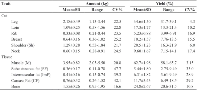
Table 1 summarizes the basic statistic (mean, standard deviation, range and coefficient of variation) for the amount and yield of cuts and carcass composition. The carcass cuts and the composition obtained herein
9
5
agreed to the current light lamb carcass in the northern region of Portugal18,19 and in other countries of southern Europe20,21,15.
Table 1. Mean, standard deviation (SD), range and coefficient of variation (CV) of amount and yield of cuts and carcass composition in tissues
Trait Amount (kg) Yield (%)
Mean±SD Range CV% Mean±SD Range CV%
Cut Leg 2.18±0.49 1.13-3.44 22.5 34.6±1.50 31.7-39.1 4.3 Loin 1.09±0.25 0.58-1.56 22.8 17.3±1.77 13.3-21.3 10.2 Rib 0.33±0.08 0.21-0.44 23.5 5.23±0.88 3.99-6.91 16.9 Breast 0.64±0.16 0.36-1.02 25.2 10.2±1.57 7.76-13.5 15.5 Shoulder (Sh) 1.29±0.28 0.53-1.84 21.7 20.5±1.23 16.3-21.9 6.0 Neck 0.60±0.15 0.28-0.91 24.5 9.60±1.67 7.15-14.1 17.4 Tissue Muscle (M) 3.95±0.82 2.05-5.50 20.8 62.7±1.98 58.1-65.7 3.15 Subcutaneous fat (SF) 0.36±0.17 0.11-0.78 47.7 5.44±1.80 2.75-9.49 33.0
Intermuscular fat (ImF) 0.41±0.16 0.15-0.74 39.3 6.31±1.82 3.61-9.49 28.9
Carcass Fat (CF) 0.76±0.32 0.26-1.52 42.1 11.7±3.43 6.49-18.5 29.2
Bone 1.55±0.26 0.95-1.95 16.6 24.8±2.67 20.6-31.5 10.8
The proportion of the cuts largely depends on the type of dressing carcass that is made. In general, the cut having a higher proportion in carcasses of light lambs are the leg (34.6%) and shoulder (20.5%) that in the current work represents about 55% (Table 1). In general, the light lambs carcasses presents small amounts of carcass fat (11.7%), especially subcutaneous fat (5.4%), and the carcass muscle represents almost 63% of carcass weight. These results are in accordance with the biology of tissue growth mammals, which shows that fat growth occurs later, with maturity15. In general, subcutaneous fat is the component of the dissectible fat that increased the most with carcass weight22,18. These works report a small proportion of fat in light lambs carcasses. Despite the fat deposition pattern and the reduced amount of carcass fat, its variation was significant (CV between 42 and 48% and 29 to 33% for the amount and the yield respectively). These results are in agreement with those of Díaz22 and Santos18.
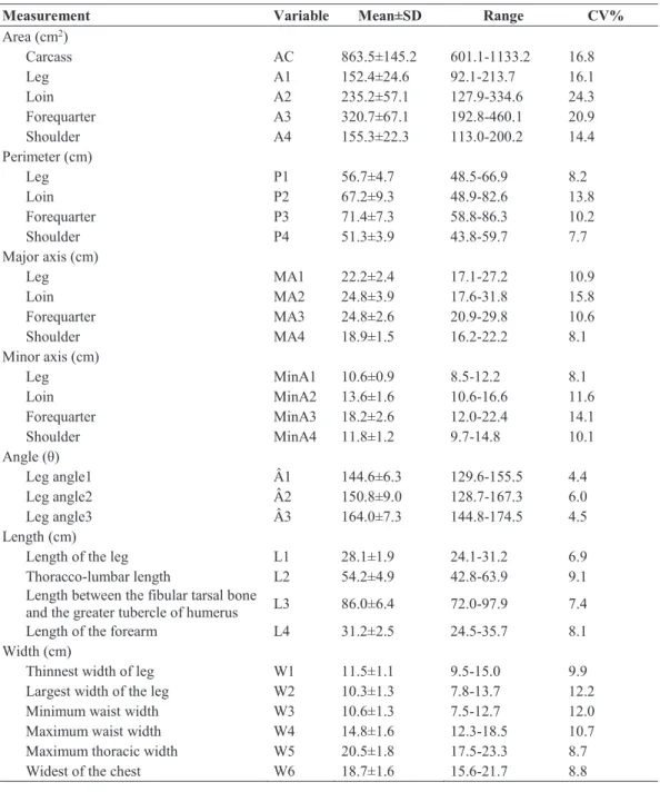
The mean, standard deviation, range and coefficient of variation of VIA measurements are presented in Table 2.
In general, the area measurements showed highest variation (CV between 14.4 and 24.3%) and the angles show smallest variation (CV between 4.4 and 6.0%). The other VIA measurements present a CV ranging from 6.9 to 15.8% (Table 2). This pattern in the variation was also reported by other authors using VIA. Rius-Vilarrasa13 using heavier lamb carcasses (18.8 kg; CV=12.9%) reported CV between 7.6 and 9.2% for area measurements and variations between 3.2 and 7.3% for the length and width measurements.
9
6
Table 2. Means, standard deviations (SD), range and coefficient of variation (CV) of VIA measurements
Measurement Variable Mean±SD Range CV%
Area (cm2) Carcass AC 863.5±145.2 601.1-1133.2 16.8 Leg A1 152.4±24.6 92.1-213.7 16.1 Loin A2 235.2±57.1 127.9-334.6 24.3 Forequarter A3 320.7±67.1 192.8-460.1 20.9 Shoulder A4 155.3±22.3 113.0-200.2 14.4 Perimeter (cm) Leg P1 56.7±4.7 48.5-66.9 8.2 Loin P2 67.2±9.3 48.9-82.6 13.8 Forequarter P3 71.4±7.3 58.8-86.3 10.2 Shoulder P4 51.3±3.9 43.8-59.7 7.7 Major axis (cm) Leg MA1 22.2±2.4 17.1-27.2 10.9 Loin MA2 24.8±3.9 17.6-31.8 15.8 Forequarter MA3 24.8±2.6 20.9-29.8 10.6 Shoulder MA4 18.9±1.5 16.2-22.2 8.1 Minor axis (cm) Leg MinA1 10.6±0.9 8.5-12.2 8.1 Loin MinA2 13.6±1.6 10.6-16.6 11.6 Forequarter MinA3 18.2±2.6 12.0-22.4 14.1 Shoulder MinA4 11.8±1.2 9.7-14.8 10.1 Angle (θ) Leg angle1 Â1 144.6±6.3 129.6-155.5 4.4 Leg angle2 Â2 150.8±9.0 128.7-167.3 6.0 Leg angle3 Â3 164.0±7.3 144.8-174.5 4.5 Length (cm)
Length of the leg L1 28.1±1.9 24.1-31.2 6.9
Thoracco-lumbar length L2 54.2±4.9 42.8-63.9 9.1
Length between the fibular tarsal bone
and the greater tubercle of humerus L3 86.0±6.4 72.0-97.9 7.4
Length of the forearm L4 31.2±2.5 24.5-35.7 8.1
Width (cm)
Thinnest width of leg W1 11.5±1.1 9.5-15.0 9.9
Largest width of the leg W2 10.3±1.3 7.8-13.7 12.2
Minimum waist width W3 10.6±1.3 7.5-12.7 12.0
Maximum waist width W4 14.8±1.6 12.3-18.5 10.7
Maximum thoracic width W5 20.5±1.8 17.5-23.3 8.7
Widest of the chest W6 18.7±1.6 15.6-21.7 8.8
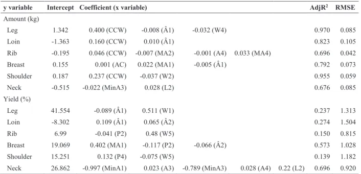
Tables 3 and 4 refer to the stepwise regressions for predicting the amount and yield of cuts and carcass composition in tissues (y variables) based on the CCW and thirty VIA measurements (x variables). The regression models based on VIA measurements and CCW confirm that it is possible to accurately predict the amount of the cuts (AdjR2 between 0.676 and 0.970, P<0.01). The best models explain 97% and 96% of leg (CCW+Â1+W4) and shoulder amount (CCW+W2), respectively (Table 3). This is a main finding because leg and shoulder are primal cuts that represent 55% of the carcass weight. Similar results were also reported by Rius-Vilarrasa23 who found higher accuracies when VIA was used in the prediction equations of the primal cuts. Relating to prediction models of cut yields, poor to moderate accuracy (AdjR2 between 0.139 and 0.696, P<0.01) was found (Table 3). Also Stanford8 presented some constraints in predicting the proportion of loin, but not of the leg (R2=0.71, RMSE = 0.66%) and shoulder (R2=0.62, RMSE = 0.88%) primal cuts. Also Brady10 corroborates those results, reporting a moderate accuracy (R2=0.561) in the prediction of yields of primal cuts using a VIA system named Lamb Vision System. These results contrast however with the work by Oliver14, undertaken with cattle, who found good predictive accuracy for commercial meat cuts yields (R2 = 0.79, 0.94, 0.84, and 0.90, respectively, for the extra, first, second, and third categories).
9
7
Table 3. Adjusted R2 (AdjR2) and residual mean square error (RMSE) for each model predicting the amount and
yield of leg, loin, rib, breast, shoulder and neck cuts by CCW and VIA measurements
y variable Intercept Coefficient (x variable) AdjR2 RMSE
Amount (kg)
Leg 1.342 0.400 (CCW) -0.008 (Â1) -0.032 (W4) 0.970 0.085
Loin -1.363 0.160 (CCW) 0.010 (Â1) 0.823 0.105
Rib -0.195 0.046 (CCW) -0.007 (MA2) -0.001 (A4) 0.033 (MA4) 0.696 0.042
Breast 0.155 0.001 (AC) 0.022 (MA1) -0.005 (Â1) 0.792 0.073
Shoulder 0.187 0.237 (CCW) -0.037 (W2) 0.955 0.059 Neck -0.515 -0.022 (MinA3) 0.028 (L2) 0.676 0.085 Yield (%) Leg 41.554 -0.089 (Â1) 0.511 (W1) 0.237 1.313 Loin -8.302 0.109 (Â1) 0.065 (Â2) 0.274 1.504 Rib 6.99 -0.041 (P2) 0.48 (W5) 0.150 0.815 Breast 19.069 0.402 (MA1) -0.117 (P2) -0.066 (Â2) 0.573 1.028 Shoulder 15.251 0.132 (P4) -0.075 (W5) 0.139 1.182
Neck 26.862 -0.997 (MinA1) 0.023 (A3) -0.789 (MinA3) 0.028 (A4) 0.22 (L2) 0.696 0.920
CCW- cold carcass weight; Â1- leg angle1; W4- maximum waist width; MA2- major axis of the loin; A4- area of the shoulder; MA4- major axis of the shoulder; AC- area of the carcass; MA1- major axis of the leg; W2- Largest width of the leg; MinA3- minor axis of the forequarter; L2- Thoracco-lumbar length; W1- Thinnest width of leg; Â2- leg angle2; P1- perimeter of the leg; W5- Maximum thoracic width; P2- perimeter of the loin; P4- perimeter of the shoulder; MinA1- minor axis of the leg; A3- area of the forequarter.
For the tissue amounts in the carcass, the models gathered in the current work show moderate to high accuracy to explain the variation of muscle and fat tissues (AdjR2 between 0.626 and 0.978, P<0.01). The best model includes CCW and one VIA measurement (W2) and it explains near 98% of the muscle variation (Table 4). Prediction models developed to estimate carcass tissue yields using CCW and VIA measurements have moderate accuracies, expressed as AdjR2 values, ranging from 0.472 to 0.731 (P <0.01). The values obtained in the present work are comparable with other reports. For example, Brady10 and Cunha24 using VIA measurements and lamb carcass weight as predictors for the carcass tissue yield achieved R2 values up to 0.71 (RMSE = 2.6%) and 0.74 (RMSE = 2.05%), respectively. In another study, the lean meat yield, which is close to the muscle yield, was also moderately predicted (R2 = 0.52, RMSE of 2.17%) using carcass measures from a VIAScan system9. These authors also assert in a broader sample of genotypes that the use of additional measurements, such as the cross-sectional area of muscle longissimus thoracis et lumborum, will be useful in order to improve the models accuracy. This orientation should also be taken into consideration for light carcasses because usually the abattoirs receive animals from different genotypes.
Table 4. Adjusted R2 (AdjR2) and residual mean square error (RMSE) for each model predicting the muscle,
subcutaneous fat, intermuscular fat and carcass fat (amount and yield) by CCW and VIA measurements
Y variable Intercept Coefficient (X variable) AdjR2 RMSE
Amount (kg)
Muscle 0.45 0.607 (CCW) -0.06 (W2) 0.978 0.110
SF -1.20 -0.034 (MinA2) 0.039 (W2) 0.018 (L3) 0.727 0.083
IF -0.875 0.001 (AC) 0.004 (Â2) 0.626 0.091
Carcass Fat -1.69 0.002 (AC) 0.006 (Â2) 0.663 0.173
Yield (%)
Muscle 2.707 0.181 (MA2) -0.473 (MinA4) 0.204 (W4) 0.551 0.647
SF -18.20 -0.296 (P3) 0.961 (MA1) 0.048 (Â2) 0.668 (W2) 0.154 (L3) 0.731 0.869
IF -2.42 -0.4 (MinA2) -0.372 (P3) 1.174 (MA3) 0.752 (W4) 0.716 0.906
Carcass Fat -14.23 0.697 (MA3) 0.529 (W4) 0.472 2.329
SF- subcutaneous fat; ImF- intermuscular fat; CCW- cold carcass weight; W2- Largest width of the leg; MinA2- minor axis of the loin; L3- Length between the fibular tarsal bone and the greater tubercle of humerus; AC- area of the carcass; Â2- leg angle2; MA2- major axis of the loin; MinA4- minor axis of the shoulder; W4- maximum waist width; P3- perimeter of the forequarter; MA1- major axis of the leg; MA3- major axis of the forequarter.
9
8
CONCLUSION
The results of the present study sustain the findings from other reports on the ability of the VIA system to predict the primal cuts and carcass composition in tissues of light lamb. Therefore, objective, rapid, non-invasive and non-destructive technologies such as VIA may offer a noteworthy opportunity to record accurate information of light lamb carcasses that can support the entire chain from the abattoir to the producer and the consumer adding value to this type of carcasses.
REFERENCES
1. Russo, C; Preziuso, G.; Verita, P. (2003). EU carcass classification system: carcass and meat quality in light lambs. Meat Sci. 64: 411–416.
2. Sanudo, C.; Alfonso, M.; Sanchez, A.; Delfa, R.; Teixeira, A. (2000). Carcass and meat quality in light lambs from different fat classes in the EU carcass classification system. Meat Sci. 56: 89-94.
3. Johansen, J.; Aastveit, A. H.; Egelandsdal, B.; Kvaal, K.; Roe, M. (2006). Validation of the EUROP system for lamb classification in Norway; repeatability and accuracy of visual assessment and prediction of lamb carcass composition. Meat Sci. 74: 497–509.
4. Craigie, C. R.; Navajas, E. A.; Purchas, R. W.; Maltin, C. A.; Buenger, L.; Hoskin, S. O.; Ross, D. W.; Morris, S. T.; Roehe, R. (2012). A review of the development and use of video image analysis (VIA) for beef carcass evaluation as an alternative to the current EUROP system and other subjective systems.
Meat Sci 92: 307-318.
5. Bunger, L.; Menezes, A. M.; McLean, K. A.; Gordon, J.; Yates, J.; Moore, K.; Lambe, N. R. (2015). Selecting terminal sire breed rams for lean meat percentage – effects on their crossbred lambs. FAIM 56-61.
6. Scholz, A. M.; Buenger, L.; Kongsro, J.; Baulain, U.; Mitchell, A. D. (2015). Non-invasive methods for the determination of body and carcass composition in livestock: dual-energy X-ray absorptiometry, computed tomography, magnetic resonance imaging and ultrasound: invited review. Animal 9: 1250-1264.
7. Craigie, C. R.; Fowler, S.; Knight, M.; Stuart, A.; Hopkins, D.; Reis, M. M. (2015). Spectral imaging techniques for predicting meat quality – an Australasian perspective. FAIM 75-79.
8. Stanford, K.; Richmond, R. J.; Jones, S. D. M.; Robertson, W. M.; Price, M. A.; Gordon, A. J. (1998). Video image analysis for on-line classification of lamb carcasses. Anim Sci 67: 311–316.
9. Hopkins, D. L.; Safari, E.; Thompson, J. M.; Smith, C. R. (2004). Video image analysis in the Australian meat industry - precision and accuracy of predicting lean meat yield in lamb carcasses. Meat Sci. 67: 269–274.
10. Brady, A. S.; Belk, K. E.; LeValley, S. B.; Dalsted, N. L.; Scanga, J. A.; Tatum, J. D.; Smith, G. C. (2003). An evaluation of the lamb vision system as a predictor of lamb carcass red meat yield percentage.
J. Anim Sci. 81: 1488–1498.
11. Ngo, L.; Ho, H.; Hunter, P.; Quinn, K.; Thomson, A.; Pearson, G. (2016). Post-mortem prediction of primal and selected retail cut weights of New Zealand lamb from carcass and animal characteristics.
Meat Sci. 112: 39-45.
12. Einarsson, E.; Eythorsdottir, E.; Smith, C. R.; Jonmundsson, J. V. (2014). The ability of video image analysis to predict lean meat yield and EUROP score of lamb carcasses. Animal 8: 1170–1177.
13. Rius-Vilarrasa, E.; Buenger, L.; Brotherstone, S.; Macfarlane, J. M.; Lambe, N. R.; Matthews, K. R.; Haresign, W.; Roehe, R. (2010). Genetic parameters for carcass dimensional measurements from Video Image Analysis and their association with conformation and fat class scores. Livestock Science 128: 92-100.
9
9
14. Oliver, A.; Mendizabal, J. A.; Ripoll, G.; Alberti, P.; Purroy, A. (2010). Predicting meat yields and commercial meat cuts from carcasses of young bulls of Spanish breeds by the SEUROP method and an image analysis system. Meat Sci. 84: 628-633.
15. Santos, V. A. C.; Silva, S. R.; Azevedo, J. M. T. (2008). Carcass composition and meat quality of equally mature kids and lambs. J. Anim. Sci. 86: 1943–1950.
16. Fisher, A. V.; Deboer, H. (1994). The EAAP standard method of sheep carcass assessment. Carcass measurements and dissection procedures report of the EAAP Working Group on Carcass Evaluation, in cooperation with the CIHEAM Instituto Agronomico Mediterraneo of Zaragoza and the CEC Directorate Gneral for Agriculture in Brussels. Livest. Prod. Sci. 38: 149–159.
17. MacNeil, M. D. (1983). Choice of a prediction equation and the use of the selected equation in subsequent experimentation. J. Anim. Sci. 57: 1328–1336.
18. Santos, V. A. C.; Silva, S. R.; Mena, E. G.; Azevedo, J. M. T. (2007). Live weight and sex effects on carcass and meat quality of "Borrego terrincho-PDO" suckling lambs. Meat Sci. 77: 654–661.
19. Teixeira, A.; Cadavez, V.; Delfa, R.; Bueno, M. S. (2004). Carcass conformation and joints composition of Churra Galega Bragançana and crossbred lambs by Suffolk and Merino Precoce sire breeds. Span. J.
Agric. Res. 2: 217–225.
20. Miguelez, E.; Zumalacarregui, J. M.; Osorio, M. T.; Beteta, O.; Mateo, J. (2006). Carcass characteristics of suckling lambs protected by the PGI "Lechazo de Castilla y Leon" European quality label: Effect of breed, sex and carcass weight. Meat Sci. 73: 82–89.
21. Diaz, M. T.; de la Fuente, J., Perez, C.; Lauzurica, S.; Alvarez, I.; Ruiz de Huidobro, F.; Velasco, S.; Caneque, V. (2006). Body composition in relation to slaughter weight and gender in suckling lambs.
Small Rumin. Res. 64: 126–132.
22. Diaz, M. T.; de la Fuente, J.; Lauzurica, S.; Perez, C.; Velasco, S.; Alvarez, I.; de Huidobro, F. R.; Onega, E.; Blazquez, B.; Caneque, V. (2005). Use of carcass weight to classify Manchego sucking lambs and its relation to carcass and meat quality. Anim. Sci. 80: 61–69.
23. Rius-Vilarrasa, E.; Buenger, L.; Maltin, C.; Matthews, K. R.; Roehe, R. (2009). Evaluation of Video Image Analysis (VIA) technology to predict meat yield of sheep carcasses on-line under UK abattoir conditions. Meat Sci. 82: 94-100.
24. Cunha, B. C. N.; Belk, K. E.; Scanga, J. A., LeValley, S. B.; Tatum, J. D.; Smith, G. C. (2004). Development and validation of equations utilizing lamb vision system output to predict lamb carcass fabrication yields. J. Anim. Sci. 82: 2069–2076.
ACKNOWLEDGMENTS
Supporting this study were the Coordination of Improvement of Higher Education Personnel (CAPES) from the Brazilian government (process: 1052/13-6); The authors wish to express thanks to the Associação de Criadores de Ovinos Mirandeses (ACOM) for providing the carcasses used in this study.