R E S E A R C H A R T I C L E
Open Access
Susceptibility of neuron-like cells derived from
bovine Wharton
’
s jelly to bovine
herpesvirus
type
5 infections
Tereza C Cardoso
1*, Juliana B Novais
1, Talita F Antello
1, Camila Silva-Frade
1, Marina C Ferrarezi
1, Heitor F Ferrari
1,
Roberto Gameiro
2and Eduardo F Flores
3Abstract
Background:Bovine herpesvirus type 5 (BoHV-5), frequently lethal in cattle, is associated with significant agricultural economic losses due to neurological disease. Cattle and rabbits are frequently used as models to study the biology and pathogenesis of BoHV-5 infection. In particular, neural invasion and proliferation are two of the factors important in BoHV-5 infection. The present study investigated the potential of bovine Wharton’s jelly mesenchymal stromal cells (bWJ-MSCs) to differentiate into a neuronal phenotype and support robust BoHV-5 replication.
Results:Upon inducing differentiation within a defined neuronal specific medium, most bWJ-MSCs acquired the distinctive neuronal morphological features and stained positively for the neuronal/glial markers MAP2 (neuronal microtubule associated protein 2), N200 (neurofilament 200), NT3 (neutrophin 3), tau and GFAP (glial fibrillary acidic protein). Expression of nestin, N200,β-tubulin III (TuJI) and GFAP was further demonstrated by reverse transcriptase polymerase chain reaction (RT-PCR). Following BoHV-5 inoculation, there were low rates of cell detachment, good cell viability at 96 h post-infection (p.i.), and small vesicles developed along neuronal branches. Levels of BoHV-5 antigens and DNA were associated with the peak in viral titres at 72 h p.i. BoHV-5 glycoprotein C mRNA expression was significantly correlated with production of progeny virus at 72 h p.i. (p < 0.05).
Conclusion:The results demonstrated the ability of bWJ-MSCs to differentiate into a neuronal phenotypein vitroand support productive BoHV-5 replication. These findings constitute a remarkable contribution to thein vitrostudy of neurotropic viruses. This work may pave the way for bWJ-MSCs to be used as an alternative to animal models in the study of BoHV-5 biology.
Keywords:BoHV-5,in vitroreplication, Neuronal culture
Background
The two most basic properties of stem cells are their capacity for indefinite self-renewal and differentiation into multiple cell or tissue types [1-3]. Embryonic stem cells are totipotent and can be maintained in culture in the presence of leukemia inhibitory factor (LIF) [4]. Withdrawal of LIF induces the formation of cellular aggregates called embryonic bodies. A wide variety of cell types migrate away from embryonic bodies, includ-ing some with neuron-like morphology [5]. In addition,
embryonic stem cells may be differentiated into neurons and glial cells with retinoic acid or basic fibroblast growth factor (bFGF) [6,7].
Neurons derived from embryonic stem cells express neurofilaments, neuron-specific class III β-tubulin (TuJl) and a number of neuron-specific microtubule-associated proteins [8]. Neural stem cells (NSC) are immature, un-committed cells that exist in both the developing brain and the adult nervous system [8]. These cells can undergo expansion and differentiate into neurons, astrocytes, and oligodendrocytes [8,9]. Stem cells differentiated into neu-rons from both these sources display a limited prolifera-tion potential. Obtaining them requires invasive procedures and results in ethical limitations in their use [10]. Because
* Correspondence:tcardoso@fmva.unesp.br
1UNESP
–University of São Paulo State, Laboratory of Animal Virology and Cell Culture, São Paulo, Brazil
Full list of author information is available at the end of the article
neuroblastoma cells can mimic the morphological and biochemical characteristics of primary neurons, they have proved to be highly valuable models in analyzing the neu-ropathogenesis and neurotropism of a range of viruses [11]. To overcome ethical issues regarding the use of fetal and adult brain samples, mesenchymal cells (MSCs) iso-lated from the Wharton’s jelly (WJ) of human and animal umbilical cords (UC) represent an attractive alternative. These cells are easily obtained, proliferate rapidly in cul-ture, are immunologically compatible, and represent fetal adnexa that is usually discarded [1,5]. MSCs have been previously isolated from dogs, sheep, horses [2,3,12], and recently bovine WJ-UC [13]. The WJ-UC cell structure is embryonic in origin and encloses the yolk sac, which is the source of the primordial germ cells and the first hematopoietic stem cells [5].
The neurotropic RNA and DNA viruses that produce central nervous system (CNS) diseases employ a range of pathologic mechanisms in a variety of hosts. The use-ful biological models applied to isolate, propagate, and study biological properties of these viruses include mice, rabbits, monkeys and hamsters. Theα-herpesviruses, in-cluding human herpes simplex types 1 and 2 (HSV-1 and HSV-2), varicella-zoster virus (VZV), animal patho-gens such as pseudorabies virus (PRV), bovine herpes-virus types 1 and 5 (BoHV-1 and BoHV-5), and Marek’s disease virus are neurotropic viruses capable of invading the peripheral and central nervous systems to cause neurological disease [14]. BoHV-5 belongs to the family
Herpesviridae, subfamily α-Herpesvirinae, and genus
Varicellovirus. BoHV-5 is genetically and antigenically closely related to BoHV-1, a highly prevalent virus re-sponsible for respiratory and genital disease in cattle around the world [15].
BoHV-1, associated with respiratory and genital dis-ease, and BoHV-5, associated with neurological disdis-ease, cause significant agricultural loss. [16]. Experimentally infected cattle and/or rabbits have been used to better understand the neural invasion and spread of BoHV-1 and BoHV-5 [16-19]. Indeed, rabbits have been used as an animal model to study selected aspects of acute and latent infections caused by BoHV-5 [19]. Unlike BoHV-1, BoHV-5 can be difficult to isolate from cell culture and animals presenting neurological symptoms after experi-mental inoculation [20-23]. These difficulties in viral isola-tion might be related to low virus titres present in tissue sections [18].
In contrast with in vivo studies, in vitro studies with Madin-Darby bovine kidney (MDBK) cells and primary cell cultures of bovine origin represent adequate substrates for viral in vitro studies [24,25]. In cell culture, BoHV-1 and BoHV-5 share similar phenotypes and produce similar cytopathic effects (CPE). CPE are characterized by a round cell shape and presence of multinucleated and syncytial
cells [25]. In a previous report, the CPE observed in bo-vine lung cells infected with BoHV-1 were elongated and spindle-shape cells; in contrast, BoHV-5 infected cells showed syncytial-like CPE [26]. In spite of many reports describing the difficulty of re-isolating the virus from field samples and experimentally infected rabbits, the MDBK cell line has been used as a standard substrate for BoHV-5 isolation and propagationin vitro [25]. To date, no effort has been made to develop a betterin vitroculture system for virus isolation/propagation. Optimizing viral culture would reduce the need for the use of animal models. In this context, the aim of the present study was to gener-ate a novel in vitro biological model/substrate that would differentiate bovine WJ-MSC cells into neuron-like cells susceptible to neurotropic BoHV-5 replication. In addition, such a model would provide a means for rapidly examining key aspects of the interactions be-tween BoHV-5 and bovine neuron-like cells. In particu-lar, this model system may overcome many of the limitations of live animals, primary neuronal culture, or neural mesenchymal cells from fetuses with less costly experimentation and ethical constraints.
Results
Differentiation of stem cells into neuron-like cells
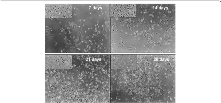
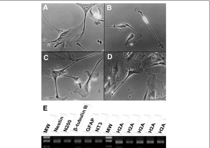
The main objective of the present study was to assess the full neural potential of bWJ-MSCs after culture in a retinoic acid-deprived reprogramming medium. Continuous culture for 28 days resulted in cells exhibiting varied morphologies, including cells with multiple long thin-axon-like processes at 7, 14, 21 and 28 days (Figure 1A-D). Furthermore, we observed the presence of neurons and oligodendrocytes based on morphology of spindle-like cells with multiple, network forming (Figure 2C and 2D), long-branch projec-tions (Figure 2A). In addition, neural-like cells displayed distinct neuronal morphology, ranging from simple bipolar to large, extensively branched, multipolar cells (Figure 3). Thus, after 28 days of neuron induction, bWJ-Uc cells were capable of generating both neuron and glial-like cells.
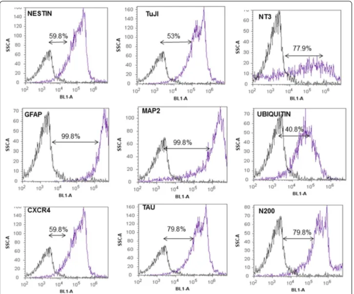
Characterization of differentiated neuron-like cells The distribution of neural/glial cell markers in differen-tiated cells is shown in Table 1. Immunostaining for neuro-differentiation markers displayed 75-100% expression of N200, MAP2, NT3, Tau, and GFAP. Approximately 50-75% of differentiated cells were positive for nestin, CXCR4, and TuJI. The ubiquitin marker was less frequently detected among differentiated cells (25-50%). Consistent with these observations, RT-PCR confirmed presence of nestin, N200,
least 70% of putative neuronal markers such as NT3, GFAP, MAP2, Tau and N200 (Figure 4). Nestin,β-tubulin III, ubiquitin, and CXCR4 were also expressed, but at lower rates (Figure 4). Our experiments confirmed previous stud-ies demonstrating the ability of human MSCs derived from Wharton’s jelly layer to generate neuron-like cells. Our results also demonstrated cross-reactivity among human and bovine monoclonal antibodies against these markers.
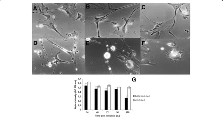
Susceptibility of neuron-like cells to BoHV-5 infection The susceptibility of neuron-like cells to BoHV-5 infec-tion was investigated by assessing the viral growth curve, cell viability, and cytopathology. The one-step kinetic growth curve profile for infected neuron-like cells was established. CPE are an important parameter in the
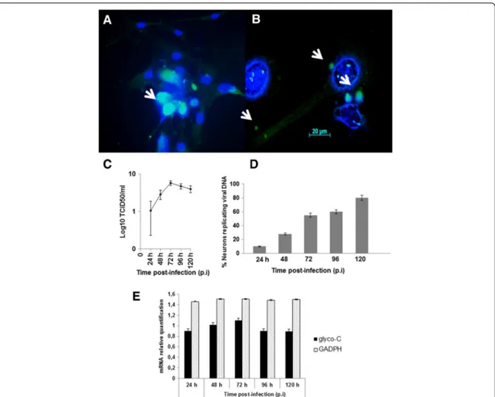
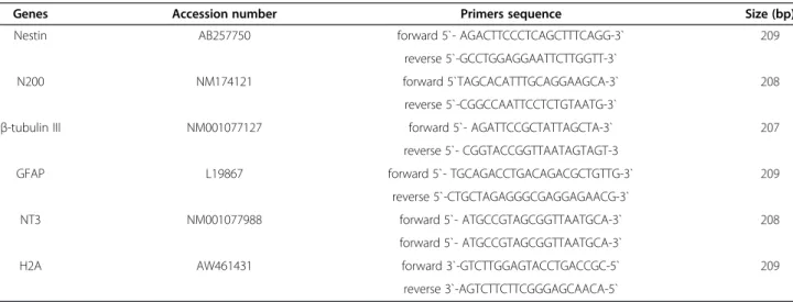
in vitro characterization of viral lytic potential. To study the CPE, an indirect measure of viral cell-to-cell spread– morphology, viral antigens, and DNA were monitored. Following infection, CPE was characterized by an enlarge-ment of the neuronal branches (Figure 5A-D) and by appearance of small and large vesicles (Figure 5E-F). BoHV-5 antigens were detected by immunofluorescence at 96 h p.i. (Figure 6A) and active BoHV-5 replication was detected by ISH (Figure 6B). The viral DNA label was observed almost exclusively in the perinuclear region of infected neurons and ranged from sporadic to dense grain accumulations along neuron branches (Figure 6B). Almost 60% of neurons demonstrated active viral DNA replication by 72–96 h p.i. (500 cells per time-point in two parallel experiments) (Figure 6D). In the replication curve, max-imum viral titre was observed at 72 h p.i. (Figure 6C) and
loss of cell viability occurred only after 96 h p.i. (Figure 5G). Altogether, these results demonstrate that differentiated bWJ-MCS cells support productive BoHV-5 replication.
Discussion
Several studies have described the ability of Wharton’s jelly cells from a variety of animal species to differentiate into neurons and/or glial cells in vitro [3]. The most commonly applied protocol to induce undifferentiated MSC cells to differentiate into neuron-like cells is based on treatment with bFGF (basic fibroblast growth factor), dimethylsulfoxide (DMSO) and butylatedhydroxyanisole (BHA) [7]. Some differences have also been described in the use of retinoic acid (RA) and/or insulin associated with hydrocortisone [26]. Retinoic acid is an oxidized derivative of retinol and is the active form of vitamin A. RA functions as a specific ligand for the retinoic acid receptor signaling pathway, which is required in embry-onic development, bone formation, and in the mainten-ance of normal epithelial structures. The addition of RA to induction medium has been shown to influence neur-onal differentiation and to improve cell survival [26].
The precise composition of the culture medium is crit-ical to avoid inconsistent results, especially when the reagents are obtained from different sources. With regard to RA, most of the neural induction methods in rodent or human MSCs employ a combination of RA with other chemical agents, cytokines, and growth factors [7-9]. In the present study, a commercially defined medium with no supplement was used to facilitate the reproducibility of the culture system. Another advantage of the culture
Figure 2Neuron-like cells characterization.After 28 days of neurogenic induction, a neuron-like morphology was visible under phase contrast microscopy.A) Neuron-like cells with triangular, round and cone-shaped morphology;B) Bi-polar neuron-like morphology;C) Network among neuron-like cells;D) Long axon of a Golgi neuron;x 100 magnification;E) RT-PCR analysis of neuronal specific markers:nestin, N200,β-tubulin III, GFAP and NT3 transcript expression in neuron-like cells after differentiation were observed. The H2A gene was used as an internal control for the reverse transcription-polymerase chain reaction. MW-molecular weight 1 kb plus.
system developed herein is the natural adherence of neuron-like cells to the plastic flasks. This phenotype is different from neuron-like cells derived from fetal brain tissue which require a specific substrate to initiate differ-entiation [10]. Collectively, the results presented here in-dicate that it is possible to derive neuron-like cells from bWJ-MSCs. These differentiated cells maintain identical morphology and cell viability after 28 days ofin vitro cul-ture with a commercial medium. Moreover, there are no ethical issues in using cells in vitrofrom umbilical cords versus fetal brains. Following neuronal differentiation, the neuron-like cells clearly exhibited their neurogenic potential by expressing classic neuronal markers such N200, MAP2, NT3, Tau and GFAP. Nestin, CXCR4, and TuJI were also detected, but at lower intensities. Import-antly, none of these neuronal markers were detectable inundifferentiated cells, suggesting the lack of spontan-eous neuronal potential. However, a previous study using porcine `WJ-MSCs described similar expression levels of neuron-specific enolase (NSE) in differentiated and undifferentiated cells [9]. This finding has also been reported for bone marrow stromal cells isolated from rats, pigs, and humans [7,8]. Neurofilaments and GFAP, both considered important neuronal/glial proteins, were also found to be expressed in undifferentiated and differ-entiated MSCs using RA as a neuronal inducer for human bone marrow stromal cells [8]. Furthermore, flow cytometric analysis found that differentiated bWJ-MSCs expressed NT3, GFAP, MAP2, Tau, and N200. This indicates the presence of neural precursors, mature neurons, and glial cells in the same culture. Similar results have been reported with human neural progeni-tors obtained from umbilical cord blood; however, no GFAP expression could be detected in this experiment
[7,8]. Thus, phenotyping of neuron-like cells revealed that more than 80% of the initial cell population became differentiated and expressed neurological receptors after 28 days of induction. This time period was inferior when compared to neural stem cells produced from fetal brain in humans [10], but similar to one study performed in dogs using Wharton’s jelly source [12].
The neurotropic RNA and DNA viruses that produce CNS diseases in humans and animals use a variety of patho-logical mechanisms. For members of the α- Herpesvirinae
subfamily, almost all types and areas of the nervous sys-tem have been identified as targets for viral replication [16,18]. Although many studies have been performed
in vivousing rabbits and cattle, it is difficult to examine BoHV-1 replication in vivo. anin vitro culture of rabbit sensory neurons and ganglionic non-neuron cells to examine BoHV-1 replication [27].
Because of this difficulty, suitablein vitrosystems imple-menting the three R’s principle of Russel and Burch (1959) are needed to study primary host-virus interactions [27]. Because of the clinical relevance of theα-subfamily Her-pesvirinae,in vitromodels would be highly appreciated to replace animal studies. In addition to ethical considera-tions,in vitrostudies would eliminate potential confound-ing factors such as individual animal variation and environmental factors [28].
Both herpes simplex virus 1 (HSV-1) and BoHV-1/5 establish lifelong latent infections in rabbit and cow sen-sory nerve ganglia [18,27]. Several days after infection by HSV-1 and BoHV-1/5, some neurons are productively infected (acute phase) [27]. Other neurons, however, be-come latently infected [18,23]. In the present study, the absence of floating neuron-like cells following BoHV-5 infection suggests a low rate of cell death. This was con-firmed by MTT assay results showing high cell viability and 60% ISH positivity. These findings can be explained by the expression of a latency-associated region (LTR) gene that is involved in neuronal survival during HSV-1 infectionin vitro[17]. In addition,in vitroreplication does not always correlate within vivoviral behavior. Thus, it is particularly important to note that data obtained from
in vitro studies are not necessarily representative of viral host interactions occurringin vivo[27].
However, the molecular mechanisms by which pro-ductive and persistent viruses produce nervous system diseases are diverse and not completely understood. The results obtained in this investigation open the door to questions about neuronal death/survival mechanisms fol-lowing BoHV-5 infection and replication.
Conclusion
This report provides the first characterization of neuron-like cells derived from bWJ-MSCs. We have demonstrated that these cells are susceptible to BoHV-5 infection. These Table 1 Incidence of various markers that neuron-like
cells derived from bWJ-UC express after 28 days of differentiation visually counting under UV microscopy
Marker Neuron-like cells bWJ-UC
Nestin +++a _
MAP2 ++++ _
GFAP ++++ _
N200 ++++ +
NT3 ++++ _
TuJI +++ _
Tau ++++ _
Ubiquitin ++ ++
CXCR4 +++ _
Bovine IgG _ _
Bovine IgM _ _
aValues were determined by visually counting positive cells (
−, 0%; +, 0-25%; +
results open the door to replace animal models to study particular aspects of BoHV-5 biology and pathogenesis. Our in vitro model reduces the need for animal experi-mentation. The neuron-like cells developed herein can be used as a viable alternative to investigate several aspects of BoHV-5 biology including neurotropism, neurovirulence, and gene expression.
Methods
Bovine Wharton’s jelly-derived multipotent mesenchymal stromal cells (bWJ-MSCs)
The bWJ-MSCs were isolated and characterized as pre-viously described [13] according to the Animal Care Committee at the University of São Paulo State, Brazil. The cell monolayers have been maintained in the
laboratory of Animal Virology and Cell Culture since 2010 following standard procedures for seeding, freeze-thawing, and propagation. Passage 30 undifferentiated bWJ-MSCs were kept at 3.5 × 10⁵ cells/mL in 25-cm2
tissue culture flasks (Falcon, BD) at 38.5°C in 5% CO2
in a humidified incubator. The cells were seeded with Advanced-DMEM (InvitrogenW
, Life Technologies™, Carlsbad, CA, USA) with the addition of 10% fetal bo-vine serum (FBS; Sigma-AldrichW
, St. Louis, MO, USA), 2 mM glutamine and 10 mM non-essential amino acids (Sigma-AldrichW
pictures were taken to observe the morphology of undif-ferentiated bWJ-MSCs.
Differentiation of bWJ-MSCs
The differentiation protocol was divided into three stages. In stage I, bWJ-MSCs were resuspended at 2.8 × 10⁴cells/ mL in Neurobasal™medium supplemented with 10% fetal bovine serum (FBS, Sigma-AldrichW
) for 24 h. Stage II consisted of replacing the culture medium with Neuroba-sal™and B27 supplement (InvitrogenW). The medium was replaced after 24 h of induction; and stage III was started by replacing the old medium with Neurobasal™with the addition of a neuro-differentiation supplement (Invitro-genW
). Stage III was sustained for 15 consecutive days. The neuron-like cells were cultured both in 25-cm2flasks for the viability assay and on Lab-TekW well chamber slides for immunostaining (Nunc™, Rochester, NY, USA). All of the media were supplemented with penicillin (100 IU/mL) and streptomycin (100μg/mL). All cultures were incubated at 38.5°C in 5% CO2with 95% humidity.
Immunostaining for neural cell markers
Differentiated cells at 28 days of culture were fixed with 4% paraformaldehyde for 15 min in Lab-TekW
chamber slides (Nunc™). The cells were permeabilized for 10 min at room temperature in 0.4% Triton X-100 diluted in phosphate buffered solution (PBS). The fixed neuron-like
cells were incubated overnight at 4°C with each of the primary antibodies anti-Nestin (cat # N5413, diluted at 1:100), anti-NT3 (neurotrophin 3; cat # SAB3300030, 1:50), anti-CXCR4 (chemokine receptor 4; cat # C3116, 1:20), GFAP (cat # WH0002670M1, 1:50), anti-MAP2 (microtubule associated protein 2; cat # M1406, 1:20), anti-Tau (cat # T6402, 1:200), anti-ubiquitin (cat # GW10073F, 1:200), anti-βtubulin III (neuronal TuJI, cat # T8578, 1:100), and anti-N200 (neurofilament200k; cat # N4142, 1:200), all purchased from Sigma-AldrichW. On the next day, after three washes, cells were incubated with the respective goat secondary antibody (1:100) anti-mouse, anti-rabbit and anti-chicken FITC (Sigma-AldrichW
). For nuclear staining, DAPI (1 mg/ml) was diluted in Fluormount™ aqueous medium and loaded onto samples for 15 min. The images were collected under an AxioImagerW
A.1 light and an ultraviolet (UV) microscope connected to an AxioCamW
MRc (Carl Zeiss, Oberkochen, Germany). The images were processed using AxioVisionW
4.8 software (Carl Zeiss) for each antigen, whereas values were determined by visually counting positive cells (−, 0%; +, 0-25%; ++, 25-50%; +++, 50-75%; ++++, 75-100%). The undifferentiated cells underwent the same staining procedure to assess for cross reactions among antibodies. The negative controls consisted of in-cubation of slides with bovine IgG and IgM isotypes as the primary antibodies.
Flow cytometric analysis for neural cell markers
Following differentiation at 28 days of culture, 1 × 10⁶ differentiated cells were harvested after being detached with 0.25% trypsin (Sigma-AldrichW
), washed with PBS, permeabilized in 4% of paraformaldehyde, and incubated for 18 h at 4°C with the monoclonal antibodies at the same dilutions described for immunostaining in 1% Tri-ton X-100 and 0.5% bovine serum albumin (BSA). After incubation with the primary antibodies, the cells were washed three times with PBS plus 0.1% Triton X-100. Next, a 1:50 dilution of the secondary antibody was added to 100 μl of the cell suspension and then incubated at 37°C for 30 min. The cell suspension was washed as previously described. After the final wash, the cells were fixed with 4%
paraformaldehyde. Data were acquired with an Attune™ acoustic focusing cytometer system (Applied Biosystems™, Foster City, CA, USA) and at least 50,000 events were counted. The negative pattern was examined by applying the same cell suspension with the initial incubation with bovine IgG and IgM isotypes, and the result was included in the global compensation to exclude auto-fluorescence. A BL1-A (488 nm) filter was used in each analysis.
Reverse transcription-polymerase chain reaction
Reverse transcription-polymerase chain reaction (RT-PCR) analysis was used to evaluate the differentiation of bWJ-MSCs into neuron-like cells. Briefly, total RNA was extracted from the cell culture at 28 days using the
TRizol™method (InvitrogenW). mRNA expression of the nestin, N200, β-tubulin III (TuJI), GFAP and NT3 genes was reverse transcribed using an enhanced avian RT first strand synthesis kit (STR-1, Sigma-Aldrich). The PCR re-action was conducted following JumpStart™Taq ready mix according to manufacturer’s instructions (Sigma-AldrichW
) with the specific primers described in Table 2. A house-keeping gene, H2A (bovine histone), was used as an in-ternal control. All of the primers were designed to amplify 207 to 209 bp and were electrophoresed on a 1.5% agarose gel. Samples were visualized by ethidium bromide staining and photographed.
Susceptibility of neuron-like cells to viral infection After differentiation, neuron-like cells were maintained in a Neurobasal™ medium (Invitrogen™) plus B27 without FBS. The BoHV-5 strain used in the experiment was iso-lated from an outbreak of meningoencephalitis [22]. This strain has 90% sequence similarity to EVI99 based on the US9 region (GenBank accession number AY064172). The strain was kept for 9 consecutive cell passages in our la-boratory [29]. For the infection, neuron-like cells were grown to 70% confluence in Lab-TekW
chambers slides (Nunc™) and infected with 100μl of a BoHV-5 suspen-sion (103.2TCID50/ml), corresponding to a multiplicity of
infection (m.o.i.) of 0.1. The viral suspension was allowed to adsorb for 1 h at 38.5°C. After incubation, the viral suspension was removed and the monolayers were sup-plemented with the same medium. Inoculated cells were monitored thereafter for cytopathic effect (CPE).
Viability assay and viral growth kinetics
The cell proliferation analysis was performed using an In Vitro Toxicology AssayWKit, an MTT-based assay, in both infected and non-infected neuron-like cells at 0, 24, 48, 72, 96 and 120 h post-infection (p.i.) (TOXI-1 Kit; Sigma-AldrichW). For each p.i. time point, the culture
supernatant was removed and 2 mL of MTT (tetrazo-lium salts) were added following the manufacturer’s recommendations (Sigma-AldrichW
). Absorbance was measured at 600 nm with a Biophotometer (EppendorfW, Hamburg, Germany). All reported values are means of triplicated samples.
One-step growth curve of the BoHV-5 in neuron-like cells was assayed in a multiplication kinetics experiment, followed by infection at 80% of monolayer confluence with m.o.i of 1. After 90 min of adsorption at 38.5°C, the inoculum was removed and fresh medium was added. The neuron-like cells were incubated at different time intervals (24, 48, 72, 96 and 120 h p.i). After incubation, both supernatant and lysate cells were harvested and assayed for the presence of the virus by infecting (Madin-Darby bovine kidney cells) MDBK cells as described previously [30]. All experiments were performed in triplicate. Infectious virus titres were calculated accord-ing to the Spearmann-Kärber method and expressed as Log10TCID50/mL.
Viral nucleic acid detection by in situ hybridization assay To estimate the number of neurons actively replicating viral DNA, neuron-like cells grown on Lab-TekW
(Nunc™) chamber slides were infected with at least 1 TCID50 per
cell, incubated for 24, 48, 72, 96 and 120 h p.i. and fixed with 95% ethanol-glacial acetic acid (3:1) for 30 min in an ice bath.In situhybridization assay (ISH) was performed as described previously [21,31,32]. To perform in situ
hybridization, the DNA probe was prepared from PCR amplicons of BoHV-5 DNA glycoprotein-C gene accord-ing to previous study [28,29,32]. The 159 base pair (bp) amplicons were produced by primary forward PCR primer gI + (5`- GTG CTC TTC TCC ATC GCC-3`) and reverse primer gI- (5`-GCG GAG GAGGAG TTG TCG G-3-bio`) (Invitrogen™, Brazil). After DNA purification from agarose gel, the PCR product was linked into the TA-vector
Table 2 Cell surface neurogenic markers analysed by RT-PCR
Genes Accession number Primers sequence Size (bp)
Nestin AB257750 forward 5`- AGACTTCCCTCAGCTTTCAGG-3` 209
reverse 5`-GCCTGGAGGAATTCTTGGTT-3`
N200 NM174121 forward 5`TAGCACATTTGCAGGAAGCA-3` 208
reverse 5`-CGGCCAATTCCTCTGTAATG-3`
β-tubulin III NM001077127 forward 5`- AGATTCCGCTATTAGCTA-3` 207
reverse 5`- CGGTACCGGTTAATAGTAGT-3
GFAP L19867 forward 5`- TGCAGACCTGACAGACGCTGTTG-3` 209
reverse 5`-CTGCTAGAGGGCGAGGAGAACG-3`
NT3 NM001077988 forward 5`- ATGCCGTAGCGGTTAATGCA-3` 208
forward 5`- ATGCCGTAGCGGTTAATGCA-3`
H2A AW461431 forward 3`-GTCTTGGAGTACCTGACCGC-5` 209
(pGEMTEasy, Promega, Madison, WI, USA) and ligation products were introduced intoE. coliby heat shock. Posi-tive colonies were confirmed by DNA sequencing. A con-firmed positive colony was cultured and plasmid DNA was prepared using a commercially available kit (Promega Mini- PrepW
, Promega, Madison, WI, USA). The probe was generated by PCR reaction [31] targeting the gC gene located in the plasmid as described before using the biotin labeled reverse primer (Invitrogen™).
Briefly, for ISH assay the infected and uninfected neuron-like cells were placed on glass slides (EasyPathW
), pre-treated with pure poly-L-lysine (Sigma-AldrichW
) and fixed with 4% (w/v) paraformaldehyde (Sigma-AldrichW) in phosphate-buffered saline (PBS) for 24 h at 4°C. Slides were treated with proteinase K(10 μg/mL, Invitrogen™) for 10 min at room temperature and washed in PBS. A 159-bp denatured biotin labeled probe consisting of 2
μL (2 ng/mL) of probe and 98 μL of pre-hybridization buffer (50% formamide, 5% bovine seroalbumin, 1% N-lauroylsarcosine and 0.02% sodium dodecyl sulphate, Sigma-AldrichW
) was applied the slides. The slides were incubated overnight at 37°C under a plastic coverslip in a humidified chamber. The slides were then washed and excess probe was removed by washing in increasingly stringent solutions consisting of 1x SSC (saline sodium citrate) and 0.1x SSC for 10 min at 42°C. The detection system consisted of incubation with monoclonal anti-body against biotin labeled to FITC (Sigma-AldrichW).
The ISH positive reaction was visualized after the slides were covered using FluormountW
(Sigma-AldrichW
) medium and observed under a UV microscope. The images were collected under an Axio Imager A.1 light microscope connected to an AxioCamMRc (Carl Zeiss Oberkochen, Germany), and the micrographs were pro-cessed with AxioVision 4.7 software (Carl Zeiss). The results were expressed as the mean percentage of neurons positively marked.
Total RNA isolation and quantitative real time polymerase chain reaction (qPCR)
Upon harvesting at 24, 48, 72, 96 and 120 h p.i., the cells, monolayers and respective supernatants, and total RNA was extracted using the Trizol LS™protocol according to manufacturer’s instructions (InvitrogenW
). An average of 150 ng of total RNA was used for first-strand cDNA syn-thesis with Enhanced Avian RT First Strand Synsyn-thesis (Sigma-AldrichW
). The qPCR was carried out and ana-lyzed by the software on a StepOnePlusW
real time instru-ment (Applied Biosystems™). The real time PCR mixtures (50 μl) contained 1.2μg of cDNA, 400 nM primers and 200 nM probes. The probes were FAM-label customized for glyco-C gene sequences according to a previous report [33]. The PCR was initiated by sequential amplification of 40 cycles at 95°C (15 s) and 60°C (60 s). The results were
obtained from three replicates of each sample to ensure representative and accuracy pipetting. The expression of bovine GAPDH (glyceraldehyde-3-phosphate dehydro-genase) gene was also quantified in a similar way for normalization. The comparative delta-delta Ct method
was used to analyze the results. The expression level of the BoHV-5glyco-Cgene at the corresponding time point in infected and uninfected neuron-like cells was quantified in comparison to GAPDH Ctvalues.
Statistical analysis
All of the experiments were performed in triplicate or greater. Results of representative experiments are pre-sented. Descriptive statistics include the mean ± standard deviation (s.d.). Statistical analysis was performed using ANOVA to evaluate the results. Student’st-test was used for comparison of uninfected (control) and BoHV-5 infected cells. Two-tailed p values < 0.05 were considered significant.
Competing interests
The authors declare that they have no competing interest.
Authors’contributions
TCC, JBN and TFA designed and directed studies, and were involved in the interpretation of the data. MCF, CSF and HFF participated in
immunofluorescence assay. RG and HFF performed cytometry analysis. EFF helped in the manuscript design and discussion of results. All authors read and approved the final manuscript.
Acknowledgements
This work was supported by FAPESP (Grants 2009/17635-3 and 2010/50782-7). The authors are grateful for the donation of the bovine umbilical cords. TC Cardoso and EF Flores are recipients of funding from the CNPq (Brazilian Council for Research).
Author details
1UNESP
–University of São Paulo State, Laboratory of Animal Virology and Cell Culture, São Paulo, Brazil.2UNESP
–University of São Paulo State, Embryology Laboratory, Faculty of Veterinary Medicine, São Paulo, Araçatuba 16050-680, Brazil.3UNESP
–University of São Paulo State, Federal University of Santa Maria, Virology Section, Santa Maria, RS 97115-900, Brazil.
Received: 2 August 2012 Accepted: 3 December 2012 Published: 10 December 2012
References
1. Mitchell KE, Weiss ML, Mitchell BM, Martin P, Davis D, Morales L, Helwig B, Beerenstrauch M, Abou-Easa K, Hildreth T, Troyer D:Matrix cells from Wharton’s jelly from neurons and glia.Stem Cells2003,21:50–60. 2. Corradetti B, Lange-Consiglio A, Cremonesi F, Bizzaro D:Isolation, in vitro
culture and characterization of foetal umbilical cord stem cells at birth.
Vet Res Comm2008,32:139–142.
3. Cremonesi F, Corradetti B, Lange-Consiglio A:Fetal adnexa derived stem cells from domestic animal: progress and perspectives.Theriogenology
2011,75:1400–1415.
4. Guan K, Rohuedel J, Wobus AM:Embryonic stem cell differentiation models: cardiogenesis, myogenesis, epithelial and vascular smooth muscle differentiation in vitro.Cytotechnology1999,30:211–226. 5. Troyer DL, Weiss ML:Concise review: Wharton’s Jelly-derived cells are
primitive stromal cell population.Stem Cells2008,26:591–599. 6. Bi Y, Gong M, Zhang X, Zhang X, Jiang W, Zhang Y, Chen J, Liu Y, He T-C, Li T:
7. Salgado AJ, Fraga JS, Mesquita AR, Neves NM, Reis RL, Sousa N:Role of human umbilical cord mesenchymal progenitors conditioned media in neuronal/glial cell densities, viability, and proliferation.Stem Cells Dev
2010,19:1067–1074.
8. Chua SI, Bielecki R, Wong CJ, Yamanaka N, Rogers IM, Casper RF:Neural progenitors, neurons and oligodendrocytes from human umbilical cord blood cells in a serum-free, feeder-free cell culture.Biochem Biophy Res Comm2009,379:217–223.
9. Choong P-F, Mok P-L, Cheong S-K, Leong C-F, Then K-Y:Generating neuron-like cells from BM-derived mesenchymal stromal cells in vitro.
Cytotherapy2007,9:170–183.
10. Pugazhenthi S, Nair S, Velmurugan K, Liang O, Mahalingam R, Cohrs RJ, Nagel MA, Gilden D:Varicella-zoster virus infection of differentiated human neural stem cells.J Virol2011,85:6678–6686.
11. Christensen J, Steain M, Slobedman B, Abendroth A:Differentiated neuroblastoma cells provide a highly efficient model for studies of productive varicella-zooster virus infection of neuronal cells.J Virol2011,
85:8436–8442.
12. Uranio MF, Valentini L, Lange-Consiglio A, Caira M, Guaricci AC, Lábbate A, Catacchio CR, Ventura M, Cremonesi F, Dell’Aquila ME:Isolation, proliferation, cytogenetic, and molecular characterization and in vitro differentiation potency of canine stem cells from foetal adnexa: a comparative study of amniotic fluid, amnion, and umbilical cord matrix.
Mol Reprod Dev2011,78:361–373.
13. Cardoso TC, Ferrari HF, Garcia AF, Novais JB, Silva-Frade C, Ferrarezi MC, Andrade AL, Gameiro R:Isolation and characterization of wharton’s jelly-derived multipotent mesenchymal stromal cells obtained from bovine umbilical cord and maintained in a defined serum-free three-dimensional system.BMC Biotechnol2012,12:8.
14. Pellet PE, Roizman B:The family Herpesviridae: a brief introduction. In
Fields Virology. Edited by Knipe DM, Howley PM. Philadelphia: Lippincott Williams & Williams; 2007,734–736.
15. Delhon GA, Moraes MP, Lu Z, Afonso CL, Flores EF, Weiblen R, Kutish GF, Rock DL:Genome of Bovine Herpesvirus 5.J Virol2003,77:10339–10347. 16. Chowdhury SI, Onderci M, Bhattacharjee PS, Al-Mubarak A, Weiss ML, Zhou
Y:Bovine herpesvirus 5 (BHV-5) US9 is essential for BHV-5 neuropathogenesis.J Virol2002,76:3839–3851.
17. Delhon GA, Gozález MJ, Murcia PR:Susceptibility of sensory neurons to apoptosis following infection by bovine herpesvirus type 1.J Gen Virol
2002,83:2257–2267.
18. Flôres FS, Flôres V, Caron L, Flores EF, Weiben R, Winkelmann ER, Mayer SV, Bastos RG:Distribution of Bovine Herpesvirus type 5 DNA in the central nervous systems of latently experimentally infected calves.J Clin Microbiol2003,41:4512–4520.
19. Valera AR, Pidone CL, Massone AR, Quiroga MA, Riganti JG, Corva SG, Galosi CM:A simple method of infecting rabbits with bovine herpesvirus 1 and 5.J Virol Meth2008,150:77–79.
20. Cardoso TC, Ferrari HF, Garcia AF, Bregano LC, Andrade AL, Nogueira AHC:
Immunohistochemical approach to the pathogenesis of clinical cases of Bovine Herpesvirus type 5 infections.Diag Pathol2010,5:57.
21. Cardoso TC, Gomes DE, Ferrari HF, Silva-Frade C, Rosa ACG, Andrade AL, Luvizotto MCR:A novel in situ polymerase chain reaction hybridization assay for the direct detection of bovine herpesvirus type 5 in formalin-fixed, paraffin-embedded tissues.J Virol Meth2010,163:509–512. 22. Ferrari HF, Luvizotto MCR, Rahal P, Cardoso TC:Detection of bovine
Herpesvirus type 5 in formalin-fixed, paraffin-embedded bovine brain by PCR: a useful adjunct to conventional tissue-based diagnostic test of bovine encephalitis.J Virol Meth2007,146:335–340.
23. Meyer G, Lemaire M, Ros C, Belák K, Gabriel A, Cassart D, Coignoul E, Belák S, Thiry E:Comparative pathogenesis of acute and latent infections of calves with bovine herpesvirus types 1 and 5.Arch Virol2001,
146:633–652.
24. Ladelfa MF, Del MédicoZajac MP, Kotsias F, Delgado F, Muylkens B, Thiry J, Thiry E, Romera SA:Comparative study on the in vitro and in vivo properties of two bovine herpesvirus-5 reference strains.Acta Vet Scan
2011,53:37.
25. Marin MS, Leunda MR, Verna AE, Faverin C, Pérez SE, Odeón AC:In vitro replication of bovine herpesvirus types 1 and 5.J Virol Meth2012,
181:80–85.
26. Seal BS, Whetstone CA:Immediate early gene expression and gene mapping comparison among isolates of bovine herpesvirus 1 and 5.
Vet Microbiol1994,38:369–384.
27. Perez SE, Bretschheider G, Leunda MR, Osorio FA, Flores EF, Odeón AC:
Primary infection, latency, and reactivation of Bovine Herpesvirus type 5 in bovine nervous system.Vet Pathol2002,39:437–444.
28. Stekers L, Vandekerckhove AP, Van den Broeck AP, Glorieux S, Nainynck HJ:
Characteristics of BoHV-1 subtypes in bovine respiratory and genital mucosa explants: a phylogenetic enlightenment.Vet Res2001,42:33. 29. Vogel FSF, Caron L, Flores EF:Distribution of bovine herpesvirus type 5 in
the central nervous systems of latently, experimentally infected calves.
J Clin Virol2003,77:10339–10347.
30. Silva-Frade C, Martins Júnior A, Borsanelli AC, Cardoso TC:Effects of bovine Herpesvirus type 5 on development of in vitro produced bovine embryos.Theriogenology2010,73:324–331.
31. Silva-Frade C, Gameiro R, Martins Júnior A, Cardoso TC:Apoptotic and developmental effects of bovine Herpesvirus type-5 infection onin vitro -produced bovine embryos.Theriogenology2010,74:1296–1303.
32. Silva MS, Brum MCS, Loreto ELS, Weiben R, Flores EF:Molecular and antigenic characterization of Brazilian bovine herpesvirus type 1 isolates recovered from the brain of cattle with neurological disease.Virus Res
2007,129:191–199.
33. Diallo IS, Corney BG, Rodwell BJ:Detection and differentiation of bovine herpesvirus 1 and 5 using multiplex real-time polymerase chain reaction.
J Virol Meth2011,175:46–52.
doi:10.1186/1746-6148-8-242
Cite this article as:Cardosoet al.:Susceptibility of neuron-like cells derived from bovine Wharton’s jelly to bovineherpesvirustype 5 infections.BMC Veterinary Research20128:242.
Submit your next manuscript to BioMed Central and take full advantage of:
• Convenient online submission
• Thorough peer review
• No space constraints or color figure charges
• Immediate publication on acceptance
• Inclusion in PubMed, CAS, Scopus and Google Scholar
• Research which is freely available for redistribution