Open Access Article│www.njcmindia.org pISSN 0976 3325│eISSN 2229 6816
National Journal of Community Medicine│Volume 4│Issue 1│Jan – Mar 2013 Page 182
Case Report ▌
HERPES ZOSTER IN CHILDREN AND ADOLESCENTS:
CASE SERIES OF 8 PATIENTS
Pragya A Nair1, Pankil H Patel2
1Professor; 2Tutor, Skin & VD, Pramukhswami Medical College, Karamsad, Anand
Correspondence: Dr Pragya A Nair, Email: drpragash2000@yahoo.com
ABSTRACT
Herpes zoster can occur at any age but is rare in childhood and adolescents. Zoster can occur at any time after primary varicella infection or varicella vaccination. Recent studies have shown its increasing incidence in children. Maternal varicella infection during pregnancy and varicella occurring in the newborn represent risk factors for childhood herpes zoster. As varicella vaccine is a live attenuated virus, herpes zoster can develop in a vaccine recipient, but its incidence is less than natural infection. It is usually diagnosed clinically as unilateral vesicular eruption following a dermatome or dermatomes. Zoster in children is frequently mild, post herpatic neuralgia occurs rarely if ever. We present eight cases of zoster in children and adolescents.
Keywords: Herpes zoster, Varicella Zoster Virus, HIV, Children, Adolescents
INTRODUCTION
Herpes zoster (HZ) or shingles is an acute vesiculobullous cutaneous infection in dermatomal distribution, predominantly in adults and older persons. It is caused by reactivation of latent varicella-zoster virus (VZV) that resides in a dorsal root ganglion.1 Children
are infrequently affected with HZ. In cases
where past history of varicella was not obtained, it is suggested that the initial contact with the
virus may result in zoster.2 HZ occurs at an
overall rate of 3.40 cases per 1000 persons.
Hope-Simpson's field study showed an incidence of
0.74 cases per 1,000 population per annum among the 0 to 9 and 1.38/1000 in 10 to 19 year-old age group. The attack rate during the first two decades is approximately seven times less
than the seventh decade.3 The earliest age
reported is in a 3-month old infant.4 However,
the true incidence of HZ in children may be even higher since some patients do not seek medical attention because of the benign course. We present eight cases of zoster in children and adolescents.
CASE REPORT
Case 1: A 4-year-old girl had a three day h/o asymptomatic papulovesicular eruption on the left side of the thorax and upper limb involving C7-8 dermatome.
Case 2: A 10-year-old boy, serologically positive for Human Immunodeficiency Virus (HIV), had six day h/o multiple pus filled lesion over right abdomen, back and lower limb with mild burning pain. Multiple pustules were present involving right T9-10, L1-5 dermatomes with few discrete lesions on the left side of the body(Fig. 1a &1b).
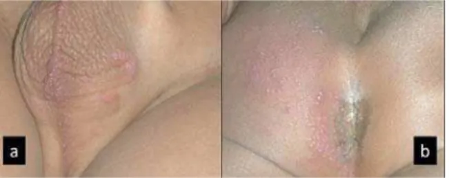
Case 3: A 5-year-old boy had a two day h/o asymptomatic vesicular eruption over genitals on the left side involving S2 dermatome (Fig.2a & 2b).
Case 4: A 10-year-old girl presented with two days history of fluid filled lesions with burning pain on back & abdomen below umbilicus on right side involving right T11-T12 dermatome. She had varicella at the age of 4 years.
Open Access Article│www.njcmindia.org pISSN 0976 3325│eISSN 2229 6816
National Journal of Community Medicine│Volume 4│Issue 1│Jan – Mar 2013 Page 183
Case 6: A 16-year-old male had pain on the right buttock and thigh for two days followed by the onset of vesicular eruption involving S1 dermatome. He had varicella at the age of 5 years.
Case 7: A 16-year-old female had grouped vesicular eruption on the right side of thorax for 2 days associated with burning sensation.
Vesiculobullous lesions on erythematous base were present in the distribution of T4 dermatome. She had varicella at the age of 6 years.
Case 8: A 7 year old girl had 3 day history of fluid filled lesions over lower abdomen involving right T9-10 dermatome. P/h/o varicella at the age of 3 years was present.
Table 1: Summary of 8 Herpes zoster cases
Case Age (Yrs)
Sex Side Derma- tome
Associated symptoms
Known Exposure to
Varicella
Age(yrs) at Previous Varicella
Sequalae Immune suppression
1 4 F L C7-8 No No -- None No
2 10 M R T9-10 & L1-5
Mild burning pain No -- Secondary infection & scarring
Yes Seropositive
3 5 M L S2 No No -- None No
4 10 F R T11-12 Burning pain Yes 4 None- No 5 4 F R T8 Fever& burning pain Yes 2½ None No
6 16 M R T12-L1 Pain Yes 5 None No
7 16 F R T4 Burning sensation Yes 6 None No
8 7 F R T9-10 No Yes 3 None No
Fig 1: 10 year old HIV positive boy with multiple pus filled lesion; (a) abdomen, lower limb involving Right T9,T10,L1,L2,L3,L4 dermatomes; and (b) back involving Right T9,T10,L5 dermatomes
Fig 2: Five year old boy with vesicular eruption; (a) genitals involving left S2 dermatome; and (b) buttock involving left S2 dermatome
None of the children were immunized against varicella. No P/h/o varicella in first 3 cases, other five gave definite past history. Cases were
diagnosed clinically as HZ and supplemented by Tzanck smear preparation. Scrapings from the floor of the vesicles, performed in 6 cases revealed multinucleated giant cells in 2 cases. HIV ELISA (Enzyme Linked Immunosorbent Serologic Assay) was negative in 7 cases except one patient. Hemogram and peripheral smear was normal in all cases. Herpes simplex virus (HSV) antigen detection and viral culture was not done due to lack of facility. All the children were treated with oral acyclovir 20mg/kg, 4 times a day for five days along with symptomatic treatment for pain and burning with topical silver sulfadiazine.
DISCUSSION
Our cases ranged from 4 to 16 years of age (Table. 1). Majority of the cases were females (5 cases), female preponderance was also seen in
Prabhu et al5 study also. The thoracic
dermatomes were affected in five children
comparable with study by Prabhu et al5, Bharija
et al2 and Hope-Simpson's3 studies, while Leung
et al6 noted predilection of cervical and sacral
Open Access Article│www.njcmindia.org pISSN 0976 3325│eISSN 2229 6816
National Journal of Community Medicine│Volume 4│Issue 1│Jan – Mar 2013 Page 184
pregnancy and no recent history of family member having chicken pox noted in any case.
There are only few case reports of childhood HIV
patients acquiring zoster is reported5.
Disseminated VZV is more commonly seen in
HIV infected individuals.7 Our study reports one
HIV positive boy, who had multi-dermatomal herpes zoster with secondary infection and dissemination, no complications were noted in other 7 cases
Following initial exposure to VZV, the virus may become latent and lie dormant in the dorsal nerve root or in the extramedullary cranial nerve root ganglion cells. HZ is caused by the reactivation of latent VZV. HZ arises, years or
decades following primary infection with VZV.1
HZ cases present with a characteristic unilateral, dermatomal, vesicular eruption preceded or accompanied by pain. Lesions heal within 2 to 3 weeks, but postherpetic neuralgia (PHN) can
persist for months or years thereafter 8 and may
be intractable. In infants and children it is more common in girls, usually not accompanied by pain or PHN but fever, headache and regional lymphadenopathy can occur. Zoster in children is frequently mild. The probability of PHN in children and adolescents is extremely low, rarely
if it ever occurs.9 Differential diagnosis for
herpes zoster particularly in infants and children includes irritant contact dermatitis, insect bite and bullous impetigo which needs to be kept in mind.
The occurrence of zoster in childhood is related to exposure to VZV postnatal, perinatal or intrauterine. Herpes zoster in children probably represents the result of an immature immune
response to the transplacentally acquired VZV3.
Low levels of lymphocytes, natural killer cells, cytokines characterize this poor response, and virus-specific immunoglobulins may result in inability to maintain the latency of VZV leading to early appearance of zoster in children.10
Chickenpox in the first year of life was found to be a risk factor for childhood zoster, with a relative risk between 2.8 and 20.9. Neither chickenpox in the second year of life nor recent vaccinations were found to be risk factors for
childhood zoster.11 Such observation was not
seen in our study as none of our cases had history of chickenpox in first year of life.
Childhood HZ was thought to be an indicator for an underlying malignancy, whereas recent studies have shown no increase in the incidence of malignancy in children with HZ. Approximately 3% of the pediatric zoster cases occur in children with malignancies.
CONCLUSION
HZ is an infrequent, but not a rare, disease of children. Its infrequent recognition could be explained by its benign clinical course. For this reason, patients so affected may never reach the physician. Because of a low index of suspicion, the eruption is often treated casually as a local cutaneous problem, and its actual nature remains unrecognized. The probability of postherpetic neuralgia in children and adolescents is extremely low. Zoster is seldom associated with undiagnosed malignancy in the primary care setting.
REFERENCE
1. Gnann JW Jr., Whitley RJ. Clinical practice. Herpes zoster. N Engl J Med. 2002;347:340–6.
2. Bharija SC, Kanwar AJ, Belhaj MS. Herpes zoster. Ind J Pedia. 1988;55(2):301-3.
3. Hope-Simpson RE: The nature of herpes zoster: A long term study and a new hypothesis. Proc Roy Soc Med. 1965; 58:9-20.
4. Handa S. Herpes zoster in a 3-month-old infant. Paed Dermatol. 1997; 14:133.
5. Prabhu S, Sripathi H, Gupta S, Prabhu M. Childhood herpes zoster: A clustering of ten cases. Indian J Dermatol. 2009;54:62-4.
6. Leung AKC, Robson WLM, Leong AG. Herpes Zoster in Childhood. J of Pediatr Health Care. 2006 Sept;20(5):300-3.
7. Archana Singal, Shilpa Mehta, Deepika Pandhi. Herpes zoster with dissemination. Indian
Pediatrics:April.2006;43:353-56.
8. Helgason S, Petursson G, Gudmundsson S, Sigurdsson JA. Prevalence of postherpetic neuralgia after a first episode of herpes zoster: prospective study with long term follow up. BMJ. 2000;321:794–6.
9. Feder HM Jr,Hoss DM.Herpes zoster in otherwise healthy children. Pediatr Infect Dis J . 2004 May;23(5);451- 7.
10. Huang JL, sun PC, Hung IJ. Herpes zoster in infancy after intrauterine exposure to varicella zoster virus: report of two cases. J Formos Med Assoc 1994 Jan;93(1):75-7.
