JULIANA CHAMORRO RENGIFO
PARAGRYLLINI (ORTHOPTERA: GRYLLOIDEA: PHALANGOPSIDAE) BRASIL: DESCRIÇÕES DE NOVOS TÁXONS
Dissertação apresentada à Universidade Federal de Viçosa, como parte das exigências do Programa de Pós-Graduação em Entomologia, para obtenção do título de Magister Scientiae.
VIÇOSA
Ficha catalográfica preparada pela Seção de Catalogação e Classificação da Biblioteca Central da UFV
T
Chamorro Rengifo, Juliana, 1979-
C453p Paragryllini (Orthoptera: Grylloidea: Phalangopsidae) 2009 Brasil : descrições de novos táxons / Juliana Chamorro Rengifo. – Viçosa, MG, 2009.
vi, 84p.: il. 29cm.
Inclui anexo.
Orientador: Carlos Frankl Sperber.
Dissertação (mestrado) - Universidade Federal de Viçosa. Inclui bibliografia.
1. Grilo - Classificação. 2. Morfologia. 3. Inseto - Habitat. 4. Entomologia. 5. Zoologia - Classificação. 6. Amazônia I. Universidade Federal de Viçosa. II. Título.
JULIANA CHAMORRO RENGIFO
PARAGRYLLINI (ORTHOPTERA: GRYLLOIDEA: PHALANGOPSIDAE) BRASIL: DESCRIÇÕES DE NOVOS TÁXONS
Dissertação apresentada à Universidade Federal de Viçosa, como parte das exigências do Programa de Pós-Graduação em Entomologia, para obtenção do título de Magister Scientiae.
“Somos do tamanho dos nossos sonhos” (Fernando Pessoa)
“Perder tempo em aprender coisas que não interessam, priva-nos de descobrir coisas interessantes”.
AGRADECIMENTOS
À Universidade Federal de Viçosa e em especial ao Programa de Pós-Graduação em Entomologia.
Aos meus colegas do Laboratório de Orthopterologia. Em especial à Lucimar.
Ao meu orientador Carlos Frankl Sperber por ter me recebido no seu laboratório, por seus conselhos e apoio.
À Carina Marciela Mews por ter compartilhado comigo seu conhecimento sobre taxonomia de Grylloidea, por ter me ensinado a usar a câmara clara, por todo seu apoio e ajuda.
Ao Cristiano Lopes Andrade, sem ele não teria sido possível esta dissertação, por seu apoio, por tudo o que me ensinou, por ser um exemplo de trabalho constante e
sistemático, porque através de seu comportamento, ensinar o que é respeitar as diferenças.
À Karina Furieri e Holger Braun que me apoiaram e co-orientaram. Ao Marcos Lhano por sua ajuda inicial, desde antes de chegar a Viçosa.
A todos os professores que me deram aula durante o mestrado: Zhé, Fiúza, Serrão, Lino, Terezinha, Carlos e Og. À Flávia embora não tenha me dado aula.
À Orthoptera Species File, pelo apoio financeiro indireto para meu mestrado. Às minhas amigas, Zulma e Yenis, pelo apoio e força.
ÍNDICE
RESUMO ...v
ABSTRACT...vi
1. INTRODUÇÃO GERAL ...1
1.1. A genitália de Grylloidea ...3
1.2. A tribo Paragryllini Desutter...10
2. OBJETIVOS ...13
3.1. Locais de coleta dos espécimes...13
3.2. Coleta dos espécimes ...14
3.3. Procedimentos técnicos e taxonômicos...14
3.3.1. Dissecação da genitália ...16
3.4. Fotografias e ilustrações...16
3.5. Nomenclatura utilizada ...17
3.6. Glossário de termos...17
3.7. Estrutura dos capítulos da dissertação...17
4. REFERÊNCIAS BIBLIOGRÁFICAS...19
5. ARTIGO 1...23
6. ARTIGO 2...57
7. CONCLUSÕES ...82
RESUMO
CHAMORRO-RENGIFO, Juliana. M.Sc., Universidade Federal de Viçosa, junho de 2009. Paragryllini (Orthoptera: Grylloidea: Phalangopsidae) Brasil: descrições de novos táxons. Orientador: Carlos Frankl Sperber. Co-orientadores: Cristiano Lopes Andrade, Holger Braun e Karina Schimdt Furieri.
ABSTRACT
CHAMORRO-RENGIFO, Juliana. M.Sc., Universidade Federal de Viçosa, june, 2009. Paragryllini (Orthoptera: Grylloidea: Phalangopsidae) Brazil: descriptions of new taxa. Advisor: Carlos Frankl Sperber. Co-Advisors: Cristiano Lopes Andrade, Holger Braun and Karina Schmidt Furieri.
1. INTRODUÇÃO GERAL
Grylloidea Laicharding 1781, o grupo dos chamados grilos verdadeiros, pertence à
Subordem Ensifera, junto com sete superfamílias extintas (Elcanoidea Handlirsch 1906, Permoraphidioidea Tillyard 1932, Oedischioidea Handlirsch 1906, Triassomanteoidea Tillyard 1923, Xenopteroidea Riek 1955, Gryllavoidea Gorochov 1986, Phasmomimoidea Sharov 1968) e cinco superfamílias com representantes vivos atualmente: Hagloidea Handlirsch 1906, Rhaphidophoroidea Walker, F. 1871, Schizodactyloidea Blanchard 1845, Stenopelmatoidea Burmeister 1838 e Tettigonioidea Krauss 1902. Esta classificação foi disponibilizada por Orthoptera Species File (Eades & Otte, 2009), através de revisão e compilação da literatura.
Existem registros de Orthoptera desde o Carbonífero Superior, há aproximadamente 300 milhões de anos (Sharov, 1971). Para a América do Sul não se conhecem registros de Ensifera do Paleozóico e do Mesozóico Inferior. No Brasil e na Eurásia foram encontrados fósseis de Grylloidea identificados como pertencentes a três subfamílias da extinta família Baissogryllidae, e dois fósseis da subfamília dos Gryllinae. A evolução de Grylloidea é considerada mais lenta que a dos Tettigonioidea e Stenopelmatoidea. Possivelmente, a radiação adaptativa do Cenozóico deu como resultado a aparição das tribos atualmente reconhecidas (Gorochov, 2001).
As espécies de Grylloidea apresentam uma grande variação de tamanhos, formas e cores (Desutter, 1990). Caracterizam-se por terem três tarsômeros em todas as pernas, cercos longos e não especializados (contrario aos Tettigonioidea), com pêlos clavados na área interna. A placa subgenital da fêmea e do macho não têm estiletes e o ovipositor
é usualmente comprido e fino, com valvas internas reduzidas (Desutter-Grandcolas, 1998). A genitália do macho é assimétrica dorso-ventralmente e, segundo a mais recente interpretação, é constituída por três camadas como nos demais Orthoptera: a camada mais externa, a epifálica; a do meio, a ectofálica; e a mais interna, a endofálica (Desutter, 1987).
anal da tégmina e pode também corresponder à veia anal modificada (Montealegre et al., 2009). O aparelho estridulador é atualmente considerado uma característica ancestral (Alexander, 1962; Otte, 1977, 1992). Supõe-se que o sistema de som apareceu nos Grylloidea e Tettigonioidea no final do Permiano. Os modelos evolutivos consideram a estridulação uma característica ancestral dos grilos e que foi perdida independentemente em diversos grupos (Otte, 1977, 1992; Walker & Masaki, 1989).
Grylloidea está amplamente distribuída no mundo, com uma alta diversidade nas regiões tropicais. Aproximadamente 3.000 espécies de grilos são conhecidas atualmente (Eades & Otte, 2009). Sobre a classificação do grupo, não existe consenso sobre qual classificação é a mais adequada. Bruner (1916) agrupou os grilos em 11 famílias, excluindo Gryllotalpidae. Chopard sugeriu 12 famílias em 1956 e 1965, logo no período entre 1967 e 1968 três famílias (Gryllotalpidae, Oecanthidae e Gryllidae) e dez subfamílias de Gryllidae (Gryllinae, Cacoplistinae, Eneopterinae, Mogoplistinae, Myrmecophilinae, Pentacentrinae, Phalangopsinae, Ptyeroplistinae, Scleropterinae e Trigonidiinae). E finalmente 12 famílias em 1969.
Vickery (1976) e Kevan (1982) sugeriram a separação de Gryllotalpidae em uma superfamília à parte, e a classificação de Grylloidea em 11 famílias e 16 subfamílias: Gryllidae (Brachytrupinae, Gryllinae e Nemobiinae), Pentacentridae (Pentacentrinae, Lissotrachelinae e Aphemogryllinae), Cacoplistidae, Pteroplistidae, Scleropteridae,
Phalangopsidae (Luzarinae e Heterogryllinae), Oecanthidae, Mogoplistidae (Bothriophylacinae e Mogoplistinae), Trigonidiidae (Trigonidiinae e Phylloscirtinae), Eneopteridae (Eneopterinae, Itarinae, Podoscirtinae e Prognathogryllinae) e Myrmecophilidae. Alexander & Otte (1967) e Otte & Alexander (1983) consideraram apenas a família Gryllidae, com 14 subfamílias, incluindo entre elas Gryllotalpinae.
Desutter (1987, 1988) classificou os gêneros neotropicais de Grylloidea em três superfamílias e 13 famílias: Gryllotalpoidea (Gryllotalpidae), Myrmecophiloidea (Malgasiidae, Myrmecophilidae e Mogoplistidae), Grylloidea (Pteroplistidae, Oecanthidae, Neoaclidae, Paragryllidae, Phalangopsidae, Trigonidiidae, Podoscirtidae, Eneopteridae e Gryllidae) além de considerar dois grupos sem representantes na América do Sul: Pentacentridae e Scleropteridae.
Phalangopsinae, Landrevinae, Gryllomiminae, Itarinae, Gryllomorphinae, Gryllinae, Phaloriinae, Cacoplistinae, Oecanthinae e Pteroplistinae) e Mogoplistidae. Mas Gorochov (2001) novamente sugeriu outra classificação, aparentemente sem nenhuma evidência conclusiva sobre as características que definiriam cada grupo. Ele dividiu a superfamília em cinco famílias: Trigonidiidae, Podoscirtidae, Eneopteridae, Gryllidae e Phalangopsidae.
Segundo Otte (1994) e Otte & Naskrecki (1997) há quatro famílias de Grylloidea: Gryllidae, Mogoplistidae, Myrmecophilidae e Gryllotalpidae. Gryllidae é dividido em 15 subfamílias: Gryllinae, Brachytrupinae, Nemobiinae, Trigonidiinae, Pentacentrinae, Sclerogryllinae, Phalangopsinae, Malgasiinae, Cacoplistinae, Itarinae, Eneopterinae, Euscyrtinae, Podoscirtinae, Pteroplistinae e Oecanthinae.
1.1. A genitália de Grylloidea
A genitália dos machos não é um órgão intromitente, mas tem duas funções: a
formação do espermatóforo e a transferência dele para a genitália da fêmea. As estruturas envolvidas na formação do espermatóforo estão na metade ventral do complexo fálico genital (Randell, 1961). Já a estrutura envolvida na transferência do espermatóforo encontra-se na porção dorsal: o pseudepifalo (Chopard, 1920a), que tem como função pressionar a placa subgenital da fêmea e expor o poro genital. Por outro lado, Randell (1961) também sugeriu que a função dos parâmeros pseudepifálicos é a de servir de guia, suportando os escleritos endofálicos, os apódemas endofálicos (virga) e a dobra ectofálica durante a inserção do ducto do espermatóforo na fêmea.
A proposta mais recente sobre a estrutura da genitália do macho, mas não completamente aceita por todos os atuais taxônomos e sistematas, é a de Desutter (1987, modificada pela mesma autora em 2003). No geral, a interpretação é muito semelhante à de Chopard (1920), mas diferindo muito da de Ander (1939, 1956, 1970). Também Difere da de Snodgrass (1937) pela identificação do esclerito dorsal em Grylloidea e Gryllotalpoidea (ectofalo, pseudepifalo), e na maioria dos Rhaphidophoridae e Tettigoniidae (verdadeira esclerotização, par e um epifalo somente), e pela identificação das invaginações presentes na parte dorsal do complexo fálico (invaginação epi-ectofálica e epi-ectofálica, cavidade endofálica) (Desutter-Grandcolas 2003).
A interpretação de Desutter-Grandcolas (2003) considera para os Ensifera três componentes maiores desenvolvidos e modificados em três diferentes clados:
1. Anel membranoso, rodeando o gonóporo (corresponderia ao ectofalo em Snodgrass (1937) e Ander (1956)). Subdividido em lóbulos ventral, dorsal e lateral. Pode estar usualmente diferenciado dorsalmente e muito conspícuo na genitália dos Ensifera, crescendo a partir de modificações do anel ectofálico. Dentre estas estruturas, a mais importante é o pseudepifalo (sensu Chopard 1920a), originando-se a partir de uma esclerotização e diferenciando-se a partir da porção mais dorsal do ectofalo; é uma
invaginação em forma de T, que aparece na metade mais baixa da parte dorsal do ectofalo (invaginação ectofálica) (Desutter-Grandcolas, 2003).
2. Prega dorsal localizada entre o ectofalo e os paraproctos. Essa dobra é variavelmente prolongada lateralmente. Sua localização corresponde ao epifalo de Chopard (1920a) e Desutter-Grandcolas (2003).
3. Invaginação separando as duas estruturas anteriores, separando a dobra epifálica do anel ectofálico. Essa invaginação epi-ecto está sempre presente dorsalmente, é variavelmente desenvolvida e pode ser completamente circular ao redor do ectofalo, embora a parte ventral seja sempre estreita. Dorsalmente, ela pode ser comprida e plana, algumas vezes prolongada, como uma bolsa mediana tubular estreita, ou muito limitada (Desutter-Grandcolas, 2003).
Em alguns grupos, além da cavidade, apresenta diversos escleritos e apódemas (cavidade endofálica, esclerito endofálico e apódema endofálico) (Desutter, 1987, 1990).
A genitália dos grilos tem algumas características semelhantes à dos Gryllotalpoidea: o epifalo é limitado a uma pequena dobra membranosa, a invaginação ectofálica é comprida e desenvolvida dorsalmente, e a parte superior do ectofalo é esclerotizada, formando um pseudepifalo. As autoapomorfias nos Grylloidea são justamente a forte assimetria dorsoventral e o complexo pseudepifalo (Desutter, 1987). Também, a invaginação ectofálica é fechada e forma dois apódemas, conectados mediante uma parte esclerotizada da invaginação, chamada arco ectofálico (Desutter, 1987), estrutura responsável pelo movimento de alguns elementos apicais do pseudepifalo (especialmente os escleritos localizado na parte baixa, mais reconhecidos como parâmeros) (Desutter-Grandcolas, 2003).
A seguinte sesão é um resumo das principais interpretações da morfologia da genitália de machos e a nomenclatura acerca da estrutura da genitália em Ensifera.
Chopard (1920 a, b)
A região genital compreende a placa subgenital, as quatro valvas genitais e o pseudepifalo. As valvas genitais são inferiores e membranosas; cada um dos costados do orifício do canal ejaculador apresenta uma membrana vertical dobrada, muito extensível, estas se amoldam sobre o espermatóforo. O pseudepifalo é a estrutura considerada homologa ao epifalo dos Phasgonuridae (atualmente subordinados a Tettigoniidae). O epifalo é uma camada adicional quitinosa, que pode estar presente
entre os paraproctos e as valvas genitais, e pode ser simples (Rhaphidophoridae) ou estar em pares (titilador: Tettigoniidae).
Walker (1922)
de espermatóforo, no qual o ducto ejaculatório se abre, e de cuja base e paredes crescem os parâmeros. O autor considera um pseudoesternito desenvolvido e esclerotizado somente em Gryllidae (para Walker igual a Grylloidea) e em alguns Rhaphidophoridae; em outros grupos, este tem a forma de uma dobra membranosa pequena.
Walker usou para exemplificar a estrutura dos Grylloidea o gênero Gryllus. Identificou um pseudoesternito muito semelhante ao do gênero Ceuthophilus (Rhaphidophoridae) que termina em três pontas, com a mesma função que os titiladores, e estão conectadas lateralmente com o rami. Identificou lóbulos ventrais longos e flexíveis que encerram a cavidade na qual a está o saco do espermatóforo. O saco é uma bolsa profunda e redonda, formada por uma só camada de quitina fina, curvada ao redor do final do saco e projetado sobre sua saída em um par de espinhas finas, localizadas juntas. Sobre o saco de espermatóforos e embaixo do pseudoesternito encontram-se duas barras, uma junto à outra na linha média, e continuando lateralmente com um par de barras, as quais aparecem na superfície interna do saco, perto da saída. Estas duas curvas formam uma evaginação do teto do saco de espermatóforos. Desde a conexão muscular, eles parecem representar a base dos parâmeros, e podem ser chamados endoparâmeros. Junto aos parâmeros podem também estar associadas as barras laterais anteriormente mencionadas. Talvez também possam estar associados aos dois lóbulos
suportados pelo esclerito trirradiado, o qual está situado justo embaixo das prolongações laterais do pseudoesternito, e que está também em conexão estreita com as barras laterais (ectoparâmeros).
Snodgrass (1937)
Tettigonioidea). Concluiu que todos os Ensifera têm uma cavidade dorsal, e que nos grilos, converte-se numa bolsa interna e comprida onde o filamento do espermatóforo é formado (Desutter-Grandcolas, 2003).
Ander (1939, 1956 e 1970)
Este autor re-considerou a interpretação de Snodgrass (1937), para quem o falo consiste em vários lóbulos (um dorsal, um par ventral e um lateral facultativo), eventualmente subdivididos em lóbulos secundários. Na parede superior dos lóbulos dorsais, estão desenvolvidas bandas esclerotizadas ou distintamente esclerotizadas [= titiladores], freqüêntemente com projeções livres. Os titiladores são visíveis quando os lóbulos fálicos estão invertidos, e na posição de descanso e são mais o menos cobertos dentro da cavidade dorsal. Esta cavidade está situada embaixo dos lóbulos dorsais e desaparece quando o falo é evertido. Entretanto, Ander considerou que o esclerito dorsal, em Raphidophoridae. O que ele chamou de pseudesternito está localizado no tegumento, entre os paraproctos e os lóbulos fálicos dorsais, e assim não seria homologado com o esclerito presente nos grilos. Neste grupo, os lóbulos dorsais são muito especializados e formam um esclerito apical e dorsal, o pseudoepifalo com apódemas e pequenos escleritos adjacentes. Depois Ander (1970) reconsiderou sua hipótese e diferenciou uma “cavidade do titilador”, correspondente à cavidade dorsal de Snodgrass, e uma cavidade dorsal. Esta cavidade dorsal foi restrita ao saco de espermatóforos (Ander 1939, 1956, 1970 apud Desutter-Grandcolas, 2003).
Chopard (1961)
Esta interpretação é novamente baseada em uma publicação com informação para
pseudepifalo e dirigem-se para o interior do corpo, contribuindo para a sustentanção do saco de espermatóforos.
Randell (1964)
Randell realizou uma adaptação da terminologia usada por Chopard (1961), com algumas modificações e adições. Identificou o pseudepifalo como epifalo, e o par de parâmeros pseudepifálicos como parâmeros ectofálicos. Identificou os escleritos endofálicos e os parâmeros endofálicos como virga, os escleritos ectofálicos como endoparâmeros, e aparentemente o arco ectofálico como lóbulo médio, associado à articulação dos endoparâmeros (escleritos ectofálicos) e os ectoparâmeros (parâmeros pseudepifálicos), em alguns casos, aparece como um lóbulo pequeno e afilado, na margem média dos ectoparâmeros.
Mesa (1999 em Mesa & Garcia 1999)
A interpretação do autor está em desacordo com a nomenclatura proposta por Chopard (1961), Randell (1964) e Desutter (1987,1988, 2003). Mesa propôs uma nomenclatura simplificada, agrupando os escleritos em placas ectofálica e endofálica,
cada uma delas dividida em áreas proximal e distal, unidas por um tecido conector. Cada um destes quatro escleritos básicos podem aparecer como uma peça simples ou dividida em dois escleritos simétricos. Os escleritos ectofálicos ocupam o campo dorsal e em algumas espécies também os lados laterais do lóbulo dorsal. O nome de cada esclerito e a nomenclatura é como se segue: 1) esclerito ectofálico proximal (PECS); 2) esclerito ectofálico distal (DECS), chamado de “epifalo” por Randell (1964) e “pseudepifalo” por Chopard (1961) e Desutter (2003); 3) esclerito endofálico proximal (PENS), que está acomodado dentro do lóbulo dorsal sustentado pela musculatura; 4) esclerito endofálico distal (DENS); e um quinto esclerito, longo, que ocupa a linha media ventral do lóbulo dorsal e possivelmente serve como suporte ao espermatóforo.
A terminologia usada por Gorochov encontra-se em suas publicações de 1995a,b e 2002. É uma modificação da terminologia usada por Randell (1964), com adições realizadas por outros autores (Alexander & Otte, 1967).
Gorochov (2002) defende a terminologia de Randell (1967) devido a seu fundamento funcional o que segundo o autor, permite fazer mais comparações com a terminologia de outros autores, e permite usar poucos termos para estruturas convergentes de origem mais ou menos similar. Assim o mesmo termo pode ser usado tanto para estruturas convergentes como para estruturas homólogas, o que é especialmente importante quando a origem e a homologia das estruturas estão mais ou menos confusas (situação usual em Grylloidea). Embora Gorochov use nomes iguais para algumas estruturas em grupos diferentes, ele reconhece que tem origens diferentes, e não como foi exposto por Dessuter-Grancolas (2003), onde ela afirma que Gorochov considera homologias entre Grylloidea, Tettigoniidae e Rhaphidophoridae, especialmente com o pseudepifalo (Gorochov 2007). Gorochov não rejeita a interpretação e nomenclatura de Desutter (2003), mas também não a usa em seus artigos.
Desutter-Grandcolas (2003)
1.2. A tribo Paragryllini Desutter
Paragryllidae foi inicialmente proposto por Desutter (1988), sendo um grupo separado na categoria de família, formado por Paragryllinae (Paragryllini + Benoistellini) e a subfamília monogenérica Rumeinae. Logo depois este táxon supra-genérico foi transferido e subordinado a Phalangopsidae (Desutter-Grandcolas, 1992), onde tanto Paragryllini como Benoistellini foram mantidos em seus status, e Rumeinae ganhou o status de tribo. No entanto, essas três tribos não foram designadas a nenhuma subfamília.
Gorochov (1995a,b apud Gorochov 2007) subordinou todos os gêneros a Paragryllini, dentro da subfamília Cacoplistinae. Posteriormente Gorochov (2007) re-avaliou a mudança anterior e incluiu Neoaclini em Paragryllini e transferiu a tribo completa para Phalangopsinae. Neste trabalho consideramos Phalangopsidae como uma família separada incluindo Paragryllini sensu stricto (não incluindo Neoaclini).
O caráter diagnóstico mais importante da tribo segundo Gorochov (2007) é a forma do ovipositor levemente afilada no ápice, com a área subapical das valvas
conspicuamente alargada (em vista lateral).
Paragryllini encontra-se subordinado a Phalangopsidae pelas seguintes características compartilhadas: tamanho das pernas com relação ao corpo; forma da cabeça (embora seja um pouco mais globulosa); características gerais da tíbia posterior e dos tarsômeros médios; as asas truncadas no ápice e tégminas desenvolvidas; largura dos cerci; pequeno tamanho das tíbias posteriores, curvatura do ovipositor; redução dos palpos e do número de esporões dorsais da tíbia posterior (jamais superior a três sobre o lado interno e externo) (Desutter-Grandcolas 1992).
Paragryllini (Grylloidea: Phalangopsidae), como é usada nesta dissertação, inclui os seguintes gêneros:
Paragryllini Desutter-Grandcolas 1988
Rumea Desutter-Grandcolas, 1988 3 spp.
A classificação dos gêneros é baseada na morfologia externa e na genitália de machos e fêmeas, ou em um dos dois sexos, dependendo do grupo. À seguir são descritas as características principais dos gêneros:
Paragryllus Guérin-Méneville, 1844: Este apresenta uma distribuição maior comparado com os outros cinco gêneros, encontrea-se registrado desde a parte Sur de Norte America até o Brasil. Se define pelos caracteres de machos e fêmeas. Os indivíduos apresentam fortes pernas anteriores e médias. Asas bem desenvolvidas, aparelho estridulador é grande e com um espelho igualmente desenvolvido, com numerosas nervuras arqueadas. Os machos apresentam o terceiro esporão apical interno da tíbia posterior inchado; provavelmente de natureza glandular e a placa supra-anal com ornamentações. Na genitália, o esclerito endofálico apresenta forma de “V” invertida, com dois apódemas terminais, a dobra endofálica é membranosa e mais ou menos comprida. Os apódemas ectofálicos, os rami, os parâmetros epifálicos e o epifalo são bem desenvolvidos. As fêmeas apresentam uma nervura separando a câmara dorsal da câmara lateral, a câmara dorsal com nervuras não regulares. A papila copulatória é comprida, mais ou menos retangular e geralmente curvada no ápice. O ápice pode ser
reto ou tripartido (Desutter-Grandcolas 1992, Gorochov 2007).
Rumea Desutter, 1988: O gênero é conhecido para a Guyana Francesa e para o Perú. Os caracteres taxonômicos deste grupo encontram-se na genitália do macho: a dobra ectofálica é esclerotizada dorsalmente, e evidente hiper-desenvolvimento ventralmente, formando um canal membranoso ventral ou semelhando-se a dois escleritos compridos. Os apódemas ectofálicos são grossos e muito curtos; os parâmeros epifálicos, o pseudoepifalo e os rami são regredidos. As fêmeas apresentam a genitália completamente membranosa. Pela estrutura do complexo fálico, Rumea é o taxa mais derivado da tribo (Desutter, 1988; Desutter-Grandcolas, 1992).
Silvastella Desutter-Grandcolas, 1992: Este grupo é conhecido unicamente pra a Guyana Francesa. Para este grupo somente são conhecidas as fêmeas. Compartilha algumas características com os Beinostella, como o tamanho do fastígio, o pronoto, as nervuras dos élitros, e os esporões das tíbias posteriores. A genitália apresenta uma papila copulatória comprida, pouco esclerotizada e com pregas em toda a superfície (Desutter-Grandcolas, 1992).
Oaxacla Gorochov, 2007: Este gênero é conhecido unicamente para o México. Dentre as características mais importantes estão: as pernas anteriores sem tímpano, as tégminas tanto nos machos como nas fêmeas são muito curtas e sem aparelho
estridulador. O abdômen dos machos tem dois tubérculos glandulares sobre o sétimo e oitavo tergito e numerosos pêlos na parte distal do sexto tergito. A genitália do macho apresenta a ponte pseudepifálica (epifálica para Gorochov) dividida em três partes e um par de fortes lóbulos posteriores laterais esclerotizados. Os escleritos e apódemas endofálicos são semi-membranosos (placa molde do espermatóforo para Gorochov), e os parâmeros pseudepifálicos (ectoparâmeros para Gorochov) não desenvolvidos (Gorochov, 2007).
parte distal, é fortemente esclerotizada, e tem dois lóbulos laterais posteriores como espinhos. O braço pseudepifálico é grande (tanto em largura como em comprimento), pouco esclerotizado, tem forma de um lóbulo e a parte apical é estreita. Os parâmeros pseudepifálicos (ectoparâmeros de Gorochov) não são desenvolvidos, os escleritos endofálicos e apódemas endofálicos (molde do espermatóforo de Gorochov) estão rodeado por uma estrutura lateral membranosa ligada com os braços pseudepifálicos laterais (Gorochov, 2007).
Paragryllini é exclusivamente Neotropical. Duas espécies do gênero Paragryllus foram descritas com base em espécimes coletados na África (Chopard, 1968). Contudo, há dúvidas sobre a identificação e determinação dessas espécies. Acerca das características ecológicas, assim como para muitas espécies de grilos, ainda são pouco conhecidas. Por meio de observações fortuitas, sabe-se que os indivíduos deste grupo estão estreitamente relacionados com ramos, cascas e cavidades de árvores (Gorochov, 2007; Desutter-Grandcolas, 1992).
2. OBJETIVOS
O principal objetivo desta dissertação é contribuir com o conhecimento tanto taxonómico como da ampliação da distribuição geográfica dos gêneros da tribo Paragryllini. Também constitui objetivo desta dissertação utilizar uma nova técnica desenvolvida para a divulgação de imagens da genitália de grilos,. Aqui, esta técnica é proposta como uma forma complementar de ilustrar a genitália em publicações de taxonomia de Orthoptera.
3. MATERIAIS E MÉTODOS
3.1. Locais de coleta dos espécimes
(TM) e outra próxima ao Lago Janauarí (LJ) (-60.03, -3.21). Uma das áreas de estudo, a floresta inundável de água preta (“inundation-forest, Black-water” = “Igapó” (Sioli, 1956, Apud Adis, 1981)) está situada na área baixa do curso do rio TM, perto da nascente do Rio Negro, a uma distância de aproximadamente 20 km de Manaus rio-acima. O LJ se situa entre o Rio Negro e o Rio Solimões, sendo que a profundidade das águas desses dois últimos rios varia durante o ano todo, inundando anualmente as áreas vizinhas (Adis 1981).
3.2. Coleta dos espécimes
Os espécimes constituem parte do material coletado pelo pesquisador Joachim Adis, do Instituto Max-Planck (Alemanha), que realizou coletas sistemáticas na Amazônia Central entre os anos de 1974 e 1987. Com as coletas prestendiou-se realizar um inventário da fauna de artrópodes, com análises de dominância, fenologia e dinâmica da comunidade. Um dos objetivos específicos foi pesquisar sobre os efeitos das flutuações periódicas sobre esse grupo de animais.
Os grilos, assim como outros grupos de artrópodes, foram capturados com armadilhas “arboreal photo-eclectors”. Este método detecta a migração de indivíduos sobre o tronco das árvores, tanto para animais que apresentam vôo, quanto para os que não, inclusive pode detectar o movimento de animais que se aproximem voando para o tronco (para mais detalhes consultar Adis, 1981).
Tem-se especimens coletados para as seguintes datas: fevereiro, março, agosto, setembro e dezembro de 1976; janeiro, abril de 1977, novembro de 1978 e setembro de 1987). O material foi depositado originalmente na coleção do Instituto Nacional de Pesquisas da Amazônia (INPA) Brasil. Logo depois foi emprestado por aproximadamente 20 anos ao Max Planck Institute of Limnology na Alemanha. Atualmente, encontra-se emprestado ao Laboratório de Orthopterologia (Universidade Federal de Viçosa, Brasil).
3.3. Procedimentos técnicos e taxonômicos
Para observar as características das asas posteriores, removeu-se a asa direita de alguns machos; do macho designado holótipo no caso onde só existia um macho, ou dos machos parátipos onde existiam mais de um macho. Para esticar as asas, estas foram colocadas em uma placa de Petri sobre papel filtro, pouco a pouco se adicionou solução alcoólica a 70%. A medida em que elas iam sendo esticadas com um pincel, se retirava o excesso de solução. Depois de ter a asa totalmente aberta, se colocavam junto com o papel filtro num lugar quente (uma caixa com uma lâmpada) por aproximadamente uma hora. Transcorrido esse tempo as asas foram transferidas para as lâminas histológicas. As láminas das asas foram fotografadas e incluídas nas pranchas.
As medidas das diferentes partes externas do corpo dos indivíduos foram realizadas usando um paquimetro digital, sob lupa. As seguintes medidas foram registradas (Figura 1):
a. Comprimento do corpo, medida dorsalmente desde o fastígio até a placa supra-anal;
b. Menor distância entre os olhos, medido dorsalmente; c. Comprimento do pronoto ao longo da linha média;
d. Maior largura do pronoto, mensurada na margem posterior, ao nível da margem ventral dos lobos laterais;
e. Maior comprimento do fêmur posterior; mensurado pela face externa; f. Maior comprimento da tíbia posterior, mensurado pela face externa;
g. Comprimento do ovipositor, medido lateralmente, desde a extremidade esclerozada, acima da placa subgenital até o ápice.
A contagem de estruturas como espinhos e esporões foi feita sob lupa. Todo o material foi etiquetado com o nome da nova espécie e o nome dos autores. Usaram-se etiquetas de cor vermelha para os holótipos e de cor azul para os alótipos e demais parátipos.
3.3.1. Dissecação da genitália
As genitálias dos machos foram dissecadas colocadas em solução aquosa de KOH 10% durante aproximadamente 48 horas. O tempo dependeu da ação da solução sobre os tecidos membranosos, segundo-se de lavajem em água. A genitália das fêmeas não foi submetida à solução aquosa de KOH, devido a ser de natureza membranosa. As genitálias foram guardadas em recipientes pequenos de plástico com glicerina, dentro dos vidros dos espécimes correspondentes.
3.4. Fotografias e ilustrações
As fotografias usadas tanto para as pranchas como para fazer os desenhos das partes do corpo, excetuando os da genitália, foi usado o método de superposição de imagens com diferentes planos de foco e unidas através do programa computacional livre (freeware) CombineZM (Hadley 2006). Este programa foi utilizado pela primeira vez em taxonomia por Lopes-Andrade (2007a,b) em estudos com coleópteros da família Ciidae, e nesta dissertação está sendo usado pela primeira vez para grilos. Há outros
softwares disponíveis e mais comumente utilizados em taxonomia, mas que são pagos e de alto custo. Decidiu-se usar este procedimento já que embora os desenhos das genitálias transmitam muita informação sobre esta estrutura, são interpretações que dependem da pessoa que faz o desenho e podem não refletir a realidade (Dreisbach, 1952) além de muitos dos detalhes e a perspectiva tridimensional da estrutura serem perdidos.
com menor distorção e de alta qualidade. Cada genitália foi colocada dentro de uma placa de Petri pequena e submersa até ficar totalmente coberta pela glicerina. Com a estrutura na posição desejada, e sem movimento, tirarou-se as fotografias. Utilizou-se uma câmara Canon S70 adaptada à lupa Leica MZ16 e duas lâmpadas halógenas de 150 Watts. As imagens finais do corpo e da genitália foram o resultado da união de 4 a 10 fotos. Os desenhos nas pranchas das outras partes do corpo foram realizados usando as fotografias e o programa CorelDraw X3. Os desenhos da genitália foram feitos através de uma câmara clara acoplada à lupa Leica MZ16.
3.5. Nomenclatura utilizada
A nomenclatura da genitália dos machos foi baseada em Desutter (1987, 1988), tendo em conta as modificações da mesma autora posteriormente (Desutter-Grandcolas, 2003). Entretanto, realizaram-se algumas modificações e novas interpretações depois de comparar as genitálias dos machos entre os diferentes gêneros de Paragryllini. A
nomenclatura das tégminas foi baseada em Ragge (1955) e Sellier (1954), apud Desutter-Grandcolas (1995).
3.6. Glossário de termos
Para facilitar a compressão de alguns dos termos usados acerca das estruturas da genitália dos machos, se elaborou um glossário (Anexo 1), construído com diferentes fontes de informação, sobretudo tiveram-se em conta os principais autores consultados para a elaboração da introdução geral e os artigos.
3.7. Estrutura dos capítulos da dissertação
Capítulo 1: The first report of Rumea Desutter, 1988 (Orthoptera: Phalangopsidae: Paragryllini) from Brazil, with the description of three new Amazonian species.
Juliana Chamorro-Rengifo, Cristiano Lopes-Andrade & Carlos Frankl Sperber Seguindo as normas da Zootaxa. http://www.mapress.com/zootaxa/
Artigo já submetido à Zootaxa.
Capítulo 2: Marciela and Sperberus, two new genera of Paragryllini crickets (Orthoptera: Phalangopsidae) from the Amazonian forest.
Juliana Chamorro-Rengifo, Cristiano Lopes-Andrade & Carlos Frankl Sperber
Seguindo as normas da Entomological Science. http://www.wiley.com/bw/journal.asp? ref=1343-8786
4. REFERÊNCIAS BIBLIOGRÁFICAS
Adis, J. 1981. Comparative ecological studies of the terrestrial arthropod fauna in Central Amazonian inundation-forest. Amazoniana, 7(2): 87–173.
Alexander, R. D. 1962. The role of behavioral study in cricket classification. Systematic Zoology, 11: 53-72.
Alexander, R. D., Otte, D. 1967. The evolution of genitalia and mating behavior in crickets (Gryllidae) and other Orthoptera. Miscellaneous Publications Museum of Zoology, University of Michigan 133:1-62.
Ander, K. 1939. Vergleichend Anatomische und Phylogenetische Studien Über die Ensifera (Saltatoria). Lund: Berlingska Boktryckeriet, 314 pp.
Ander, K. 1956. Orthoptera Saltatoria. In TUXEN, S. L. (Ed.) Taxonomist’s Glossary of Genitalia of Insects, 2nd edition. Copenhagen: Ejnar Munksgaard, 53-62 p.
Ander, K. 1970. Orthoptera Saltatoria. In TUXEN, S. L. (Ed.) Taxonomist’s Glossary of Genitalia of Insects, 2nd edition. Copenhagen: Ejnar Munksgaard, 61-71 p.
Bruner, L. 1916. South American crickets, Gryllotalpoidea, and Achetoidea. Annals of the Carnegie Museum, 10: 344-428.
Chopard, L. 1920a. Recherches sur la conformation et le développement des derniers segments abdominaux chez les Orthoptères. Thése de la Faculté des Sciences de Paris, Imprimerie Oberthur, Rennes, 325 pp.
Chopard, L. 1920b. Diagnose de Gryllides (Orthoptéres) nouveaux, de la Guyana française, recueillis par M. R. Benoist. Bulletin du museum national d'histoire naturelle, Paris, 310-315.
Chopard, L. 1956. Some crickets from South America (Grylloidea and Tridactyloidea). Proceedings of the United States National Museum, 106: 241-93.
Chopard, L. 1961. Les divisions du genre Gryllus basées sur l'étude de l'appareil copulateur (Orth. Gryllidae). Eos, 37: 267-287.
Chopard, L. 1965. Ordre des Orthoptères. In: GRASSÉ, P. P. Traité de zoologie. Paris: Masson et Cie Éditeurs, Libraires de L'Académie de Médecine, 617-722 p.
Chopard, L. 1967. Gryllides. Fam. Gryllidae: Subfam. Gryllinae (Trib. Gymnogryllini, Gryllini, Gryllomorphini, Nemobiini). In: BEIER, M. Orthopterum Catalogus. Gravenrage, Netherlands: Dr. W.Junk, 10 :1-211 p.
Chopard, L. 1969. Orthoptera: Grylloidea. 2. (The Fauna of India, s/ n.). Calcuta: Government of India, 421 pp.
Desutter, L. 1987. Structure et évolution du complexe phalique dês Gryllidae (Ortoptéres) et classification des genres néotropicaux de Grylloidea: Premiere partie. Annales de la Societé Entomologic de France, 23: 213 – 240.
Desutter, L. 1988. Structure et évolution du complexe phalique des Grylloidea (Ortoptéres) et classification des genres néotropicaux de Grylloidea: deuxiéme partie. Annales de la Societé Entomologic de France, 24: 343 – 373.
Desutter, L. 1990. Etude phylogénétique, biogéographique et écologique dés Grylloidea néotropicaux (Insectes, Orthoptéres). Université Paris-Sud, Centre d’Orsay, Tese de Doutorado, 347 pp.
Desutter-Grandcolas, L. 1992. Les Phalangopsidae de Guyana Française (Orthoptères, Grylloidea): systématique, éléments de Phylogénie et de biologie. Bulletin du museum national d'histoire naturelle, Paris, 4, serie 14. Section A, n 1: 93-177.
Desutter-Grandcolas, L. 1995. Functional forewing morphology and stridulation in crickets (Orthoptera, Grylloidea). Journal of Zoology: 236, 243-252.
Desutter-Grandcolas, L. 1998. Grylloidea. In: JUBERTHIE, C. & DECU, V. (eds), Encyclopedia biospeleologica, tome II: 989-1001 p.
Desutter-Grandcolas, L. 2003. Phylogeny and the evolution of acoustic communication in extant Ensifera (Insecta, Orthoptera). Zoologica Scripta, 32 (6): 525-561.
Dreisbach, R. R. 1957. Preparing and photographing slides of insect genitalia. Systematic Zoology, 1 (3): 134-136.
Eades, D. C. & Otte, D. 2009. Orthoptera Species File Online. Version 2.2. Accessible at < http://osf2.orthoptera.org/HomePage.aspx>. (16/janeiro/2009).
Gorochov, A. V. 1984. On the higher classification of recent Ensifera (Orthoptera). Verh. Sympos. Internat. Entomofaunist. Europ. Centr., 10, Budapest 1983(1984): 187-189 p.
Gorochov, A. V. 1986. System and morphological evolution of crickets from the family Gryllidae (Orthoptera) with description of new taxa: Communication 2. Zoologicheskii Zhurnal, 65: 851-8.
Gorochov, A. V. 1995a. System and evolution of the suborder Ensifera (Orthoptera). Part 1. Proceedings of the Zoological Institute, 260: 1-224.
Gorochov, A. V. 1995b. System and evolution of the suborder Ensifera (Orthoptera). Part 2. Proceedings of the Zoological Institute, 260: 1-212.
Gorochov, A. V. 2002. Taxonomy of Podoscirtinae (Orthoptera: Gryllidae). Part 1: the male genitalia and Indo-Malayan Podoscirtini. Zoosystematica Rossica, 10(2): 303-350.
Gorochov, A. V. 2007. Taxonomic study of Mexican Phalangopsinae (Orthoptera: Gryllidae). Zoosystematica Rossica, 16: 177-200.
Guérin-Méneville, 1844. Iconographie du règne animal de G. Cuvier. J. B. Baillère, Paris, 1829-1844 pp.
Hadley, A. 2006. CombineZM public domain image processing software. Available from: http://www.hadleyweb.pwp.blueyonder.co.uk/CZM/combinezm.htm. (10/Março/2009).
Kevan, D. K. McE. 1982. Orthoptera. In: PARKER, S.P. Synopsis and classification of living organisms. New York: McGraw-Hill, 352-383 p.
Lopes-Andrade, C. 2007a. Neoapterocis, a new genus of apterous Ciidae (Coleoptera: Tenebrionoidea) from Chile and Mexico. Zootaxa, 1481: 35–47.
Lopes-Andrade, C. 2007b. Notes on Falsocis Pic (Coleoptera: Tenebrionoidea: Ciidae), with the description of an endangered Brazilian species. Zootaxa: 1544, 41–58.
Mesa, A. García P. C. 1999. Paranurogryllus, a new genus of Cricket with two new species (Orthoptera, Grylloidea, Gryllidae). Journal of Orthoptera Research, 8: 65-72.
Montealegre-Z., F. Windmill F. C. J., Morris, G. K. & Robert D. 2009. Mechanical phase shifter for coherent acoustic radiation in the stridulating wings of crickets: the plectrum mechanism. The Journal of Experimental Biology, 212: 257-269.
Otte, D. 1992. Evolution of cricket songs. Journal of Orthoptera Research, 1: 25-49.
Otte, D. 1994. Orthoptera species file. Number 1. Crickets (Grylloidea). Journal of Orthoptera Research, 1: 1-120.
Otte, D. 1977. Communication in Orthoptera. In: T. A. Sebeok, how Animals Communicate. Bloomington, Indiana University Press, 336-361.
Otte, D. & Alexander, R. D. 1983. The Australian crickets (Orthoptera: Gryllidae). Monographs of the Academy of Natural Sciences of Philadelphia, 22: 1-477.
Otte, D. & Naskrecki, P. Orthoptera species online. (1997) http://viceroy.eeb.uconn.edu/Orthoptera.
Ragge, D.R. 1955. The wing-venation of the Orthoptera Saltatoria. London: British Museum (Natural History), London: i-vi + 1-159.
Sales, G. D. & Pye, J. D. 1974. Ultrasonic communication in animals. London: Chapman and Hall, 281 pp.
Sellier, R. 1954. Recherches sur la morphogenèse et le polymorphisme alaires chez les Orthoptères Gryllides. Annales des Sciences Naturelles, 16: 595-739.
Sharov, A. G. 1971. Phylogeny of the Orthopteroidea. Transactions of the Paleontology Institute Academy of Science, 118: 1-216.
Sioli, H. 1956. Über natur und mensch im Brasilianischen Amazonasgebiet. Erdkunde, 10(2), 89-109.
Snodgrass, R. E. 1937. The male genitalia of Orthopteroid insects. Smithsonian Miscellaneous Collections 96(5): 1-107.
Uvarov, B. P. 1939. Twenty-four generic names in Orthoptera. Annals and Magazine of Natural History 3(2): 457-459, 637.
Vickery, V. R. 1976. Taxon ranking in Grylloidea and Gryllotalpoidea. Notes of Lyman Entomological Museum & Research Laboratory, 3: 1-20.
Walker, E. M. 1922. The terminal structures of orthopteroid insects: a phylogenetic study. Part II. Annals of the Entomological Society of America, 15: 1-88.
5. ARTIGO 1:
The first report of Rumea Desutter, 1988 (Orthoptera:
Phalangopsidae: Paragryllini) from Brazil, with the
description of three new Amazonian species
JULIANA CHAMORRO-RENGIFO
,
CRISTIANO LOPES-ANDRADE
The first report of Rumea Desutter, 1988 (Orthoptera: Phalangopsidae: Paragryllini) in Brazil, with the description of three new Amazonian species
JULIANA CHAMORRO-RENGIFO1,4,5, CRISTIANO LOPES-ANDRADE2 & CARLOS FRANKL SPERBER1,3,4
1
Programa de Pós-Graduação em Entomologia, Departamento de Biologia Animal, Universidade Federal de Viçosa, 36570-000 Viçosa MG, Brazil; E-mail: julianachamorro@yahoo.es
2
Departamento de Biologia Animal, Universidade Federal de Viçosa, 36570-000 Viçosa MG, Brazil; E-mail: ciidae@gmail.com
3
Departamento de Biologia Geral, Universidade Federal de Viçosa, 36570-000 Viçosa MG, Brazil; E-mail: sperber@ufv.br
4
Laboratório de Orthopterologia, Departamento de Biologia Geral, Universidade Federal de Viçosa, 36570-000 Viçosa
5
Corresponding author
Abstract
We describe three new cricket species of Rumea Desutter collected at lowland
rainforests in Central Amazonia, Brazil: R. zebra sp. nov., R. manauensis sp. nov. R. tigris sp. nov. We provide detailed descriptions of each species, together with measurements, drawings and photographs of external morphological features. We compared the male genitalia of the examined Rumea species with the ones of other Paragryllini, briefly discussing the gross morphology of this structure in the tribe. We also provide a brief discussion on the geographic distribution of the Rumea species.
Key words: Grylloidea, crickets, genitalia, Neotropical Region, Central Amazonia, Manaus, inundation forest, new photograph technique.
Resumen
características morfológicas externas. Comparamos la genitalia de los machos de las especies de Rumea examinadas, con la genitalia de otros Paragryllini, discutimos brevemente la morfología general de esta estructura dentro de la tribu. Presentamos una discusión acerca de la distribución geográfica de las especies de Rumea.
Palabras clave: Grylloidea, grillos, genitalia, Región Neotropical, Amazonia Central, Manaus, bosque inundable, nueva tecnica de fotografía.
Introduction
Paragryllidae was originally proposed by Desutter (1988) as a separate family comprising Paragryllinae (Paragryllini + Benoistellini) and the monogereric subfamily Rumeinae. Subsequently, these suprageneric taxa were transferred to Phalangopsidae (Desutter-Grandcolas 1992), in which Paragryllini and Benoistellini were maintained with their status, and Rumeinae gained the status of tribe. However, these three tribes were not assigned to any subfamily by that time. Gorochov (1995, apud Gorochov 2007) joined all the genera of these tribes in a single tribe, Paragryllini, inside the subfamily Cacoplistinae. Gorochov (2007) re-evaluated his opinion, included the taxa of Neoaclini in Paragryllini and transferred the whole tribe to Phalangopsinae. Phalangopsidae has been treated either as a family (e.g. Desutter-Grandcolas 1992;
Gorochov 2001) or a subfamily (e.g. Gorochov 2007) . Recent authors working on the taxonomy of the group have kept Phalangopsidae as a separate family (e.g. Sperber et al. 2003; Mews & Sperber 2008; Mews et al. 2008). Here, we consider Phalangopsidae as a separate family and Paragryllini (not including Neoaclini) as a tribe inside it, without considering any subfamily-level classification for the family. Our decision is arbitrary, with the sole intention of being practical. The suprageneric relationships within Grylloidea are far from being fully understood, and additional opinion on the matter without a phylogenetic framework or comparative studies of character sets will not improve the debate. Focuses of future works should be not merely on the taxonomic status of the group (family, subfamily or tribe), but on whether or not it constitutes a clade.
Guérin-Méneville, 1844 (12); Rumea Desutter, 1988 (3); and Silvastella Desutter-Grandcolas, 1992 (2).
The name “Paragryllidae” was first mentioned by Desutter (1987). However, the author has not provided either diagnostic characters or a type-genus for the group, making the name unavailable at that time. Shortly after, Desutter (1988) formally diagnosed the suprageneric taxon Paragryllidae, with the following combination of characters: palpi very short; external surface of the hindtibia with no more than three feeble spurs at the outer and three at the inner margins; hindwings always present; each hindwing with the apical area truncated, with the same length or barely exceeding the length of the tegmina; right tegmen with stridulatory apparatus complete and well developed, with numerous strong oblique veins, the veins being parallel to each other at the harp and forming concentric arcs at the mirror; base of each cercus thick; ovipositor usually curved. However, Gorochov (2007) gave tribal status for the group (Paragryllini), mentioning an outstanding feature: the shape of the distal part of the ovipositor, which is slightly thickened, acute at the apex, with distinctly widened (high) subapical part of upper valves and well exposed apical part of lower valves in lateral view. Desutter-Grandcolas (1992) considered Neoaclini and Paragryllini (as Paragryllidae) as separate taxa, while Gorochov (2007) includes the species of Neoaclini in Paragryllini.
Rumea is a Neotropical cricket genus and currently comprises three described species: R. gaschei Desutter, 1988 (type species, by monotypy); R. guyanensis Desutter-Grandcolas, 1992; and R. micra Desutter-Desutter-Grandcolas, 1992. Specimens of Rumea are characterized by their small to median size (16.0 to 20.5mm), body usually brownish, legs bearing alternate light and dark incomplete rings, base of the antennal scape bearing a fringe of robust black bristles, each hindtibia with two internal and three external spurs, and the first tarsus of each hindtibia with two rows of spines (Desutter-Grandcolas 1992). The male genitalia of Rumea species are extremely different from the ones found in phalangopsid crickets of other genera, and the sclerites of the female genitalia are barely sclerotized (Desutter-Grandcolas 1992).
Rumea species are known from few localities in the French Guyana and Peru. It was supposed that Rumea crickets live in tree trunks, tree hollows and bushes in the forest (Desutter-Grandcolas 1992), possibly associated with the canopy.
manauensis sp. nov.; and R. tigris sp. nov. In addition, we briefly discuss the gross morphology of the male genitalia of Paragryllini and also the current biogeographic knowledge on the genus Rumea.
Material and methods
Specimens, examination and dissection
The examined specimens of Rumea were all collected by Joachim Adis during his ecological surveys on terrestrial arthropods at seasonal inundation forests in the Brazilian Central Amazonia. The crickets were captured with arboreal photo-eclectors (see Adis 1981) between 1974 and 1987. These specimens, all preserved in 70% ethanol solution, were originally deposited at the collection of the Instituto Nacional de Pesquisas da Amazônia (Brazil) and then held in trust for around twenty years at the Max Planck Institute of Limnology (Germany). Recently, these and other specimens from the same field collections were loaned to the Laboratório de Orthopterologia (Universidade Federal de Viçosa, Brazil) for identification. Besides the new species of Rumea, several other new cricket species were already recognized. They are already under description and will be presented in forthcoming articles by the authors or colleagues.
We examined three males and three females of R. zebra sp. nov., one male and two females of R. manauensis sp. nov., and one male of R. tigris sp. nov.
The examination and comparison of specimens, and the description of the new species, were made under a Leica MZ16 stereomicroscope. The new species of Rumea were compared with the descriptions and drawings of the known described species (Desutter 1988; Desutter-Grandcolas 1992). The six Rumea species are easily distinguished by the external morphological characters shown in Table 1. We do not provide an identification key to species, since the genus is possibly highly diversified, and any tentative identification key would not work well at the moment.
Ragge (1955) and Sellier (1954), apud Desutter-Grandcolas (1995), for the male forewings (tegmina).
We extracted the male genitalia, left it for 48h in 10% aqueous solution of KOH, and then washed it under running water. We extracted the female genitalia but did not leave it in KOH, since the structure was extremely membranous in the dissected specimens. Male and female genitalia were kept in vials with glycerine, inside the same bottle of the specimen. We used the nomenclature of Desutter (1987, 1988, 2003) for female and male genitalia, with few modifications. We compared the genitalia of the examined species of Rumea with the ones observed in other Paragryllini species. However, in the present work we provided photographs only of the genitalia of a Rumea and a Paragryllus not described species collected in Viçosa, Minas Gerais, Brazil (Fig. 1). We propose a new interpretation to the morphology of the male genitalia of Rumea species (see the Discussion section for an explanation).
Photographing and drawing
The genitalia of crickets, especially the male genitalia, are not easily photographed due to their great depth and consequent loss of focus. Slide mounting of the genitalia would permit an examination of details of the parts, but it would not be possible to photograph the entire structure mounted in such a way. Drawing the
Digital photographs of the crickets and their extracted genitalia were taken with a Canon S70 mounted to a Leica MZ16 stereomicroscope. Final images of the body and the genitalia were the result of joining 4 to 10 photographs in different focus using the image stacking freeware CombineZM (Hadley 2006). This freeware was first used in taxonomy by Lopes-Andrade (2007a,b) in studies on ciid beetles. It is a non-cost alternative for other similar softwares, and is being used for the first time in taxonomic studies of crickets.
Drawings were made with a "camara lucida” adapted to the stereomicroscope, then digitalized and edited. Colour photographs of the specimens are available at the Orthoptera Species File (Eades & Otte 2008).
Abbreviations, labels and depositories
We use the following abbreviations for measurements (in mm) and counting of parts: TL, total body length; HF, length of the hindfemur; HT, length of the hindtibia; sHT, number of spines of the HT; sHts, number of spines of the first tarsomere of the hindtibia; PL, pronotal length at midline; PW, maximum pronotal width; EyeW, minimum eye width; TegL, maximum tegmina length; TegW, maximum tegmina width; SL, length of the stridulatory file of the male tegmen; NT, number of teeth in the stridulatory file of the male tegmen; OL, length of the ovipositor. We took all the
measurements with an electronic digital calliper. We counted the number of spines (sHT and sHts) of the inner and outer margins of the left and right legs, providing the range of the variation found for either the holotype and allotype, and the range of variation observed for males and females of the whole type series.
Each specimen of the type series received an additional label containing the name of the species, authors and its status in the type series (holotype, allotype or paratype). We used red labels for holotypes and blue labels for paratypes, if any. We based the description of each species on the morphology of the male holotype and the female allotype, if any. We provided the observed variation, if any, in a separate section after each description.
Specimens have been deposited in or belong to the following institutional collections (with acronyms used in this paper):
Descriptions
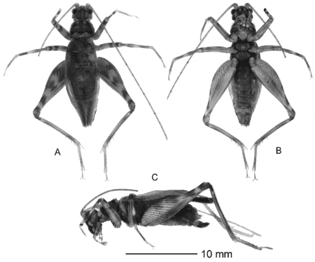
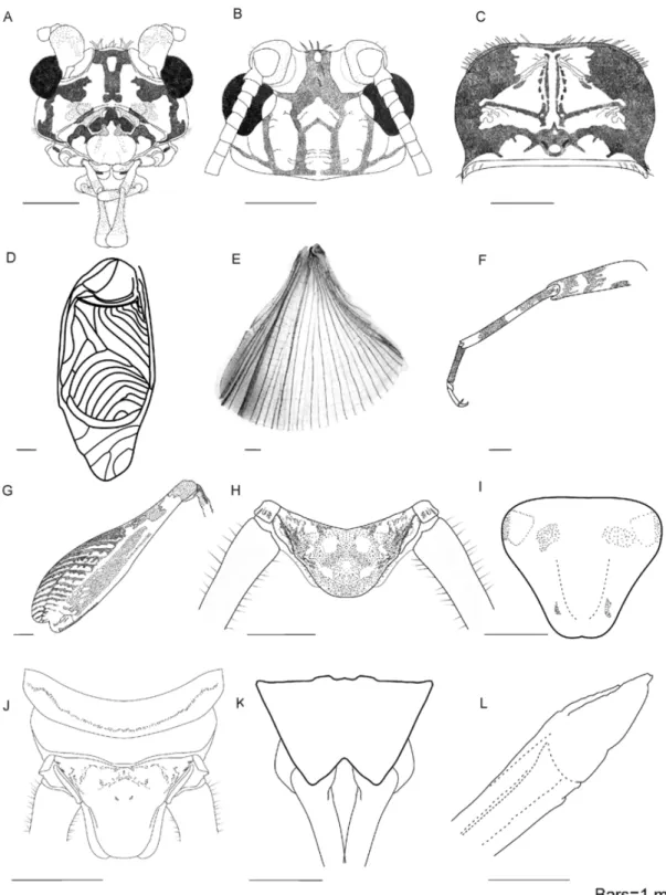
Rumea zebra Chamorro-Rengifo, Lopes-Andrade & Sperber, sp. nov. (Figs 2–4)
Etymology. The specific epithet refers to the coloration pattern of the legs, which consists of alternating dark yellowish and dark brownish stripes, resembling the coloration pattern of a zebra.
Diagnosis. The body coloration of R. zebra sp. nov. is lighter than that of R. manauensis sp. nov. and R. tigris sp. nov., so the contrast between the brownish and yellowish stripes is comparatively more distinctive. This new species is also smaller than the latter two species. It can also be distinguished from the other described Rumea by the following combination of characters: (i) face with striped coloration, the brownish stripes being narrower than that of R. manauensis sp. nov.; (ii) pronotum with anterior and posterior sides broadly rounded; male genitalia with (iii) dorsal tip of the longitudinal midline of the pseudepiphallic arm with two small lobes (conspicuous in lateral view), (iv) one lateral lobe (of the pseudepiphallus) visible in dorsal view, (v) each lateral lobe of the pseudepiphallic arm directed outwards, and not laterally pronounced, (vii) ectophallic apodemes slightly curved inwards.
either side of the longitudinal midline of the rostrum. Labrum greyish with two faint lateral stripes, none reaching the ventral margin. Mandibles yellowish with a few dark spots; cutting edge dark brownish. Maxillary and labial palpi with whitish bases and several distinctive dark spots at the base and tip of each palpus. Last palpomere of each maxillary palp dark brownish. Mouthparts in ventral view, excepting the appendages, yellowish with several black spots. Antennal scape light yellowish, with an irregular dark brownish spots; antennomeres light yellowish. Dorsal surface of the head (Fig. 3B) with four brownish longitudinal stripes: two stripes originating at the posterior margin and jointed at the eyes level, so that only one thick stripe reaches the anterior margin; two lateral stripes, one at each side, originating at the posterior margin and ending at the ventral margin of each eye. Additionally, there is one stripe, originating at the posterior base of each lateral stripe, which is bifurcated forming one stripe that reaches the inner margin of the eye and another faint stripe extending to the anterior ventral surface but disappearing, not reaching the face. Eyes dark brownish, with a whitish oval area at the dorsal inner surface; central ocellus oval, almost three times larger than the other two ocelli together, the latter being very small and inconspicuous. Thorax with the anterior and posterior pronotal angles rounded (Fig. 3C). Pronotum with disc mostly dark yellowish, bearing dark brownish marks; posterior area light yellowish; dark brownish lateral lobes and strong bristles at the anterior and lateral margins. Longitudinal midline
one at the tip and the other at the middle. Each hindleg with the femur (Fig. 3G) bearing four distinct brownish areas intercalated with yellowish areas; hindtibia dark yellowish, with a dark brownish mark at the dorsal tip. First and second tarsomeres of fore- and midlegs darker than the third tarsomere and darker than the tarsus of the hindleg. Tarsus claws of all legs whitish, with black tips. Abdomen with the tergites light brownish, with irregular dark brownish spots like shadows. Sternites light brownish, the first and second ones lighter than the others. Penultimate tergite of the female (Fig. 3J) with a transversal division, with the anterior portion lifted up and leaving the posterior portion in low-relief (possibly a glandular area). Cerci dark yellowish. Posterior margin of the supra-anal plate broadly rounded (Figs 3H,J), larger in female (Fig. 3J) than in male (Fig. 3H), the plate in male being almost black, and in the female dark yellowish with irregular brownish spots like shadows. Subgenital plate of male (Fig. 3I.) with an indistinct emargination at the middle of the posterior margin (Fig. 3I); in female (Fig. 3K), this emargination is deep and broad, leading a V-shape cut of almost one-third the length of the plate at the longitudinal midline. Ovipositor (Fig. 3L) 0.72X the body length, with two little lobes at the ventral side before the apex. Male genitalia (Fig. 4) with the dorsal tip of the pseudepiphallic arm with two small lobes at the midline, visible in lateral view (Fig. 4B); pseudepiphallic arm with lateral lobes directed outwards (Fig. 4A,C), not projected in lateral view (Fig. 4B); each pseudepiphallic
lateral lobe with only one projection, well developed, visible in dorsal view (Fig. 4A); ectophallic apodemes slightly curved inwards (Fig. 4A); both rami reduced, each one with a small undulation. Female genitalia (Fig. 4D) with a bell form; spermatheca tubular, broadly curved inwards, clockwise when seen from above, increasing in size from the basal to the apical portion.
Comments: Both antennas of the holotye are broken, the right one longer than the left one.
EyeW 1.37–1.52; TegL 13.39–14.07; TegW 4.64–4.97; OL 12.16–12.62. Right tegmen with six veins on the harp, and six to seven veins on the mirror; the last vein of the mirror being either ramified into two veins or unramified.
Type series. Holotype ♂ (INPA) labelled /Brazil, Manaus, Tarumã Mirim River. Joachim Adis [handwritten on white paper] / 47A TM BE. 22.3.1976 [handwritten on white paper] / Rumea zebra Chamorro-Rengifo, Lopes-Andrade & Sperber. 2009 [handwritten on red paper]/. Allotype ♀ (INPA), same locality data as the holotype but collected in /49A TM BE 22.3.76 [handwritten on white paper]/ and additionally labelled /Rumea zebra Chamorro-Rengifo, Lopes-Andrade & Sperber 2009 [handwritten on blue paper]/. Paratypes, same locality data as the holotype: 2♂♂ (1 INPA, 1 INPA) one collected in /48C TM BE 8.12.1976 [handwritten on translucent white paper]/ and the other in /49E TM BE 12-1-1976 [handwritten on white paper] /; 2♀♀ (1 INPA, 1 INPA) one collected in /47B TM BE 6.4.1977 [handwritten on white paper]/ and the other in /TM 48A TM BE 13.4.1977 [handwritten on white paper]/.
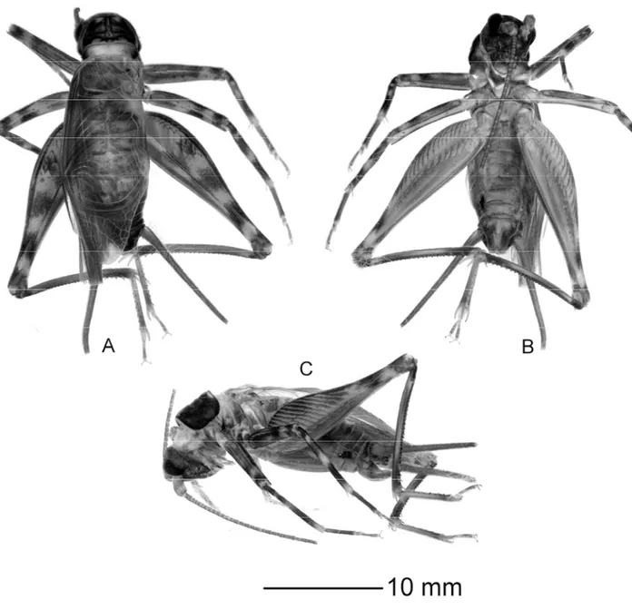
Rumea manauensis Chamorro-Rengifo, Lopes-Andrade & Sperber, sp. nov. (Figs 5–7)
Etymology. The specific epithet refers to Manaus, terra typica of this new species.
Description (holotype ♂ & allotype ♀). Holotype ♂: TL 20.45; HF 14.52; HT 11.82; sHT, inner margin 24–25, outer 23–25; sHts, inner margin 3–7, outer 8–9; PL 3.11; PW 3.88; EyeW 1.73; TegL 14.81; TegW 6.21; SL 3.68; NT 153. Allotype ♀: TL 21.09; HF 14.23; HT 11.29 (left hindleg lacking); sHT, inner margin 23, outer 22; sHts, inner margin 9, outer 9; PL 3.32; PW 3.97; EyeW 1.71; TegL 15.46; TegW 5.69; OL 17.97. General coloration consisting of dark brownish and dark yellowish areas (Fig. 5). Head with robust bristles on the vertex (Fig. 6A). Face with dark yellowish coloration, with brownish stripes. Frons with a broad brown stripe from the vertex to the frontoclypeal suture, narrowing from the dorsal to the ventral surface, with a yellowish oval area at the level of each eye; two oblique lateral stripes from below the antennal socket to the frontoclypeal suture, interrupted approximately in the first third above the frontoclypeal suture; each gena with two brownish stripes, one extending from below the eye to the ventral surface of the head, and the second stripe bordering the ventral surface of the head and reaching the postgena. Clypeus whitish, with two greyish lateral marks. Labrum whitish with two indistinct greyish lateral stripes (like shadows), not reaching the ventral margin. Mandibles yellowish, cutting edge dark brownish. Maxillary and labial palps light yellowish. Last palpomere of each maxillary palp dark brownish. Mouthparts in ventral view, excepting the appendages, light yellowish. Each antenna
marks on each side of the longitudinal midline, near the very middle of the disc, the outermost marks being dark brownish and the innermost marks being yellowish; posterior portion with an indistinct dark yellowish oval mark on each side of the midline, both marks being parallel to the posterior margin, and a dark yellowish mark with a half oval shape at the midline; posterior portion with a dark yellowish border along the margin. Tegmina translucent, brownish; right tegmen (Fig. 6D) with the mirror bearing seven curved veins, the last vein of the anterior margin being divided into three; harp with six veins. Hindwings (Fig. 6E) translucently brown. Pro- and mesosternum dark yellowish, metasternum dark brownish. Fore- and midlegs (Fig. 6F) with the same coloration pattern, as follows: each coxa and trochanter dark brownish; dorsal area of each femur light brownish with a dark brownish mark near the apex; tibia with two inconspicuous dark areas, one at the tip and the other at the middle. Hindleg with femur (Fig. 6G) bearing four distinctive brownish areas, each one between yellowish stripes; hindtibia dark yellowish, with a dark brownish area at the dorsal tip. First and second tarsomeres of fore- and midlegs darker than the third, all darker than the tarsomeres of the hindleg. Tarsus claws of all legs whitish, with black tips. Abdomen with tergites dark brownish; ninth and tenth tergites with low-relief areas (possibly glandular) in either side (Fig. 6J), these areas being close to the posterior margin in the former and close to the anterior margin in the latter. Sternites dark
arranged on the left side of the genitalia, with two lateral ducts growing from the genital chamber.
Comments: Both antennas of the holotype are broken, the left one longer than the right one.
Variation. Measurements of males (n=2, including the holotype): TL 19.52–20.45; HF 14.52–15.11; HT 11.69–11.82; sHT, inner margin 24–25, outer 23–25; sHts, inner margin 3–8, outer 8–9; PL 3.11–3.20; PW 3.88–4.21; EyeW 1.70–1.73; TegL 14.81 (measured only in the holotype; paratype with incomplete tegmina); TegW 6.21–6.57; SL 3.68–4.01; NT 153–184. Females: besides the female allotype, no other female specimen was available. Right tegmen of the male paratype similar to that of the holotype, with six veins in the harp and eight in the mirror, without ramification.
Type series. Holotype ♂ (INPA) labelled /Brazil, Manaus, Tarumã Mirim River. 08-03-1976. Joachim Adis [handwritten in white paper] / 48A TM BE 8.3.76 [handwritten on white paper] / Rumea manauensis Chamorro-Rengifo, Lopes-Andrade & Sperber 2009 [handwritten on red paper]/. Allotype ♀ (INPA), same locality data as the holotype but collected in /47B TM BE 17.2.1976 [written on white paper]/ and additionally labelled
/Rumea manauensis Chamorro-Rengifo, Lopes-Andrade & Sperber [handwritten on blue paper]/. Paratype ♂ (INPA), same locality data as the holotype but collected in /48D TM BE 06-01-1977 [handwritten on blue paper]/ and additionally labelled /Rumea manauensis Chamorro-Rengifo, Lopes-Andrade & Sperber [handwritten on blue paper]/.
Rumea tigris Chamorro-Rengifo, Lopes-Andrade & Sperber, sp. nov. (Figs 8–10)
Etymology. The specific epithet refers to the coloration pattern of the legs, which consists of alternating lines of light and dark brownish areas, similar to the colour pattern of a tiger.
dark brownish and more ventrally elongated than those of R. zebra sp. nov. and R. manauensis sp. nov., each eye bearing a whitish oval area at the dorsal inner surface that is smaller than those of R. zebra sp. nov. and R. manauensissp. nov.; (iv) anterior and posterior pronotum edges broadly rounded, giving a suboval appearance to the pronotal surface when seem from above; male genitalia with (v) dorsal tip of the pseudepiphallic arm with a rounded small lobe on the midline, (vi) lateral lobes of the pseudepiphallic bridge with two projections, one being reduced and barely visible from above, (vii) lateral lobes of the pseudepiphallic arm curved ventrad-outwards, ventrad pronounced as seen in lateral view, (viii) ectophallic apodemes slightly curved inwards.
Description (holotype ♂; female unknown). Holotype ♂: TL 20.03; HF 15.28; HT 12.66; sHT, inner margin 25–30, outer 22–25; sHts, inner margin 6–8, outer 8–9; PL 3.43; PW 4.22; EyeW 1.84; TegL 16.05; TegW 7.04; SL 4.35; NT 124. General coloration consisting of dark brownish and dark yellowish areas (Fig. 8). Head bearing robust bristles at the vertex (Fig. 9A). Face with coloration dark brownish with small light brownish areas, without of striped pattern. Frons bearing an indistinct light yellowish oval mark on the longitudinal midline at eye level; two semi-rounded light yellowish oval areas at eye level, close to the antennal socket; two oblique light yellow stripes at the middle of each gena; a light yellow undefined areas above the subgenal
another faint stripe extending to the anterior ventral surface but disappearing, not reaching the face. Eyes dark brownish, each with a whitish oval area at the dorsal inner surface; central ocellus oval, almost three times bigger than the two other ocelli, the latter been almost indiscernible. Thorax with the pronotal margins broadly rounded (Fig. 9C), giving a semicircular appearance to the pronotum when seen from above. Anterior and lateral margins bearing robust bristles; lateral surface dark brownish. Pronotum with disc dark yellowish and bearing dark brownish areas; anterior margin with dark brownish border along it, the border bearing a small subtriangular mark at the longitudinal midline; a distinctive triangular mark on each side of the midline, with one outer mark being dark brownish and the inner mark dark yellowish; posterior margin with two indistinct oval marks parallel to it, one mark at each side of the longitudinal midline; two ovals dark yellowish marks at the midline, one close to the posterior margin and the other above it; posterior portion with a light yellowish border along the margin. Tegmina translucent, brownish; right tegmen (Fig. 9D) with mirror bearing eight curved veins; harp with five veins. Hindwings (Fig. 9E) translucent, brownish. Sternum light yellowish. Metasternum darker than sternum. Fore- and midlegs (Fig. 9F) as follows: each coxa and trochanter yellowish with indistinct dark spots like shadows; femur light yellowish, with two conspicuous brownish rings, one near the apex and the other below it, being separated from each other by a yellowish space, the rings being
small lobe, being barely visible in dorsal view (Fig. 10A); each median lobe with a small projection (Fig. 10B); ectophallic apodemes directed outward (Fig. 10A); each ramus not reduced, bearing two undulations.
Comments: both antennas are broken, right one longer than the left one.
Type series. Holotype ♂ (INPA) labelled /Brazil, Manaus, Lago Janauari. Joachim Adis [handwritten on white paper] / BE 49C Lj 15.9.87 [handwritten on white paper] / Rumea tigris Chamorro-Rengifo, Lopes-Andrade & Sperber 2009 [handwritten on red paper]/.
Taxonomic key for the Rumea species
1. General coloration of the face without dark stripes………...2
1'. General coloration of the face with dark stripes ………..3
2. Genitalia of the male with the lateral lobes of the pseudepiphallic arm outward directed; lateral lobes of the pseudepiphallic arm not pronounced in lateral view………...R. gaschei
2'. Genitalia of the male with lateral lobes of the pseudepiphallic arm ventro-outward directed; lateral lobes of the pseudepiphallic arm pronounced in lateral view…..R. tigris
3. Two completed lateral lobes on the pseudepiphallic bridge………..4
3'. Two lateral lobes on the pseudepiphallic bridge, but one completed and the other
reduced………..R. micra
4. Genitalia of the male with the lateral lobes of the pseudepiphallic arm outward directed...5