Article
Gene Expression Regulation by Agonist-Independent
Constitutive Signaling of Melanocortin-1 Receptor
Ikjoo Seong, Jaesang Kim
Department of Life Science and Ewha Research Center for Systems Biology, Ewha Womans University, Seoul, Korea
Background: Melanocortin-1 receptor (Mc1r), a key signaling receptor for melanogenesis, has been reported to mediate migra-tion of B16F10 melanoma cells. Interestingly, this activity appears to be a part of the constitutive signaling of Mc1r.
Methods: We carried out small interfering RNA-mediated knock-down of Mc1r on murine melanoma B16F10 cells and per-formed microarray analysis to characterize changes in the gene expression profile.
Results: We isolated 22 and four genes whose expression decreased and increased, respectively, by 2.5-fold or higher as the re-sult of Mc1r knock-down. Several down-regulated genes have been proposed to be involved in cell migration. Among these genes are several members of the chemokine gene family.
Conclusion: We provide a gene set for further functional analyses of Mc1r. The Mc1r target genes we present may be particularly relevant for understanding the ligand-independent activity of Mc1r. Further examination of the mode of action may lead to novel strategies in regulating the migration and metastasis of melanoma cells.
Keywords: Receptor, melanocortin, type 1; Melanoma; Migration; Chemokines
INTRODUCTION
Melanocortin-1 receptor (Mc1r), a G-protein-coupled receptor expressed in melanocytes and melanoma, plays a key role in the pigmentation of hair and skin [1]. Several lines of recent evidence indicate that Mc1r also mediates cell signals regulat-ing migration of melanoma cells. First, agouti signal protein, a well-known ligand for Mc1r has been shown to stimulate the migratory activity of melanoma cells [2]. Second, another li-gand, α-MSH, inhibits migration and metastasis of melanoma cells [3,4]. Finally, we have recently reported that down-regu-lation of Mc1r via RNA interference inhibits both migration and metastasis of B16F10 murine melanoma cells [5]. That
simple knock-down of Mc1r inhibited migration in turn sug-gests that constitutive signaling by Mc1r may be pro-migrato-ry. Thus far, the promigratory signaling network down-stream of Mc1r has not been characterized at the molecular level, un-like that of promelanogenic signals. This applies to identifica-tion and funcidentifica-tional characterizaidentifica-tion of both the cytoplasmic components of the signaling network and the ultimate targets of transcriptional activation or repression.
Here, we report gene expression profiling of highly migra -tory B16F10 melanoma cells following inhibition of Mc1r ex-pression. We present several Mc1r-target genes that are poten-tially involved in cell migration. It appears that constitutive signaling of Mc1r positively regulates expression of several
Received: 31 July 2013, Accepted: 20 February 2014
Corresponding author: Jaesang Kim
Department of Life Science and Ewha Research Center for Systems Biology, Ewha Womans University, Science Building, 52 Ewhayeodae-gil, Seodaemun-gu, Seoul 120-750, Korea
Tel: +82-2-3277-3414, Fax: +82-2-3277-3760, E-mail: jkim1964@ewha.ac.kr
Copyright © 2014 Korean Endocrine Society
chemokines. The implications of our data are discussed.
METHODS
Cell culture
Murine melanoma cell line B16F10 was purchased from American Type Culture Collection. Cells were cultured in Dulbecco’s Modification of Eagle’s Medium supplemented with 10% fetal bovine serum and 1% penicillin and strepto-mycin. Transwell migration assay was carried out according to our previously published protocol [5].
RNA interference
A synthetic 21-nucleotide RNA duplex specific for Mc1r (wild-type [WT]-Mc1r) and a control RNA duplex with a 5 nucleotide mismatch (mutant [MT]-Mc1r) were purchased from Dharmacon Research Inc. (Lafayette, CO, USA). The sequences are illustrated in Fig. 1A. For migration assay or microarray screen, siRNAs were used at a concentration of 50 nM. Transfection was carried out with oligofectamine (Invit-rogen, Carlsbad, CA, USA) following the manufacturer’s pro-tocol. Details are available upon request.
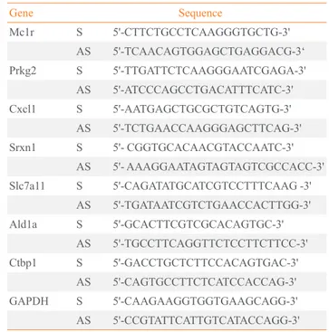
Real-time reverse transcription polymerase chain reaction Total RNA was prepared with RNeasy Mini Kit (QIAGEN, Valencia, CA, USA) following the manufacturer’s instruc-tions. cDNA synthesis was carried out with the SuperScriptII First-Strand Synthesis System for reverse transcription poly-merase chain reaction (RT-PCR, Invitrogen) using oligo-dT primers according to the manufacturer’s instructions. Quanti-tative analyses of gene expression level were performed by re-al-time PCR with SYBR Green Master mix (Applied Biosys-tems, Foster City, CA, USA). The expression level of glycer-aldehyde 3-phosphate dehydrogenase was used for normaliza-tion, and the oligonucleotide primers used are listed in Table 1.
Immunoblotting
Cells were washed twice with phosphate buffered saline and lysed in cell culture lysis buffer with protease inhibitor cocktail (Roche, Mannheim, Germany) and phenylmethylsulfonyl fluo-ride. Then, 20 µg of protein was separated by 12% sodium do-decyl sulfate polyacrylamide gel electrophoresis, transferred to nitrocellulose membrane, and immunoblotted with specific an-tibodies against mouse Srxn1 (a kind gift from professor W.
Jeong at Ewha Womans University, Seoul, Korea) and β-actin
Fig. 1. (A) Sequences of siRNAs, wild-type melanocortin-1 receptor
(WT-Mc1r), and mutant (MT)-Mc1r. (B) B16F10 cells treated with siRNAs were analyzed via transwell migration assay. Transfection of WT-Mc1r at 50 nM led to a significant reduction in migration of the cells compared to transfection of MT-Mc1r at 50 nM. (C) Confirma-tion of Mc1r down-regulaConfirma-tion by reverse transcripConfirma-tion polymerase chain reaction (RT-PCR). Two control genes (Ald1a and Ctbp1) were used to confirm the specificity of the siRNAs, and glyceraldehyde 3-phosphate dehydrogenase expression levels were used for normal-ization. Values represent the average of three independent real-time PCR experiments, each carried out in duplicate, and error bars repre-sent standard deviation. aSignificant difference with a
P value of 0.05. Mc1r RNAi duplex
Mc1r RNAi duplex
A
1.5
1.0
0.5
0.0
Mc1r
Re
la
ti
ve
e
xpre
ssi
on l
eve
l
Ald1a Ctbp1
MT-Mc1r
WT-Mc1r
a
C
MT-Mc1r WT-Mc1r
(LF-PA0066, AbFRONTIER, Seoul, Korea).
Microarray screen
Total RNA was prepared with the RNeasy Mini Kit (QIA-GEN) according to the manufacturer’s instructions and was subsequently processed to yield biotinylated cRNA using the Ambion Illumina RNA amplification kit (Ambion, Austin, TX, USA) according to the manufacturer’s instructions. The labeled cRNA preparations were applied to a MouseRef-8 v2 Expression BeadChip (Illumina, San Diego, CA, USA). De-tails of subsequent detection and quantitative analyses are avail-able upon request.
RESULTS
We used B16F10 murine melanoma cells to investigate the role of Mc1r in cell migration. B16F10 cells are highly migra-tory and have been used to demonstrate that knock-down of Mc1r leads to inhibition of migration and metastasis [5]. In or-der to identify down-stream target genes of Mc1r, we carried out microarray-based gene expression profiling in combina-tion with siRNA-mediated knock-down of Mc1r. We selected a siRNA-targeting murine Mc1r (WT-Mc1r) known to
effi-ciently inhibit migration of B16F10 cells (Fig. 1A). A mutant siRNA with five mismatches in nucleotide sequence (MT-Mc1r) was used as the control (Fig. 1A). Migration was effec-tively inhibited in WT-Mc1r-treated cells compared to MT-Mc1r-treated cells (Fig. 1B). RT-PCR assay indicated that WT-Mc1r reduced the mRNA level to about 50% of the level seen in MT-Mc1r treatment (Fig. 1C). This change in expression was not seen with the two negative control genes, Ald1a and Ctbp1, consistent with the specific activity of WT-Mc1r (Fig. 1C). Microarray data were in agreement with RT-PCR results. Mc1r was down-regulated 2.15-fold by WT-Mc1r siRNA while Ald1a and Ctbp1 were not affected (data not shown).
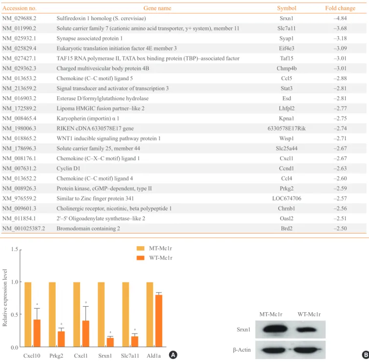
Tables 2, 3 show genes whose expression levels were up- and down-regulated, respectively, by more than 2.5-fold after averaging values from duplicate microarray assays. We con-firmed the microarray data by RT-PCR on a subset of genes down-regulated by WT-Mc1r (Fig. 2A). We also examined the down-regulation of sulfiredoxin 1 (Srxn1), which exhibited the largest reduction in expression, by immunoblotting. Con-sistent with the results from RT-PCR, Srxn1 protein demon-strated visible down-regulation by WT-Mc1r (Fig. 2B). Srxn1 is a member of an antioxidant protein family contain-ing a ParB-like nuclease domain. At least two recent studies reported that Srxn1 promotes cell migration [6,7]. Another tar-get gene, Stat3, a member of the signal transducer and activa-tor of transcription family of transcription facactiva-tors, is also known to promote cell motility via multiple mechanisms [8,9]. Notable among the genes down-regulated by WT-Mc1r are three chemokines, Ccl5, Cxcl1, and Ccl4. Ccl5, also known as RANTES, promotes the migration of multiple cell types [10-12] and is expressed in multiple metastatic human melanomas [13]. Cxcl1, also known as melanoma growth-stimulating
activity-α, is involved in melanoma pathogenesis and has been
Table 1. List of Real-Time Reverse Transcription Polymerase
Chain Reaction Primers
Gene Sequence
Mc1r S 5'-CTTCTGCCTCAAGGGTGCTG-3'
AS 5'-TCAACAGTGGAGCTGAGGACG-3‘
Prkg2 S 5'-TTGATTCTCAAGGGAATCGAGA-3'
AS 5'-ATCCCAGCCTGACATTTCATC-3'
Cxcl1 S 5'-AATGAGCTGCGCTGTCAGTG-3'
AS 5'-TCTGAACCAAGGGAGCTTCAG-3'
Srxn1 S 5'- CGGTGCACAACGTACCAATC-3'
AS 5'- AAAGGAATAGTAGTAGTCGCCACC-3'
Slc7a11 S 5'-CAGATATGCATCGTCCTTTCAAG -3'
AS 5'-TGATAATCGTCTGAACCACTTGG-3'
Ald1a S 5'-GCACTTCGTCGCACAGTGC-3'
AS 5'-TGCCTTCAGGTTCTCCTTCTTCC-3'
Ctbp1 S 5'-GACCTGCTCTTCCACAGTGAC-3'
AS 5'-CAGTGCCTTCTCATCCACCAG-3'
GAPDH S 5'-CAAGAAGGTGGTGAAGCAGG-3'
AS 5'-CCGTATTCATTGTCATACCAGG-3'
Mc1r, melanocortin-1 receptor; GAPDH, glyceraldehyde 3-phosphate
dehydrogenase.
Table 2. Genes Up-Regulated More than 2.5-Fold by Mc1r
siRNA
Accession no. Gene name Symbol Fold change
NM_053078.3 Neuronal regeneration related protein (Nrep)
D0H4S114 3.29
NM_010176.2 Fumarylacetoacetate hydrolase
Fah 2.87
NM_010442.1 Heme oxygenase (decycling) 1
Hmox1 2.77
NM_007584.2 Discoidin domain receptor family, member 1 (Ddr1)
shown to promote the migration of multiple lines of human melanoma cells [14,15]. Ccl4, also known as macrophage
in-flammatory protein-1β, likewise has been reported to increase
the migratory activity of human uveal melanoma cells [16].
Fig. 2. (A) Confirmation of microarray data by reverse transcription polymerase chain reaction (RT-PCR). A subset of genes that showed
down-regulation of 2.5-fold or higher by wild-type melanocortin-1 receptor (WT-Mc1r) compared to mutant (MT)-Mc1r in microarray assay and a nontarget gene (Ald1a) whose expression level was unchanged were used to validate the results from the microarray assay. Glyceraldehyde 3-phosphate dehydrogenase expression levels were used for normalization. Values represent the average of three inde-pendent real-time PCR experiments, each carried out in duplicate, and error bars represent standard deviation. (B) Immunoblotting for Srxn1 shows down-regulation by WT-Mc1r. β-Actin was used as the loading control. aSignificant difference with a
P value of 0.05. 1.5
1.0
0.5
0.0
Cxcl10 Prkg2 Cxcl1 Srxn1 Slc7a11 Ald1a
Relative expression level
MT-Mc1r
WT-Mc1r
a
a
a a
a
A
WT-Mc1r
Srxn1
β-Actin
MT-Mc1r
B
Table 3. Genes Down–Regulated by Mc1r siRNA by More than 2.5–Fold
Accession no. Gene name Symbol Fold change
NM_029688.2 Sulfiredoxin 1 homolog (S. cerevisiae) Srxn1 –4.84
NM_011990.2 Solute carrier family 7 (cationic amino acid transporter, y+ system), member 11 Slc7a11 –3.68
NM_025932.1 Synapse associated protein 1 Syap1 –3.18
NM_025829.4 Eukaryotic translation initiation factor 4E member 3 Eif4e3 –3.09
NM_027427.1 TAF15 RNA polymerase II, TATA box binding protein (TBP)–associated factor Taf15 –3.01
NM_029362.3 Charged multivesicular body protein 4B Chmp4b –3.01
NM_013653.2 Chemokine (C–C motif) ligand 5 Ccl5 –2.88
NM_213659.2 Signal transducer and activator of transcription 3 Stat3 –2.81
NM_016903.2 Esterase D/formylglutathione hydrolase Esd –2.81
NM_172589.2 Lipoma HMGIC fusion partner–like 2 Lhfpl2 –2.77
NM_008465.4 Karyopherin (importin) α 1 Kpna1 –2.75
NM_198006.3 RIKEN cDNA 6330578E17 gene 6330578E17Rik –2.74
NM_018865.2 WNT1 inducible signaling pathway protein 1 Wisp1 –2.71
NM_178696.3 Solute carrier family 25, member 44 Slc25a44 –2.67
NM_008176.1 Chemokine (C–X–C motif) ligand 1 Cxcl1 –2.67
NM_007631.2 Cyclin D1 Ccnd1 –2.63
NM_013652.2 Chemokine (C–C motif) ligand 4 Ccl4 –2.60
NM_008926.3 Protein kinase, cGMP–dependent, type II Prkg2 –2.59
XM_976559.2 Similar to Zinc finger protein 341 LOC674706 –2.57
NM_009601.3 Cholinergic receptor, nicotinic, beta polypeptide 1 Chrnb1 –2.56
NM_011854.1 2'–5' Oligoadenylate synthetase–like 2 Oasl2 –2.51
DISCUSSION
In this report, we describe the changes in gene expression fol-lowing knock-down of Mc1r by RNA interference. The genes thus identified represent regulatory targets of the ligand-inde-pendent signaling of Mc1r. To the best of our knowledge, this is the first study that identifies such a group of genes. At least five genes among those down-regulated have been reported to promote cell migration in various contexts. The most notable are the three chemokines, Ccl5, Cxcl1, and Ccl4. Although their expression and/or promigratory effect in melanoma cells have been reported previously, this is the first time that a regu-latory link has been established with Mc1r signaling.
Future experiments should examine whether these genes are functional in melanoma migration. The promigratory activities of Srxn1 and Stat3 have yet to be confirmed in melanoma. It will also be necessary to directly test if these chemokines pro-mote migration of melanoma and, if so, whether they function as autocrines. Inhibitors of Srxn1antioxidant activity or of Stat3 activation and nuclear localization could also be devel-oped and used as repressors of migration, and possibly of me-tastasis. For chemokines, competitive inhibitors for receptor binding or neutralizing antibodies can be tested for their ef-fects on cell migration. Detailed knowledge of the Mc1r sig-naling network and its promotion of melanoma migration, in-cluding down-stream effector genes, could provide the oppor-tunity for development of novel therapeutic strategies for mel-anoma.
CONFLICTS OF INTEREST
No potential conflict of interest relevant to this article was re-ported.
ACKNOWLEDGMENTS
This research was supported by a grant (2010K000803) from the Brain Research Center of the 21st Century Frontier Re-search Program and by the “Systems biology infrastructure es-tablishment grant” provided by the Gwangju Institute of Sci-ence & Technology in 2008 through the Ewha Research Cen-ter for Systems Biology (ERCSB).
REFERENCES
1. Garcia-Borron JC, Sanchez-Laorden BL,
Jimenez-Cer-vantes C. Melanocortin-1 receptor structure and functional regulation. Pigment Cell Res 2005;18:393-410.
2. Le Pape E, Passeron T, Giubellino A, Valencia JC, Wolber
R, Hearing VJ. Microarray analysis sheds light on the de -differentiating role of agouti signal protein in murine me-lanocytes via the Mc1r. Proc Natl Acad Sci U S A 2009; 106:1802-7.
3. Murata J, Ayukawa K, Ogasawara M, Fujii H, Saiki I. Al
-pha-melanocyte-stimulating hormone blocks invasion of reconstituted basement membrane (Matrigel) by murine B16 melanoma cells. Invasion Metastasis 1997;17:82-93.
4. Liu GS, Liu LF, Lin CJ, Tseng JC, Chuang MJ, Lam HC, Lee JK, Yang LC, Chan JH, Howng SL, Tai MH. Gene
transfer of pro-opiomelanocortin prohormone suppressed the growth and metastasis of melanoma: involvement of alpha-melanocyte-stimulating hormone-mediated inhibi-tion of the nuclear factor kappaB/cyclooxygenase-2 path-way. Mol Pharmacol 2006;69:440-51.
5. Seong I, Min HJ, Lee JH, Yeo CY, Kang DM, Oh ES, Hwang ES, Kim J. Sox10 controls migration of B16F10
melanoma cells through multiple regulatory target genes. PLoS One 2012;7:e31477.
6. Wei Q, Jiang H, Xiao Z, Baker A, Young MR, Veenstra TD, Colburn NH. Sulfiredoxin-Peroxiredoxin IV axis pro -motes human lung cancer progression through modulation of specific phosphokinase signaling. Proc Natl Acad Sci U S A 2011;108:7004-9.
7. Bowers RR, Manevich Y, Townsend DM, Tew KD.
Sul-firedoxin redox-sensitive interaction with S100A4 and non-muscle myosin IIA regulates cancer cell motility. Bio-chemistry 2012;51:7740-54.
8. Gao SP, Bromberg JF. Touched and moved by STAT3. Sci
STKE 2006;2006:pe30.
9. Teng TS, Lin B, Manser E, Ng DC, Cao X. Stat3 promotes
directional cell migration by regulating Rac1 activity via its activator betaPIX. J Cell Sci 2009;122(Pt 22):4150-9.
10. Long H, Xie R, Xiang T, Zhao Z, Lin S, Liang Z, Chen Z,
Zhu B. Autocrine CCL5 signaling promotes invasion and migration of CD133+ ovarian cancer stem-like cells via NF-kappaB-mediated MMP-9 upregulation. Stem Cells 2012;30:2309-19.
11. Wang SW, Wu HH, Liu SC, Wang PC, Ou WC, Chou WY, Shen YS, Tang CH. CCL5 and CCR5 interaction promotes
cell motility in human osteosarcoma. PLoS One 2012;7: e35101.
Mattoli S. The C-C motif chemokine ligands CCL5, CCL11, and CCL24 induce the migration of circulating fi-brocytes from patients with severe asthma. Mucosal Im-munol 2013;6:718-27.
13. Payne AS, Cornelius LA. The role of chemokines in
mela-noma tumor growth and metastasis. J Invest Dermatol 2002; 118:915-22.
14. Richmond A, Thomas HG. Melanoma growth stimulatory
activity: isolation from human melanoma tumors and char-acterization of tissue distribution. J Cell Biochem 1988;
36:185-98.
15. Di Cesare S, Marshall JC, Logan P, Antecka E, Faingold D,
Maloney SC, Burnier MN Jr. Expression and migratory analysis of 5 human uveal melanoma cell lines for
CXCL12, CXCL8, CXCL1, and HGF. J Carcinog 2007;6:2. 16. Woodward JK, Elshaw SR, Murray AK, Nichols CE, Cross
N, Laws D, Rennie IG, Sisley K. Stimulation and inhibition
of uveal melanoma invasion by HGF, GRO, IL-1alpha and