UNIVERSIDADE DE SÃO PAULO
FACULDADE DE ODONTOLOGIA DE BAURU
LUIZA DE PAULA SILVA CASSIANO
Effect of frequency of intake and amount of fluoride in milk on
enamel and dentine caries:
in situ
study
Efeito da frequência de ingestão e quantidade de fluoreto no leite
sobre cárie de esmalte e dentina: estudo
in situ
LUIZA DE PAULA SILVA CASSIANO
Effect of frequency of intake and amount of fluoride in milk on
enamel and dentine caries:
in situ
study
Efeito da frequência de ingestão e quantidade de fluoreto no leite
sobre cárie de esmalte e dentina: estudo
in situ
Dissertation presented to the Bauru School of Dentistry of the University of São Paulo to fulfillment of the requirements for the degree of Master in Dentistry in the area Stomatology and Oral Biology. Supervisor: Prof. Drᵃ Marília Afonso Rabelo Buzalaf
Dissertação constituída por artigo apresentada à Faculdade de Odontologia de Bauru da Universidade de São Paulo para obtenção do título de Mestre em Ciências no Programa de Ciências Odontológicas Aplicadas, na área de concentração Estomatologia e Biologia Oral.
Orientadora: Prof. Drᵃ Marília Afonso Rabelo Buzalaf
Versão Corrigida
Nota: A versão original desta dissertação encontra-se disponível no Serviço de Biblioteca e Documentação da Faculdade de Odontologia de Bauru – FOB/USP.
C273e
Cassiano, Luiza de Paula Silva
Effect of frequency of intake and amount of fluoride in milk on enamel and dentine caries: in situ study/ Luiza
de Paula Silva Cassiano. – Bauru, 2014. 59 p. : il. ; 31cm.
Dissertação (Mestrado) – Faculdade de Odontologia de Bauru. Universidade de São Paulo Orientadora: Prof. Drᵃ Marília Afonso Rabelo Buzalaf
Autorizo, exclusivamente para fins acadêmicos e científicos, a reprodução total ou parcial desta dissertação/tese, por processos fotocopiadores e outros meios eletrônicos.
Assinatura:
DEDICATÓRIA
Dedico esta dissertação aos meus familiares,
Aos meus pais Francisco e Márcia,
Que me dão todo suporte na conquista dos meus objetivos e são meus exemplos de força, garra e perseverança. A vocês devo tudo o que sou!
Aos meus avós Wanderley e Elva,
pelas orações e presença tão carinhosa em minha vida.
AGRADECIMENTOS
A Deus,
Por ser o meu defensor, protetor e em quem eu confio. Obrigada por colocar pessoas e oportunidades maravilhosas em minha vida e nunca me abandonar. Obrigada pela minha saúde e por me dar forças para seguir o meu caminho.
Ao meu pai Francisco,
Pelas horas que passamos conversando e tomando decisões. Pelo incentivo, carinho e proteção que me dá a qualquer hora do dia ou da noite. Por sempre me fazer ficar mais próxima de Deus. Obrigada também pelos e-mails, cada um deles que abro sempre sai um sorriso e apesar de não responder a maioria, você pode ter certeza pai que fazem toda diferença no meu dia, porque sinto você mais próximo. EU TE AMO!
À minha mãe Márcia,
Por realizar essa tarefa de mãe da melhor forma possível. Pela capacidade que tem de organizar a minha vida e fazer de tudo para me ver feliz. Obrigada pela dedicação, paciência, amor, carinho, conselhos e confidência. Por estar sempre ao meu lado nas minhas decisões me apoiando e se mostrando sempre presente para o que eu precisar. Agradeço a Deus todos os dias por ter uma mãe como você. EU TE AMO!
Aos meus irmãos Marcelo e Marina,
Aos meus familiares,
Avós, tios, tias e primos, pelo apoio e incentivo na realização dos meus projetos e por se fazerem sempre presentes apesar da distância. Muito obrigada por tudo!
Aos meus amigos de Goiânia, Mariana, Marcela, Marjorie, Tarihan,
Apesar de cada um ter seguido um caminho diferente, não importa o tempo nem a distância, sempre que nos encontramos é como se o tempo não tivesse passado. Uma amizade como a de vocês é muito saudável, sem cobranças, e sempre sabendo que um está ali para o outro para o que precisar. Muito obrigada pelo apoio, compreensão e cumplicidade. Carrego vocês sempre em meu coração!
À minha amiga e companheira de casa Letícia,
Por ser uma pessoa tão especial em minha vida. Quem tem uma amiga como você eu sempre digo que tem tudo na vida. Muito obrigada por me apoiar nos momentos mais difíceis, por ter me ajudado inúmeras vezes na realização dessa pesquisa. Pela sua sinceridade, lealdade e pelos momentos de confidência. Obrigada também pelos momentos de alegria, pela companhia nas festas, na hora de almoçar e jantar. Lê eu considero você como uma irmã que Deus colocou em minha vida para morar junto em Bauru. Espero que a nossa amizade dure para sempre independente do caminho que vamos seguir. E lembre-se sempre: Tudo que é bom dura o tempo necessário para ser inesquecível!
À minha amiga Flávia Barboza,
Às minhas amigas Beatriz, Juliana, Lívia, Naiana, Tamiris e Thais
Pelo apoio que vocês me deram para persistir na área acadêmica, por todos os momentos que dividimos alegrias, tristezas, preocupações, viagens, festas. Apesar da maioria de vocês já estar longe, sempre me lembro dos momentos que vivemos juntas desde a graduação. Vocês tem um lugar muito especial no meu coração. Obrigada por fazerem parte da minha vida.
À minha amiga Aline Leite,
Pela amizade conquistada ao longo desses dois anos do mestrado, por ter se mostrado tão prestativa quando precisei, pela ajuda no aprimoramento das técnicas para realização desse trabalho. Obrigada por sempre me ajudar com as questões que envolvem tecnologia e pelos momentos agradáveis no laboratório, os sustos, os apelidos, a hora do lanche, os seminários, as conversas. Sua presença e paciência foram muito importantes para que eu pudesse chegar até aqui. Muito obrigada por tudo!
À minha amiga e companheira de pesquisa Lívia,
Por ter me ensinado TODOS os passos necessários para a realização dessa pesquisa. Muito obrigada pela paciência, pelos telefonemas e ajuda prestada independente da hora ou do dia da semana. Pela amiga que se tornou e presença nos momentos de choro e risadas. Lívia saiba que você foi muito importante para a realização desse projeto e eu desejo muito sucesso na sua carreira.
Aos que colaboraram com a pesquisa Flavia Levy, Cristiane, Isabela, Larissa, Aline Dionizio, César, Karina, Michele e às alunas de pré- IC Letícia e Beatriz
Aos voluntários desta pesquisa,
Por terem sido fundamentais na realização deste trabalho. Pela paciência, atenção e imensa ajuda na execução do projeto. Obrigada por terem aceitado fazer parte da pesquisa e seguido às instruções. Sem dúvidas sem a ajuda de vocês esse trabalho não teria sido possível.
Aos colegas do laboratório de Bioquímica: Heloisa, Senda, Cíntia, Camila, Flávia Iano, Aline Castilho, Mileni, Bruno, Vinícius, Beatriz Souza, Melina, Priscila Muno, Priscila Salomão, Flávia Amadeu, Carina, Fernanda e Amanda, pela convivência diária no laboratório, conversas, risadas, aniversários e bons momentos que transformam o ambiente de trabalho em um lugar muito mais agradável.
Ao Prof. Juliano Pessan,
Pela importante contribuição com o trabalho e disponibilidade em ajudar durante várias etapas da realização dessa pesquisa, sempre atencioso e disposto a fazer o seu melhor. Muito obrigada!
À Profª Drª Ana Carolina,
Pela atenção e imensa ajuda nesse projeto, por ter sanado as minhas dúvidas e sempre se mostrar tão prestativa mesmo tendo tantos outros trabalhos. Carol admiro muito a forma como você trabalha e tenho você como um exemplo de como exercer a carreira do Ser Professor. Aprendi muito com você nas disciplinas que fiz durante o mestrado e você faz muito por merecer todas as homenagens que recebe dos alunos, pois você demonstra o quanto gosta do que faz e o prazer que tem em ensinar.
Ao Prof. Rodrigo Oliveira,
Aos funcionários do Departamento de Ciências Biolégicas Thelma, Larissa, Aline e Vera,
Por sempre atenderem as minhas dúvidas e pela ajuda prestada em vários momentos da pesquisa. Em especial muito obrigada à Larissa que fez as análises da microradiografia, sua ajuda foi de extrema importância para a realização desse trabalho.
Às secretárias da Pós-graduação Fátima, Leila, Meg e Maristela,
Pela disposição para responder prontamente a e-mails, telefonemas e ajudar com assuntos relacionados a documentação, prazos, relatórios. Muito obrigada pela atenção e esforço de vocês.
À Faculdade de Odontologia de Bauru-USP, na pessoa da diretora Prof.ª Drª Maria Aparecida Moreira Machado, e do senhor Presidente da Comissão de Pós-Graduação Prof. Dr. Guilherme dos Reis Pereira Janson
Pela oportunidade de realizar a pós-graduação em nível de Mestrado nesta instituição, que tem uma estrutura formidável e tenho tanto orgulho de fazer parte desde 2008 no curso de graduação em Odontologia. Obrigada FOB-USP por tudo que me proporcionou até hoje.
À Borrow Foundation,
Pelo auxílio financeiro fornecido à pesquisa, que viabilizou a realização e conclusão deste projeto.
À CAPES,
AGRADECIMENTOS ESPECIAIS
Agradeço em especial à minha orientadora Profª Marília Afonso Rabelo Buzalaf,
Ainda me lembro com muita clareza o dia que cheguei com tamanha timidez na sua sala para conversarmos sobre a possibilidade de fazer um mestrado. Pra mim seria uma coisa muito distante, mas você me incentivou de modo que essa conquista estaria mais perto do que eu imaginava. E assim você foi durante todo o tempo do mestrado, incentivadora. Apesar dos títulos e grande reconhecimento que você tem na área científica, é uma professora que está sempre de portas abertas, literalmente, recebendo seus alunos e fazendo de tudo para dividir a atenção entre todos.
Encontrei uma frase de Walt Disney que diz o seguinte: “O que quer que você faça, faça bem feito. Faça tão bem feito que, quando as pessoas te virem fazendo,
elas queiram voltar e ver você fazendo de novo e queiram trazer os outros para mostrar o quão bem você faz aquilo que faz”. Eu acredito que esse texto expressa muito bem a forma como você trabalha. Ter você como orientadora nos proporciona uma vontade muito grande de seguir os seus passos acreditando que estamos no caminho certo.
ABSTRACT
Effect of frequency of intake and amount of fluoride in milk on enamel and dentine caries: in situ study
This study analysed the effect of frequency of intake and amount of fluoride in milk on the remineralisation of artificial enamel and dentine caries lesions in situ. Pre-demineralised bovine enamel and dentine slabs were randomly allocated to 5 in situ phases. Twenty-three subjects wore removable appliances with 2 enamel and 2 dentine slabs for 7 days each phase (separated by a 7-day washout period), following a crossover double-blind protocol. In each phase, treatment was performed with milk containing 2.5 ppm fluoride (F) everyday (T1), 2.5 ppm F every other day (T2), 5.0 ppm F every day (T3), 5.0 ppm F every other day (T4) or no treatment (control; T5). The subjects were instructed to immerse the appliance in 100 ml of milk for 5 minutes and then drank 200 ml of the respective milk. The enamel alterations were quantified by surface hardness (%SHR) and transversal microradiography (TMR, ∆∆Z) and dentine by TMR only. Data were analysed by repeated-measures ANOVA/Tukey´s tests (p<0.05). For enamel, the highest %SHR was found for groups treated with fluoridated milk every day compared to control, without significant differences between T1 and T3. All groups showed positive values of ∆∆Z, except for T4; significant differences were seen between T1/T3 and T4. For dentine, the only group that presented remineralisation was T2. Fluoridated milk every day seems to have better remineralising effect on enamel than its use every other day, but no dose-response effect was seen. Dentine, however, does not seem to benefit from every day use of fluoridated milk.
RESUMO
O presente estudo teve como objetivo analisar o efeito do leite fluoretado com concentrações e frequências diferentes na remineralização de lesões de cárie de esmalte e dentina produzidas artificialmente in situ. Blocos de esmalte e dentina bovino previamente desmineralizados foram distribuídos aleatoriamente em cinco grupos. Vinte e três indivíduos usaram aparelhos removíveis contendo 2 blocos de esmalte e 2 blocos de dentina por 7 dias em cada fase (separadas por um período de washout de 7 dias), seguindo um protocolo duplo-cego, cruzado. Em cada fase, o tratamento realizado foi com leite contendo 2,5 ppm de flúor (F), todos os dias (T1), 2,5 ppm de F em dias alternados (T2), 5,0 ppm F todos os dias (T3), 5,0 ppm F em dias alternados (T4) ou sem tratamento (T5). Os sujeitos foram instruídos a mergulhar o aparelho em 100 ml de leite por 5 minutos e, em seguida, beberam 200 ml do mesmo leite. As alterações no esmalte foram quantificadas por dureza superficial e microradiografia transversal (TMR), e dentina apenas por microradiografia transversal. Os dados foram analisados por medidas repetidas ANOVA / Tukey (p <0,05). Para o esmalte, o mais alto valor de porcentagem de recuperação de dureza superficial foi encontrado para os grupos tratados com leite fluoretado todos os dias em relação ao controle, sem diferenças significativas entre T1 e T3. Todos os grupos apresentaram valores positivos de ∆∆Z, com exceção de T4; foram observadas diferenças significativas entre T1/T3 e T4. Para a dentina, o único grupo que apresentou remineralização foi T2. O leite fluoretado todos os dias parece ter melhor efeito remineralizante sobre o esmalte do que seu uso em dias intercalados, mas nenhum efeito dose-resposta foi visto. A dentina, no entanto, não parece se beneficiar do uso diário de leite fluoretado.
SUMÁRIO
1 INTRODUÇÃO ... 15
2 ARTIGO ... 21
3 DISCUSSÃO... 41
REFERÊNCIAS ... 49
Introdução 15
1 INTRODUCTION
Dental caries is a multifactorial disease; it depends on the interaction between the time that the biofilm is present on the tooth surface, the type of microorganism which comprises the oral environment and the diet of each individual. Thus, dental caries happens when the bacteria present in the dental plaque metabolize carbohydrates from the diet of the host, resulting in the production of acids that dissolve the hard tooth tissues (YEUNG et al., 2005).
From the understanding of disease and public awareness, dental caries showed a decline in recent decades, due to the control of etiologic factors, administration of fluoride in the water and use of fluoridated toothpaste as the main primary preventive measures for disease control (EINARSDOTTIR; BRATTHALL, 1996). Fluoride plays the role of inhibiting demineralization, accelerating remineralization and reducing bacterial metabolism. Fluoride exerts these functions through its topical action when used in vehicles such as toothpastes, gels, varnishes and restorative materials. Despite there are also several sources of systemic fluoride, such as water, supplements, salt and milk, its action is essentially topical since fluoride comes into contact with the teeth while passing through the oral cavity or when it returns to the oral cavity through saliva (BUZALAF et al., 2011).
Although there was a decline in the prevalence of caries in the past 20 years, populations in less developed countries and of lower socio-economic classes still have a significant incidence of the disease (CAMPUS et al., 2007; MASEREJIAN et al., 2009). This is due to the fact that water fluoridation as the main primary preventive measure in caries control is not possible in many areas because of political, geographical or technical reasons. Therefore, attention was focused towards the possibility of including fluoride into other products (KUNZEL, 1993), such as milk. This beverage has an important nutritional function. It is an important part of the human diet in the first years of life, containing essential nutrients like proteins, fats, carbohydrates, vitamins and minerals.
16 Introdução
population (YEUNG et al., 2005). Randomized clinical trials were conducted and showed that the increase in milk consumption in adolescent girls contributed significantly to the increase in bone mass (CADOGAN et al., 1997). In another study, supplying milk in schools for 2 years for needy children, contributed little but significantly for the growth of these children (BAKER et al., 1980).
In addition to the systemic benefits, milk has become an important vehicle for the prevention of dental caries. Studies done in the mid 40’s revealed that milk improved oral health (SPRAWSON,1932; SPRAWSON,1947), probably for having components that are able to protect the tooth structure from carious lesions. Among these components are listed lactose, a carbohydrate present in high concentration in milk (80%)that is the least cariogenic sugar in the diet. In addition there are casein, other proteins and lipids (RUGG-GUNN, 1993).
Casein is a phosphoprotein found in milk in larger percentage and is considered as one of the main components able to protect the tooth structure from the appearance of carious lesions (JOHANSSON, 2002). Studies show that casein inhibits the activity of glucosyltransferase, an enzyme produced by Streptococcus mutans, which transforms sucrose into glucose monomers. These monomers are rearranged promoting adherence of other bacteria and increasing the thickness of the dental biofilm (VACCA-SMITH; BOWEN, 1995; VACCA-SMITH et al., 1994; VACCA SMITH; BOWEN, 2000). Thus, the adhesion of bacteria and some salivary components present in the acquired pellicle on the enamel surface is compromised, preventing then the occurrence of carious lesions.
Introdução 17
Given the benefits to oral health that milk promotes, the process of milk fluoridation started in Japan, United States and Switzerland around the 1950’s (RUSOFF et al., 1962; ZIEGLER, 1956, 1964). Milk was then regarded as another vehicle for the prevention of dental caries, and mostly able to reach the population that did not have access to fluoridated water. As in water, the addition of fluoride in milk does not promote change in flavor or other characteristics of the beverage and its use allow for a more accurate control of the amount of fluoride that is ingested daily (BANOCZY; RUGG-GUNN; WOODWARD, 2013).
Several studies in vitro, in situ and clinical trials have demonstrated a very satisfactory effect of fluoridated milk in relation to caries prevention (BANOCZY; RUGG-GUNN; WOODWARD, 2013), but in some systematic reviews the results are still sketchy regarding prevention of dental caries through the consumption of fluoridated milk and more studies need to be done in order to establish more solid evidences (CAGETTI et al., 2013; YEUNG et al., 2005).
Currently there are over one million children receiving fluoridated milk in several countries such as Chile, Thailand, UK, Russia, Bulgaria and Republic of Macedonia as part of the public health program. The fluoridated milk is offered in schools. Depending on each place and on the background exposure to fluoride, the concentration varies between 0.5 and 0.85 mg per day, while the frequency of intake varies between 200 and 365 days per year (BANOCZY; RUGG-GUNN; WOODWARD, 2013).
18 Introdução
Artigo 21
2 ARTIGO
Frequency of intake and amount of fluoride in milk on enamel and dentine artificial caries: in situ study
Cassiano LPS1, Pessan JP2, Comar LP1, Levy FM1, Cardoso CAB1, Dionizio AS1,
Manarelli MM2, Grizzo LT1, Magalhães AC1, Buzalaf MAR1*
1Department of Biological Sciences, Bauru School of Dentistry, University of São
Paulo, Bauru, SP, Brazil.
2Araçatuba Dental School, Univ. Estadual Paulista (UNESP), Araçatuba, SP, Brazil
Short title: Effect of fluoridated milk on enamel and dentine caries
Keywords: Enamel; Dentine; Fluoride; Milk; Remineralisation; Caries.
22 Artigo
Abstract
This study analysed the effect of frequency of intake and amount of fluoride in milk on the remineralisation of artificial enamel and dentine caries lesions in situ. Pre-demineralised bovine enamel and dentine slabs were randomly allocated to 5 in situ phases. Twenty-three subjects wore removable appliances with 2 enamel and 2 dentine slabs for 7 days each phase (separated by a 7-day washout period), following a crossover double-blind protocol. In each phase, treatment was performed with milk containing 2.5 ppm fluoride (F) everyday (T1), 2.5 ppm F every other day (T2), 5.0 ppm F every day (T3), 5.0 ppm F every other day (T4) or no treatment (control; T5). The subjects were instructed to immerse the appliance in 100 ml of milk for 5 minutes and then drank 200 ml of the respective milk. The enamel alterations were quantified by surface hardness (%SHR) and transversal microradiography (TMR, ∆∆Z) and dentine by TMR only. Data were analysed by repeated-measures ANOVA/Tukey´s tests (p<0.05). For enamel, the highest %SHR was found for groups treated with fluoridated milk every day compared to control, without significant differences between T1 and T3. All groups showed positive values of ∆∆Z, except for T4; significant differences were seen between T1/T3 and T4. For dentine, the only group that presented remineralisation was T2. Fluoridated milk every day seems to have better remineralising effect on enamel than its use every other day, but no dose-response effect was seen. Dentine, however, does not seem to benefit from every day use of fluoridated milk.
Introduction
Artigo 23
Fluoride can be administered by community, self-applied and professional methods, which are often used in association [Pessan et al., 2011]. Among the community methods, water fluoridation has been regarded as the main primary preventive and public health measure for caries control, as it reaches most of the population, including socially deprived groups. The benefits of water fluoridation, however, are unavailable to a large proportion of the world's population, mainly due to political, geographical and technical reasons [Sampaio and Levy, 2011]. In order to overcome this problem, other methods of community fluoridation have been suggested. As milk is an important part of children’s diet, fluoridated milk has been used in school-based preventive programmes for many decades, in different parts of the world. One of the main advantages of this method is that it allows the delivery of a precise amount of fluoride under controlled conditions [Banoczy et al., 2013].
The effectiveness of fluoridated milk in caries control has been assessed by in vitro, in situ and clinical studies, which show a positive effect of its regular consumption on caries prevention [Banoczy et al., 2013]. However, while the evidence of water fluoridation in caries control has been firmly established over the last decades [McDonagh et al., 2000], systematic reviews have concluded that the number of studies with good quality that evaluated the effects of fluoridated milk in preventing caries is insufficient. Overall, the included studies suggested that fluoridated milk was beneficial to school children, to prevent caries in the permanent [Yeung et al., 2005] or primary [Cagetti et al., 2013] dentitions. However, high-quality studies are still necessary in order that unequivocal evidence can be established. In addition, many aspects involving the milk fluoridation programmes still need to be addressed.
24 Artigo
The impact of these differences in the amounts of fluoride delivered and frequencies of intake on the caries preventive potential of milk fluoridation schemes is not completely known. The influence of two different fluoride concentrations in milk (2.5 and 5.0 ppm, corresponding to amounts of 0.5 and 1.0 mg fluoride delivered in 200 mL of milk, respectively) on the prevention of demineralisation of sound enamel was recently evaluated in situ. Both fluoride concentrations significantly protected enamel from demineralisation when compared with non-fluoridated milk, but did not significantly differ from each other [Malinowski et al., 2012a]. A recent in vitro study evaluated the effect of different fluoride concentrations in milk (2.5, 5.0 or 10.0 ppm) and also of distinct frequencies of use (once daily, twice daily or on alternate days) for remineralising initial enamel carious lesions. The best remineralising effect was observed for 2.5 ppm fluoride milk used twice daily [Ongtenco et al., 2014]. However, only one study evaluated the influence of different fluoride concentrations in milk on enamel remineralisation in situ, but the amounts of fluoride used added in milk (1.5 or 3.0 mg) were higher than those typically employed in milk fluoridation schemes and the authors evaluated only surface rehardening [Lippert et al., 2014].
In addition, to date no studies evaluated the effect of distinct fluoride concentrations in milk on dentine remineralisation in situ neither the effect of frequencies of use of milk on the remineralisation of dentine and enamel caries in situ. Thus, the present study evaluated if there is any additional benefit of increasing the amount of fluoride in milk from 2.5 ppm to 5 ppm per day on in situ enamel and root dentine remineralisation. The effect of different frequencies of drinking fluoridated milk (every day or every other day) was also investigated. It was hypothesized that higher fluoride concentration and frequency of milk intake would lead to enhanced remineralisation of enamel and dentine.
Materials and Methods
Ethical Aspects and Subjects
Artigo 25
[Malinowski et al., 2012a]. The inclusion criteria were: stimulated salivary flow > 1 mL/min, unstimulated salivary flow > 0.25 mL/min; salivary pH > 6, good oral conditions (presenting full permanent dentition, no open cavities or deficient restorations, absence of gingivitis and periodontal disease). Other factors taken into consideration were related to the overall health of the volunteer. Pregnant women, patients with systemic diseases and using chronic medication were not eligible to participate. Subjects signed an informed consent document prior to the beginning of the study.
Specimen Preparation
Bovine lower incisors were freshly extracted and stored in 2% formaldehyde solution (pH 7.0) for 30 days at room temperature. Teeth were submitted to careful visual inspection in order to detect stains and cracks. If these were detected, the teeth were excluded. Selected teeth were carefully cleaned from gingival tissue using a periodontal curette (Duflex, Duflex do Brasil, Brazil) before being cut. Initially the roots were separated from the crowns using a diamond disk (Diaflex-F, Wilcos do Brasil, Petrópolis, Brazil) by sectioning the cervical portion of the tooth. The crown was used for obtaining enamel slabs and, from the root, dentine slabs were prepared. Then the crowns or roots were fixed in acrylic plaques (40 X 40 X 5 mm) that were placed in the ISOMET Low Speed Saw cutting machine (Buehler Ltda., Lake Bluff, IL, USA). Two diamond disks (XL 12205, 102 X 0.3 X 12.7 mm, Extec Corp., Enfield, CT, USA), which were separated by a 4-mm diameter spacer (7 cm diameter, 4 mm thickness and central role of 1.3 cm), were used to cut the slabs (300 rpm, under refrigeration). One enamel and one dentine slabs (4 X 4 mm) were obtained from the flattest portion of each crown or root, respectively, through double sections in the longitudinal and transversal directions. The surface of the slabs was ground flat with water-cooled carborundum discs (320, 600 and 1200 grades of Al2O3 papers;
Buehler, Lake Bluff, IL, USA) and polished with felt paper wet by diamond spray (1 µm; Buehler), resulting in removal of about 100 µm depth of the enamel. This was controlled with a micrometer. After polishing, the specimens were cleaned in an ultrasonic device with deionised water for 10 minutes.
26 Artigo
335.3±2.4). Dentine slabs (n=230) were not submitted to surface hardness analysis since previous studies have revealed that this type of analysis is not accurate for this substrate since hardness even in sound dentine is not evenly distributed [Moron et al., 2013]. After that, one third of the (enamel/dentine) surface was covered with nail varnish to create a sound control area. The slabs were subjected to the formation of artificial caries lesions by immersion in a solution containing 50 mM lactate, 3 mM CaCl2, 3 mM KH2PO4, 6 µM tetraetil methyl diphophanate and traces of thymol, pH
5.0 (10 M KOH to adjust pH) (30 mL per block), for 7 days (dentine) and 6 days (enamel), at 37 ºC [Buskes et al., 1985]. The mean (±) lesion depth was 64.0 ± 20.1 µm and 82.9 ± 18.9 µm for enamel and dentine, respectively, as evaluated by TMR. After demineralisation, surface hardness (SHD) determination was done in enamel slabs and the other outer one third of the surface (enamel/dentine) was protected with nail varnish (demineralised control area). For the in situ experiment, 230 pre-demineralised enamel and 230 pre-pre-demineralised dentine were randomly allocated to 23 subjects (n=2/subject/phase) according to SHD means.
In situ experiment
Acrylic palatal appliances were constructed individually for each volunteer and for each phase. They were made with four cavities (5 x 5 mm), distributed into 2 rows of 2 spaces each. Enamel and dentine slabs were randomly accommodated into the cavities and fixed in place with wax. The study comprised 5 crossover phases of 7 days each, separated among each other by a washout period of 7 days (no treatment) [Afonso et al., 2013]. In each phase, four-five volunteers were assigned to one of the five treatments, as follows: T1: fluoridated milk (0.5 mg F), administered every day (200 mL of milk containing 2.5 ppm F as NaF); T2: fluoridated milk (0.5 mg F), administered every other day; T3: fluoridated milk (1.0 mg F), administered every day (200 mL of milk containing 5.0 ppm F as NaF); T4: fluoridated milk (1.0 mg F), administered every other day; T5: No treatment. Fluoridated pasteurised whole milk (Batavo Total, BRF SA, Teutônia, RS, Brazil) was prepared by the addition of powdered sodium fluoride (Merck, Darmstadt, Germany). Preparation was done every day and fluoride concentrations were checked using an ion specific electrode, after hexamethyldisiloxane-facilitated diffusion [Taves, 1968].
Artigo 27
between the in situ periods. The devices were worn by the volunteers 12 h before starting the treatments and challenges, allowing the salivary pellicle to form. To perform the experiment, in the days that milk should be used (every day for T1 and T3 or every other day for T2 and T4), the volunteers were instructed as follows: in the morning, during breakfast, they were asked to remove the device from the mouth and to immerse it in a cup containing 100 mL of milk for 5 minutes. Then they were asked to drink 200 mL of milk and reinsert the device in the mouth immediately after. Fluoridated milk was freshly prepared each day. Palatal devices were used during the entire day, except when drinking or eating (in total 4h/day). The devices were kept in wet gauze inside a plastic container during the time they were not being used by the volunteers. It is important to highlight that the study was conducted in a fluoridated area (0.6-0.8 ppm) [Buzalaf et al., 2013], but the volunteers drank bottled water with low fluoride concentration (<0.2 ppm) provided by the researchers throughout the experimental period. The same water was used for cooking and preparing beverages. The volunteers were instructed to keep their usual eating habits and to perform oral hygiene using toothbrush, dental floss and non-fluoridated toothpaste provided by the researchers during all stages of the study. Only the surface of the device that was in contact with the palate could be brushed. The volunteers were also instructed not to use any type of fluoride or antibacterial product during the in situ period. At the end of each phase, the devices of all volunteers were collected and washed with deionised water and the slabs were again set on their respective acrylic disks to be prepared for hardness and transverse microradiography analysis.
Hardness measurement
After the in situ phase, final surface hardness (FSH) analysis was performed on all the enamel slabs as described above. The % surface hardness recovery (%SHR) was calculated, as follows: %SHR = [(FSH – SHD) / (BSH – SHD)]*100.
Transverse microradiography
28 Artigo
Artigo 29
∆∆Z/∆L was calculated as the difference between ∆Z/LD lesion and ∆Z/LD effect (after treatments T1/T2/T3/T4/T5).
Statistical analysis
The software GraphPad InStat (version 3.0) was used. Initially, data were tested for normality and homoscedasticity, using Kolmogorov-Smirnov and Bartlett’s tests, respectively. Data regarding %SHR for enamel, and ∆∆Z for enamel and dentine were analysed by repeated-measures ANOVA and Tukey’s post-hoc test. Data regarding lesion depth were analysed by Friedman’s test (non-parametric repeated-measures ANOVA) and Dunn’s multiple comparison test. In all conditions, the significance level was set at 5%.
Results
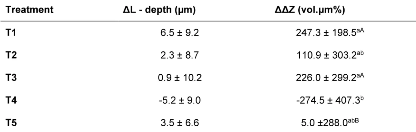
Mean %SHR of enamel slabs with artificial caries lesions after in situ remineralisation is presented in Table 1. Repeated-measures ANOVA found a significant difference among the treatments (F = 3,691, p = 0.0081). No dose-response was observed for the two fluoride concentrations in milk evaluated, regardless the frequency of intake. The highest %SHR was found for the groups treated with fluoridated milk every day, regardless the amount of F (T1 and T3) that did not significantly differ from each other but performed significantly better than T5 (no treatment). Treatment with fluoridated milk every other day (T2 and T4) presented an intermediate effect and did not significantly differ from T1, T3 and T5.
30 Artigo
For dentine, no significant differences among the groups were found for lesion depth (Fr = 4.985, p = 0.289). The results found for ∆∆Z were quite different from those obtained for enamel. A significant difference was found among the groups (repeated-measures ANOVA, F = 3.75, p = 0.010). The only group that presented remineralisation was T2 that significantly differed from all the others that suffered demineralisation (Table 3; Figure 2).
Discussion
Despite fluoridated milk has been recommended as an alternative vehicle to deliver fluoride since 1953 and some clinical studies have attested the preventive effect of this measure to prevent caries, evidence on this regard has not been firmly established so far [Cagetti et al., 2013; Yeung et al., 2005]. Recently, various mechanistic studies have attempted to contribute to a better understanding of the effect of fluoridated milk against caries, using in vitro and in situ models. These studies have evaluated different parameters, such as fluoride concentration of milk [Giacaman et al., 2012; Itthagarun et al., 2011; Lippert et al., 2012; Lippert et al., 2014; Malinowski et al., 2012b, a], temperature of milk [Lippert et al., 2012], volume of ingested milk [Lippert et al., 2014] and frequency of milk ingestion [Ongtenco et al., 2014] on the anticariogenic properties of fluoridated milk. In general, in vitro studies have reported that milk containing 2.5 ppm fluoride significantly increases enamel remineralisation and that increasing fluoride concentrations do not have an additive effect [Itthagarun et al., 2011; Malinowski et al., 2012b]. Additionally, the use of fluoridated milk twice per day remineralised enamel lesions in vitro to a greater extent when compared with the use once per day or every other day [Ongtenco et al., 2014]. Despite in vitro models are broadly employed to evaluate the anticariogenic effect of different treatments [Buzalaf et al., 2010], some very important variables such as the presence of saliva can be more precisely reproduced using in situ designs [Cochrane et al., 2012]. This is the first in situ study to evaluate the remineralising potential of milk with different fluoride concentrations and frequencies of use on both enamel and dentine.
Artigo 31
programmes, depending on the age and background exposure to fluoride [Banoczy et al., 2013]. The frequencies of milk intake (every day or every other day) were chosen to mimic the provision of milk to school children every day, as happens in some schemes [Marino et al., 2001] or on school days only (around 200 days per year), which is the case for most of the milk fluoridation programmes. Our control group received no treatment instead of milk, since there is no programme offering non-fluoridated milk in order to prevent dental caries. Additionally, plain milk (without fluoride) presented rehardening effect similar to milk containing 1.5 mg fluoride [Lippert et al., 2014]. In addition, fluoridated milk (5.0 ppm fluoride) was not significantly different from non-fluoridated milk regarding dentine demineralisation [Giacaman et al., 2012].
Since the aim of the present study was to assess the remineralising potential of fluoridated milk, enamel and dentine slabs with artificially produced caries lesions were used. The protocol of artificial demineralisation used was shown to produce satisfactory lesions both for enamel [Buskes et al., 1985; Magalhaes et al., 2009] and dentine [Moron et al., 2013]. The in situ remineralisation period chosen (7 days) was based on the study by Afonso et al. [2013]. In that study, the authors tested remineralisation periods of 3 and 7 days when fluoridated dentifrices (0-1100 ppm) were used, using surface and cross-sectional hardness as response variables. They observed more pronounced dose-response relationship for the lesions remineralised for 3 days than for those remineralised for 7 days. However, the fluoride concentrations used ranged between 0 and 1100 ppm delivered through dentifrices. In the present study, since the fluoride concentrations in milk were much lower, volunteers drank non-fluoridated water and used a fluoride-free dentifrice throughout the experimental period, we decided to use 7 days of experimental period, in order to increase the extent of remineralisation. The degree of remineralisation, however, was low.
32 Artigo
and that increasing fluoride concentrations do not have an additive effect [Itthagarun et al., 2011; Malinowski et al., 2012b]. It is important to highlight that only when milk was used every day it was possible to see significant enamel remineralisation in comparison to control (no treatment). This is in agreement with recent in vitro findings [Ongtenco et al., 2014] and is also in-line with the current knowledge regarding the mechanism of action of fluoride for caries control that emphasises the need of constant low-fluoride levels of fluoride in the oral fluids [Buzalaf et al., 2011]. It should be noted again that the degree of remineralisation, both at the surface and in deeper layers of enamel, was low. This might have been due to the low fluoride concentration in milk (2.5-5.0 ppm, compared to 1000 ppm when fluoride is included in dentifrice formulations) and low exposure to fluoride from other sources, since volunteers used fluoride-free dentifrice and drank bottled water with low fluoride concentration throughout the experimental period.
Artigo 33
al., 2012; Tjaderhane et al., 1998]. It is possible that the distinct components of milk, such as fat or proteins, have interacted with this organic layer and interfered with the fluoride , since the only group that experience remineralisation was T2, with the lowest fluoride concentration and frequency. However, this does not explain why T4 (5.0 ppm fluoride milk applied every other day) did not suffer remineralisation. Interestingly, for enamel T4 was the only group that had additional demineralisation during the in situ period. Since the present study is the only one that dealt with dentine remineralisation by fluoridated milk so far, additional studies are necessary to confirm and explain our findings.
Our data suggest that use of fluoridated milk every day seems to have better remineralising effect on enamel than its use every other day (situation that mimics use of fluoridated milk on school days only). Additional clinical studies should confirm the findings. If the results are similar, then current milk fluoridation programmes should consider the need to provide fluoridated milk to children for every day use, instead of use on school days only. Dentine, however, does not seem to benefit from every day use of fluoridated milk, which needs confirmation by further studies.
Acknowledgements
This study was supported by The Borrow Foundation. The authors thank CAPES for the concession of a Master scholarship to the first author.
34 Artigo
References
Afonso RL, Pessan JP, Igreja BB, Cantagallo CF, Danelon M, Delbem AC: In situ protocol for the determination of dose-response effect of low-fluoride dentifrices on enamel remineralization. J App Oral Sci 2013;21:525-532.
Angmar B, Carlstrom D, Glas JE: Studies on the ultrastructure of dental enamel. Iv. The mineralization of normal human enamel. J Ultrastruct Res 1963;8:12-23.
Arends J, ten Bosch JJ: Demineralization and remineralization evaluation techniques. J Dent Res 1992;71 Spec No:924-928.
Banoczy J, Rugg-Gunn A, Woodward M: Milk fluoridation for the prevention of dental caries. Acta Med Acad 2013;42:156-167.
Bratthall D, Hansel-Petersson G, Sundberg H: Reasons for the caries decline: What do the experts believe? Eur J Oral Sci 1996;104:416-422.
Buchalla W, Attin T, Roth P, Hellwig E: Influence of olive oil emulsions on dentin demineralization in vitro. Caries Res 2003;37:100-107.
Buskes JA, Christoffersen J, Arends J: Lesion formation and lesion remineralization in enamel under constant composition conditions. A new technique with applications. Caries Res 1985;19:490-496.
Buzalaf MA, Hannas AR, Magalhaes AC, Rios D, Honorio HM, Delbem AC: pH-cycling models for in vitro evaluation of the efficacy of fluoridated dentifrices for caries control: Strengths and limitations. J Appl Oral Sci 2010;18:316-334.
Buzalaf MA, Kato MT, Hannas AR: The role of matrix metalloproteinases in dental erosion. Adv Dent Res 2012;24:72-76.
Buzalaf MA, Moraes CM, Olympio KP, Pessan JP, Grizzo LT, Silva TL, Magalhaes AC, Oliveira RC, Groisman S, Ramires I: Seven years of external control of fluoride levels in the public water supply in Bauru, Sao Paulo, Brazil. J Appl Oral Sci 2013;21:92-98.
Artigo 35
Cagetti MG, Campus G, Milia E, Lingstrom P: A systematic review on fluoridated food in caries prevention. Acta Odontol Scand 2013;71:381-387.
Cochrane NJ, Zero DT, Reynolds EC: Remineralization models. Adv Dent Res 2012;24:129-132.
Giacaman RA, Munoz MJ, Ccahuana-Vasquez RA, Munoz-Sandoval C, Cury JA: Effect of fluoridated milk on enamel and root dentin demineralization evaluated by a biofilm caries model. Caries Res 2012;46:460-466.
Itthagarun A, Verma S, Lalloo R, King NM, Wefel JS, Nair RG: Effects of fluoridated milk on artificial enamel carious lesions: A ph cycling study. J Dent 2011;39:817-824.
Kato MT, Bolanho A, Zarella BL, Salo T, Tjaderhäne L, Buzalaf MA: Sodium fluoride inhibits mmp-2 and mmp-9. J Dent Res 2014;93:74-77.
Lippert F, Martinez-Mier EA, Soto-Rojas AE: Effects of fluoride concentration and temperature of milk on caries lesion rehardening. J Dent 2012;40:810-813.
Lippert F, Martinez-Mier EA, Zero DT: An in situ caries study on the interplay between fluoride dose and concentration in milk. J Dent 2014;42:883-890.
Magalhaes AC, Moron BM, Comar LP, Wiegand A, Buchalla W, Buzalaf MA: Comparison of cross-sectional hardness and transverse microradiography of artificial carious enamel lesions induced by different demineralising solutions and gels. Caries Res 2009;43:474-483.
Malinowski M, Duggal MS, Strafford SM, Toumba KJ: The effect on dental enamel of varying concentrations of fluoridated milk with a cariogenic challenge in situ. J Dent 2012a;40:929-933.
Malinowski M, Duggal MS, Strafford SM, Toumba KJ: The effect of varying concentrations of fluoridated milk on enamel remineralisation in vitro. Caries Res 2012b;46:555-560.
Marino R, Villa A, Guerrero S: A community trial of fluoridated powdered milk in chile. Comm Dent Oral Epidemiol 2001;29:435-442.
36 Artigo
Moron BM, Comar LP, Wiegand A, Buchalla W, Yu H, Buzalaf MA, Magalhaes AC: Different protocols to produce artificial dentine carious lesions in vitro and in situ: Hardness and mineral content correlation. Caries Res 2013;47:162-170.
Ongtenco KL, Anthonappa RP, Itthagarun A, King NM, Lalloo R, Nair RG: Remineralization of initial enamel carious lesions using fluoridated milk in vitro. Acta Odontol Scand 2014 (in press).
Pessan JP, Toumba KJ, Buzalaf MA: Topical use of fluorides for caries control. Monographs Oral Sci 2011;22:115-132.
Sampaio FC, Levy SM: Systemic fluoride. Monographs in oral science 2011;22:133-145.
Taves DR: Separation of fluoride by rapid diffusion using hexamethyldisiloxane. Talanta 1968;15:969-974.
Tjaderhane L, Larjava H, Sorsa T, Uitto VJ, Larmas M, Salo T: The activation and function of host matrix metalloproteinases in dentin matrix breakdown in caries lesions. J Dent Res 1998;77:1622-1629.
Artigo 37
Table 1. Mean percentage of surface hardness recovery (%SHR) of enamel slabs with
artificial caries lesions remineralised in situ, as function of treatment with fluoridated milk
containing 2.5 or 5.0 ppm fluoride every day or every other day
Treatments
T1 T2 T3 T4 T5
Mean 9.4a 8.6ab 9.7a 8.8ab 5.1b
SD 5.3 5.2 5.5 3.5 4.8
Different superscript letters indicate significant differences among the treatments (n=22).
Repeated-measures ANOVA and Tukey´s test (p<0.05). T1: fluoridated milk (2.5 ppm F) every day; T2: fluoridated milk (2.5 ppm F) every other day; T3: fluoridated milk (5.0 ppm F) every day; T4: fluoridated milk (5.0 ppm F) every other day; T5: no treatment.
Table 2. Average lesion depth (µm, ±SD; ∆L= L lesion – L treatment) and integrated mineral
loss (∆∆Z; ∆Z lesion – ∆Z treatment) for the enamel slabs with artificial caries lesions submitted to in situ remineralisation, as function of treatment with fluoridated milk containing
2.5 or 5.0 ppm fluoride every day or every other day
Treatment ∆L - depth (µm) ∆∆Z (vol.µm%)
T1 6.5 ± 9.2 247.3 ± 198.5aA
T2 2.3 ± 8.7 110.9 ± 303.2ab
T3 0.9 ± 10.2 226.0 ± 299.2aA
T4 -5.2 ± 9.0 -274.5 ± 407.3b
T5 3.5 ± 6.6 5.0 ±288.0abB
38 Artigo
Table 3. Average lesion depth (µm, ±SD; ∆L= L lesion – L treatment) and integrated mineral
loss (∆∆Z; ∆Z lesion – ∆Z treatment) for the dentine slabs with artificial caries lesions submitted to in situ remineralisation, as function of treatment with fluoridated milk containing
2.5 or 5.0 ppm fluoride every day or every other day
Treatment ∆L - depth (µm) ∆∆Z (vol.µm%)
T1 -3.22 ± 6.85 -117.9 ± 344.8a
T2 3.15 ± 16.0 350.0± 657.5b
T3 1.3 ±11.1 -182.7 ± 269.5a
T4 1.8 ± 10.0 -185.0± 360.3a
T5 -0.6 ± 7.5 -127.0± 332.39a
No significant differences were detected among the treatments for ∆L (Friedman’s test, p>0.05). For ∆∆Z, distinct letters denote significant differences among the groups (repeated-measures ANOVA and Tukey’s test, p<0.05). T1: F-milk (2.5 ppm F) every day; T2: F-milk (2.5 ppm F) every other day; T3: F-milk (5.0 ppm F) every day; T4: F-milk (5.0 ppm F) every other day; T5: no treatment. n = 13.
Figure 1 – Typical TMR images of enamel remineralised in situ for 7 days. T1 (2.5 ppm F
milk every day); T2 (2.5 ppm F milk every other day); T3 (5.0 ppm F milk every day); T4 (5.0 ppm F milk every other day); T5 (no treatment)
Figure 2 - Typical TMR images of dentine remineralised in situ for 7 days. T1 (2.5 ppm F
Discussão 41
3 DISCUSSION
The widespread use of fluoride through different vehicles has been recognized as the main responsible for the sharp decline in caries prevalence that has been observed worldwide in the last half of the past century (EINARSDOTTIR; BRATTHALL, 1996). Despite the mechanism of action of fluoride to fight caries is essentially topical, systemic vehicles of fluoride delivery, such as water, milk and salt have been also shown to be effective when implemented through community methods for caries prevention (SAMPAIO; LEVY, 2011). However, despite these methods are described as systemic, their caries preventive effect is essentially topical and happens while fluoride from these agents comes into contact with the tooth structure after being dissolved in the oral fluids (BUZALAF et al., 2011).
Water fluoridation has been regarded as the main primary preventive and public health measure for caries control, as it reaches most of the population, including socially deprived groups (MCDONAGH et al., 2000). The benefits of water fluoridation, however, are unavailable to a large proportion of the world's population, mainly due to political, geographical and technical reasons (YEUNG et al., 2005). In order to overcome this problem, other methods of community fluoridation have been suggested. As milk is an important part of children’s diet, fluoridated milk has been used in school-based preventive programmes for many decades, in different parts of the world since the 1950’s (BANOCZY; RUGG-GUNN; WOODWARD, 2013). Despite fluoridated milk has been recommended as an alternative vehicle to deliver fluoride since the 1950’s and some clinical studies have attested the preventive effect of this measure to prevent caries, evidence on this regard has not been firmly established so far (CAGETTI et al., 2013; YEUNG et al., 2005).
42 Discussão
2012), volume of ingested milk (LIPPERT; MARTINEZ-MIER; ZERO, 2014) and frequency of milk ingestion (ONGTENCO et al., 2014). Results from in vitro studies have shown that milk containing 2.5 ppm fluoride significantly increases enamel remineralisation and that increasing fluoride concentrations do not have an additive effect (ITTHAGARUN et al., 2011; MALINOWSKI et al., 2012a). Regarding the frequency of milk ingestion, the use of fluoridated milk twice per day was shown to remineralized enamel lesions to a greater extent when compared with the use once per day or every other day (ONGTENCO et al., 2014). However, in vitro models do not reproduce important variables such as the presence of saliva (BUZALAF et al., 2010) that can be mimicked using in situ models (COCHRANE; ZERO; REYNOLDS, 2012). This is the first in situ study to evaluate the interplay of two important variables (fluoride concentration in milk and frequency of milk intake) on the remineralizing potential of milk on pre-demineralized enamel and dentin slabs.
Discussão 43
fluoridated milk (5.0 ppm fluoride) was not significantly different from non-fluoridated milk regarding dentin demineralization (GIACAMAN et al., 2012).
In order that the remineralizing potential of fluoridated milk could be evaluated, enamel and dentin slabs with artificially produced caries lesions were employed. The protocol used to produce artificial caries lesions was used in previous studies and shown to produce satisfactory lesions both in bovine enamel (BUSKES; CHRISTOFFERSEN; ARENDS, 1985; MAGALHAES et al., 2009) and dentine (MORON et al., 2013). The period of in situ remineralization chosen (7 days) was based on the study by Afonso et al. (2013) that tested remineralization periods of 3 and 7 days when fluoridated dentifrices (0-1100 ppm) were used. The response variables used were surface and cross-sectional hardness. The authors observed more pronounced dose-response relationship for the lesions submitted to remineralization during 3 days than for those remineralized for 7 days. It should be pointed out, however, that the fluoride concentrations used in that study ranged between 0 and 1100 ppm delivered through dentifrices, while in the present study the fluoride concentrations in milk were much lower (2.5 or 5.0 ppm). In addition, the volunteers drank non-fluoridated water and used a fluoride-free dentifrice throughout the experimental period. Due to this low exposure to fluoride, we decided to use 7 days of experimental period, in order to increase the extent of remineralization. Even so the degree of remineralization was very low.
44 Discussão
MALINOWSKI et al., 2012a). It is important to emphasize that significant enamel remineralization in comparison to control (no treatment) was only achieved when milk was used every day. This is in agreement with recent in vitro findings (ONGTENCO et al., 2014) and is also in-line with the current knowledge regarding the mechanism of action of fluoride for caries control. This mechanism highlights the need of constant low-fluoride levels of fluoride in the oral fluids (BUZALAF et al., 2011).
Discussão 45
does not explain why T4 (5.0 ppm fluoride milk applied every other day) did not experience remineralization. In fact, for enamel T4 was the only group that had additional demineralization during the in situ period. Considering that the present study is the only one that dealt with dentin remineralization by fluoridated milk so far, additional studies are necessary to confirm and explain the present findings.
Referências 49
REFERÊNCIAS
Afonso RL, Pessan JP, Igreja BB, Cantagallo CF, Danelon M, Delbem AC. In situ protocol for the determination of dose-response effect of low-fluoride dentifrices on enamel remineralization. J Appl Oral Sci. 2013;21(6):525-32.
Amer RS, Kolker JL. Restoration of root surface caries in vulnerable elderly patients: a review of the literature. Spec Care Dentist. 2013;33(3):141-9.
Arnold WH, Cerman M, Neuhaus K, Gaengler P. Volumetric assessment and quantitative element analysis of the effect of fluoridated milk on enamel demineralization. Arch Oral Biol. 2003;48(6):467-73.
Baker IA, Elwood PC, Hughes J, Jones M, Moore F, Sweetnam PM. A randomised controlled trial of the effect of the provision of free school milk on the growth of children. J Epidemiol Comm Health. 1980;34(1):31-4.
Banoczy J, Rugg-Gunn A, Woodward M. Milk fluoridation for the prevention of dental caries. Acta Med Acad. 2013;42(2):156-67.
Buskes JA, Christoffersen J, Arends J. Lesion formation and lesion remineralization in enamel under constant composition conditions. A new technique with applications. Caries Res. 1985;19(6):490-6.
Buzalaf MA, Hannas AR, Magalhaes AC, Rios D, Honorio HM, Delbem AC. pH-cycling models for in vitro evaluation of the efficacy of fluoridated dentifrices for caries control: strengths and limitations. J Appl Oral Sci. 2010;18(4):316-34.
Buzalaf MA, Kato MT, Hannas AR. The role of matrix metalloproteinases in dental erosion. Adv Dent Res. 2012;24(2):72-6.
Buzalaf MA, Pessan JP, Honorio HM, ten Cate JM. Mechanisms of action of fluoride for caries control. Monogr Oral Sci. 2011;22:97-114.
Cadogan J, Eastell R, Jones N, Barker ME. Milk intake and bone mineral acquisition in adolescent girls: randomised, controlled intervention trial. BMJ.
1997;315(7118):1255-60.
50 Referências
Campus G, Solinas G, Cagetti MG, Senna A, Minelli L, Majori S, et al. National Pathfinder survey of 12-year-old Children's Oral Health in Italy. Caries Res. 2007;41(6):512-7.
Caroli A, Poli A, Ricotta D, Banfi G, Cocchi D. Invited review: Dairy intake and bone health: a viewpoint from the state of the art. J Dairy Sci. 2011;94(11):5249-62.
Cochrane NJ, Zero DT, Reynolds EC. Remineralization models. Adv Dent Res. 2012;24(2):129-32.
Einarsdottir KG, Bratthall D. Restoring oral health. On the rise and fall of dental caries in Iceland. Eur J Oral Sci. 1996;104(4 ( Pt 2)):459-69.
Giacaman RA, Munoz MJ, Ccahuana-Vasquez RA, Munoz-Sandoval C, Cury JA. Effect of fluoridated milk on enamel and root dentin demineralization evaluated by a biofilm caries model. Caries Res. 2012;46(5):460-6.
Itthagarun A, Verma S, Lalloo R, King NM, Wefel JS, Nair RG. Effects of fluoridated milk on artificial enamel carious lesions: a pH cycling study. J Dent. 2011;39(12):817-24.
Johansson I. Milk and diary products: possible effects on dental health. Scand J Nutr. 2002;46:119-22.
Kunzel W. Systemic use of fluoride--other methods: salt, sugar, milk, etc. Caries Res. 1993;27 Suppl 1:16-22.
Lippert F, Martinez-Mier EA, Soto-Rojas AE. Effects of fluoride concentration and temperature of milk on caries lesion rehardening. J Dent. 2012;40(10):810-3.
Lippert F, Martinez-Mier EA, Zero DT. An in situ caries study on the interplay between fluoride dose and concentration in milk. J Dent. 2014;42(7):883-90.
Magalhaes AC, Moron BM, Comar LP, Wiegand A, Buchalla W, Buzalaf MA.
Comparison of cross-sectional hardness and transverse microradiography of artificial carious enamel lesions induced by different demineralising solutions and gels. Caries Res. 2009;43(6):474-83.
Malinowski M, Duggal MS, Strafford SM, Toumba KJ. The effect of varying
Referências 51
Malinowski M, Duggal MS, Strafford SM, Toumba KJ. The effect on dental enamel of varying concentrations of fluoridated milk with a cariogenic challenge in situ. J Dent. 2012b;40(11):929-33.
Marino R, Villa A, Guerrero S. A community trial of fluoridated powdered milk in Chile. Community Dent Oral Epidemiol. 2001;29(6):435-42.
Maserejian NN, Tavares MA, Hayes C, Soncini JA, Trachtenberg FL. Prospective study of 5-year caries increment among children receiving comprehensive dental care in the New England children's amalgam trial. Community Dent Oral Epidemiol. 2009;37(1):9-18.
McDonagh MS, Whiting PF, Wilson PM, Sutton AJ, Chestnutt I, Cooper J, et al. Systematic review of water fluoridation. BMJ. 2000;321(7265):855-9.
Moron BM, Comar LP, Wiegand A, Buchalla W, Yu H, Buzalaf MA, et al. Different protocols to produce artificial dentine carious lesions in vitro and in situ: hardness and mineral content correlation. Caries Res. 2013;47(2):162-70.
Ongtenco KL, Anthonappa RP, Itthagarun A, King NM, Lalloo R, Nair RG.
Remineralization of initial enamel carious lesions using fluoridated milk in vitro. Acta Odontol Scand. 2014.
Rugg-Gunn AJ, Roberts GJ, Wright WG. Effect of human milk on plaque pH in situ and enamel dissolution in vitro compared with bovine milk, lactose, and sucrose. Caries Res. 1985;19(4):327-34.
Rugg-Gunn AJ. Nutrition, diet and dental public health. Community Dent Health. 1993;10 Suppl 2:47-56.
Rusoff LL, Konikoff BS, Frye JB, Jr., Johnston JE, Frye WW. Fluoride addition to milke and its effect on dental caries in school children. Am J Clin Nutr. 1962;11:94-101.
Sampaio FC, Levy SM. Systemic fluoride. Monogr Oral Sci. 2011;22:133-45.
Sprawson E. Preliminary Investigation of the Influence of Raw Milk on Teeth and Lymphoid Tissue. Proc R Soc Med. 1932;25(5):649-64.
52 Referências
Tjaderhane L, Larjava H, Sorsa T, Uitto VJ, Larmas M, Salo T. The activation and function of host matrix metalloproteinases in dentin matrix breakdown in caries lesions. J Dent Res. 1998;77(8):1622-9.
Vacca Smith AM, Bowen WH. The effects of milk and kappa-casein on salivary pellicle formed on hydroxyapatite discs in situ. Caries Res. 2000;34(1):88-93.
Vacca-Smith AM, Bowen WH. The effect of milk and kappa casein on streptococcal glucosyltransferase. Caries Res. 1995;29(6):498-506.
Vacca-Smith AM, Van Wuyckhuyse BC, Tabak LA, Bowen WH. The effect of milk and casein proteins on the adherence of Streptococcus mutans to saliva-coated hydroxyapatite. Arch Oral Biol. 1994;39(12):1063-9.
Yeung CA, Hitchings JL, Macfarlane TV, Threlfall AG, Tickle M, Glenny AM. Fluoridated milk for preventing dental caries. Cochrane Database Syst Rev. 2005;(3):CD003876.
Ziegler E. [Contribution to the question of fluoridation of milk]. Ann Paediatr. 1956;186(2):110-1.
Anexos 55
ANEXOS
56 Anexos
Anexos 57
ANEXO 3
INSTRUÇÕES AOS VOLUNTÁRIOS
1. Todos os materiais utilizados na pesquisa não acarretam em custo.
2. Durante o experimento, vocês deverão utilizar apenas a escova, fio dental e dentifrício não
fluoretado, fornecidos pela autora do trabalho.
3. A pesquisa será composta por cinco fases de sete dias cada uma, com 7 dias de descanso entre
elas. Vocês participarão das 5 fases experimentais (não necessariamente nesta ordem)
Fase A leite deverá ser administrado TODOS os dias consecutivamente (1x/dia)
Fase B leite deverá ser administrado de forma intercalada (um dia sim outro não)
Fase C leite deverá ser administrado TODOS os dias consecutivamente (1x/dia)
Fase D leite deverá ser administrado de forma intercalada (um dia sim outro não)
Fase E NÃO haverá exposição ao leite
4. Vocês deverão utilizar um dispositivo bucal palatino e só o removerão para as principais
refeições diárias (café da manhã, almoço, lanche e jantar, 1h cada) e para os procedimentos de aplicação de sacarose/leite.
5. Enquanto o dispositivo permanecer fora da boca durante as refeições, este deverá ficar
envolvido em gaze umedecida em água deionizada fornecida pela autora do trabalho.
6. Durante o uso do dispositivo, nenhum tipo de alimento ou bebida poderá ser ingerido, exceto
água (retirar o dispositivo para a ingestão de água e recolocá-lo imediatamente após a ingestão).
7. Evite que o dispositivo fique fora da boca por um período prolongado, restringindo-se ao tempo
necessário para a refeição (máximo de 1 hora por refeição) e um intervalo de pelo menos 2-3 h entre elas.
8. Realize sua higiene bucal normalmente, utilizando o dentifrício fornecido.
9. Durante as fases, às 8 horas da manhã, TODOS os dias (com exceção das fases específicas,
quando a exposição ao leite será intercalada – fases B e D, e quando o leite não será empregado- fase E), vocês deverão imergir o aparelho em 100 mL de leite (garrafinha marrom) por 5 minutos, e na sequência recolocar o aparelho na boca. Em seguida, deverão ingerir 200 mL de leite (garrafinha branca). NÃO lavar a boca por pelo menos 1 hora após esse procedimento.
10. Dentro das atividades, vocês deverão aplicar 1 gota de solução de sacarose (fornecida pela
58 Anexos
11. Aplicar 1 gota de solução de sacarose em cada bloco da fileira azul (4 blocos), com o aparelho
fora da boca. Aguardar 5 minutos e recolocar o aparelho na boca sem lavá-lo.
12. Muito cuidado deverá ser tomado para que a solução de sacarose aplicada na fileira vermelha
não escorra para a fileira verde. Esse cuidado é de extrema importância.
13. Uma vez ao dia, os voluntários poderão realizar a escovação do dispositivo, somente da face
voltada para o palato (céu da boca) e com o dentifrício fornecido pela autora.
14. A água com baixa concentração de flúor será fornecida pela autora e esta deverá ser utilizada
tanto para ingestão como para escovar os dentes e o dispositivo.
15. Quando qualquer material estiver acabando, entrar em contato com a autora, para que este seja
reposto.
16. Favor verificar todos os dias se os fragmentos estão em suas lojas e se a tela plástica e sua proteção em cera permanecem intactas. Caso não estejam, entrar em contato imediatamente com a autora.
17. Qualquer dúvida, entrar em contato com a autora do trabalho pelos telefones abaixo: Luiza
Cassiano
Celular: (14) 96675554
Residência Bauru: (14) 32451408