CAMILA COSTA MOREIRA
THE DOUBLE LIFE OF AN INSECT PATHOGEN: Metarhizium AS A PLANT SYMBIONT AND ITS GENETIC DIVERSITY IN COFFEE AGROECOSYSTEMS
Tese apresentada à Universidade Federal de Viçosa, como parte das exigências do Programa de Pós-Graduação em Entomologia, para obtenção do título de Doctor Scientiae.
VIÇOSA
ii
“A gente quer passar um rio a nado, e passa; mas vai dar na outra banda é num ponto muito
mais embaixo, bem diverso do em que primeiro se pensou. Viver nem não é muito
perigoso?”
iii
iv
AGRADECIMENTOS
À Universidade Federal de Viçosa e ao Programa de Pós-Graduação em Entomologia pela oportunidade e estrutura concedida para a realização deste trabalho. Ao Conselho Nacional de Desenvolvimento Científico e Tecnológico (CNPq) e à Fundação de Amparo à Pesquisa do Estado de Minas Gerais (FAPEMIG) pela bolsa de doutorado e ao Programa de Doutorado Sanduíche no Exterior da Coordenação de Aperfeiçoamento de Pessoal de Nível Superior (PDSE-CAPES) pela bolsa sanduíche. À FAPEMIG pelo financiamento de parte desta pesquisa.
Ao meu orientador Simon L. Elliot, por todos os ensinamentos, orientação e amizade ao longo desses seis anos. Sua confiança em mim e no meu trabalho foram essenciais para minha formação pessoal e científica. Você sempre será uma fonte de inspiração. Muito obrigada!
Ao meu co-orientador Eduardo S. G. Mizubuti, por toda ajuda e por ter aberto as portas do BIOPOP durante os últimos seis anos. Obrigada também pelas discussões, conversas e confiança no meu trabalho. Aprendi muito com seu exemplo.
À professora Irene Cardoso, por toda ajuda nos últimos seis anos e por ter ajudado no contato com os agricultores. Obrigada pela oportunidade de participar dos Intercâmbios, Troca de Saberes e escrever o Nossa Pesquisa na Roça. Essas experiências foram muito enriquecedoras e me ensinaram muito. Muito obrigada também pelo exemplo de dedicação ao seu trabalho e às suas nobres causas!
Ao professor Michael J. Bidochka por todos os ensinamentos sobre microbiologia e Metarhizium, por ter me recebido em seu laboratório e pelo suporte durante a minha estadia em Saint Catharines.
Ao Braz H. Junior, pela participação na defesa de doutorado e por todos os ensinamentos no BIOPOP. Esse trabalho não teria se concretizado sem a sua ajuda.
Ao professor Raul pela participação na defesa de doutorado e pela convivência na Comissão Coordenadora do PPG Entomologia.
v
Aos agricultores que gentilmente cederam suas áreas para a realização das coletas de solo e raízes: Jesus e Alcione, Clauvinei e Raquel, João dos Santos Souza e Santinha e Manoel e Graciela.
Aos amigos do Laboratório de Interações Inseto-Microrganismos: Aline, Ângela, Dani, Daniel, Tiago, Débora, Fábio, Farley, Fernanda, Marcela, Marcos, Mayara, Silma, Talitta e Thairine pela ótima convivência. À técnica Verônica por toda ajuda e por tornar vida mais fácil no laboratório. Em especial, agradeço as estagiárias e amigas Fernanda, Mayara e Thairine, sem vocês esse trabalho teria sido muito mais difícil. Muito obrigada pelo suporte, disposição e amizade. Amo vocês!
Aos amigos do Bidochka’s Lab, Emily Freeman, Irina Sementchoukova, Larissa
Barelli, Scott Behie, Soumya Moonjely e Tara Koch com quem compartilhei ótimos momentos. Muito obrigada por proporcionarem um excelente ambiente de trabalho repleto de aprendizados e amizade. Em especial agradeço ao Scott Behie por toda a ajuda e amizade durante o meu período na Brock University, aprendi mais com você do que eu mesma
imaginava. Obrigada também pelas nossas Downton Abbey’s Mondays! Agradeço também à
amiga Kristin Schaven-Behie, pela amizade e pela companhia nos almoços! Miss you guys!
Aos amigos do Laboratório de Biologia de Populações de Fitopatógenos, pela ótima convivência e por terem me recebido tão bem. Em especial agradeço ao Miller pelos ensinamentos e ajuda com os microssatélites.
Aos funcionários do Núcleo de Microscopia da UFV pela ajuda com o confocal, principalmente a Carlota e a Karla.
À Jamile Camargos e à Natália Penido do Laboratório de Genômica pela genotipagem dos microssatélites.
À amiga Raquel, que mesmo longe esteve sempre presente me encorajando durante os últimos 6 anos.
vi
Aos meus amigos de Cana Verde, especialmente as minhas Bests: Jaqueline, Marinês, Michelle e Priscila, obrigada pela nossa amizade de vida toda e por todo apoio no nosso grupo durante a minha estádia no Canadá. Amo vocês!
Às minhas amigas-irmãs mais distantes e ao mesmo tempo mais próximas: Mariana Toledo e Mônica Carvalho. Obrigada pela nossa amizade e por estarem presentes nos meus pensamentos mais felizes!
Às minhas amigas de morada em Viçosa, Dalila e Priscila pelos ótimos momentos que passamos juntas e conversas acolhedoras. À Dani pela amizade, carinho e por estar presente nos momentos mais decisivos dessa jornada!
Às minhas roommates, Bruna e Carol, por terem sido minha família no Canadá, pela amizade e por terem compartilhado momentos que vão ficar pra sempre no meu coração. À Elza, por sua presença acolhedora e pelo amor durante nosso tempo no Canadá.
À todos os amigos do CSF na Brock University em 2014, obrigada por terem feito a vida mais leve. Aos amigos da República Delícia, obrigada pelas comemorações nas datas mais significativas e pela hospedagem nos últimos dias em Saint Catharines.
Ao meu namorado Ivan, por ter sido meu companheiro, meu amigo, meu amor e
minha duplinha durante esse doutorado. Obrigada por ter “entrado” nessa comigo e me apoiado em todos os momentos. Não foi fácil chegar até aqui, mas com certeza sua presença fez tudo mais tranquilo e mais leve. Esse título também é seu! Amo-te!
Aos meus sogros, Lucília e José Maria, e à família Santana, pelo apoio, encorajamento e por terem me recebido de forma tão acolhedora.
À minha família, pela confiança e amor devotados a mim. À minha mãe, Sonia, por ser meu porto seguro, minha confidente e minha melhor amiga. Nos momentos mais difíceis, seu exemplo e seu amor me fazem seguir em frente. A cada dia que percebo algum traço seu em mim me sinto uma pessoa melhor. Obrigada Mamãe!
vii
Ao vovô Adonias e a vovó Elza pelo carinho, exemplo e pelos deliciosos domingos. À Deus pela força de todas as horas.
viii
SUMÁRIO
RESUMO ... ix ABSTRACT ... xi GENERAL INTRODUCTION ... 1 REAL TIME PCR QUANTIFICATION OF ENDOPHYTIC COLONIZATION OF METARHIZIUM
ROBERTSII AND THE DESCRIPTION OF ITS ASSOCIATION DEVELOPMENT IN PLANT ROOTS ... 17 METARHIZIUM ASSOCIATED WITH AGROFORESTRY AND FULL SUN COFFEE SYSTEMS:
COMMUNITY CHARACTERIZATION AND POPULATION GENETICS ... 42 DIVERSITY OF METARHIZIUM SPECIES IN THE RHIZOSPHERE OF COFFEE AND NON-CROP PLANTS IN AN AGROFORESTRY SYSTEM ... 83
ix
RESUMO
MOREIRA, Camila Costa, D.Sc., Universidade Federal de Viçosa, fevereiro de 2016. A vida dupla de um patógeno de insetos: Metarhizium como simbionte de plantas e sua diversidade genética em agroecossistemas de café. Orientador: Simon Luke Elliot. Coorientador: Eduardo Seiti Gomide Mizubuti.
x
xi
ABSTRACT
MOREIRA, Camila Costa, D.Sc., Universidade Federal de Viçosa, February, 2016. The double life of an insect pathogen: Metarhizium as a plant symbiont and its genetic diversity in coffee agroecosystems. Adviser: Simon Luke Elliot. Co-adviser: Eduardo Seiti Gomide Mizubuti.
xii
1
2
This thesis concerns an insect-pathogenic fungus, Metarhizium Sorokin, that is well known as an entomopathogen but is also endophytically associated with plant roots and is rhizosphere competent. More specifically, the thesis focuses on means to evaluate Metarhizium ability to colonize plant roots and on its genetic diversity in agroecosystems.
This general introduction is divided into 6 parts. First I briefly introduce the fungus Metarhizium. Secondly, I discuss the importance of the new discoveries regarding Metarhizium-plant associations. Third, I discuss Metarhizium diversity in agroecosytems, means to detect diversity and the importance of the characterization of Metarhizium communities and populations for biological control and promotion of plant health. Finally, I offer a conclusion of the thesis.
THE FUNGUS METARHIZIUM
3
The species included in the Metarhizium anisopliae species complex (Bischoff et al. 2009) are the most common species of the Metarhizium genus in soils worldwide and have been used for biological control against various insect pests (Shah & Pell 2003). The former M. anisopliae is now recognized as ten distinct phylogenetic species (Bischoff et al. 2009; Kepler et al. 2014). This species were shown to be effective for controling malaria vectors (Scholte et al. 2005; Kanzok & Jacobs-Lorena 2006), spittle bugs (Roberts & St. Leger 2004; Tiago et al. 2011) and locusts (Lomer et al. 2001). However, in other cases Metarhizium biological control potential has been inconsistent (St. Leger & Screen 2001). In part, the failure to explore Metarhizium’s entomopathogenic ability for biological control purposes came from the expectation that it will have equivalent performance to synthetic pesticides (Roy et al. 2010). Yet, the most the important cause of Metarhizium´s poor performance can be attributed to a lack of understanding of its response to ecological variables and how naturally occurring species impact ecosystem functioning through its ability to associate with plants and kill insects (Meyling & Eilenberg 2007). Expanding the knowledge of the ecological aspects is necessary to promote feasible and consistent management of Metarhizium as a biocontrol agent and plant health promoter.
METARHIZIUM AND ITS VERSATILE LIFE STYLE
A critical question in understanding an insect pathogen’s life cycle is to understand how they
4
One of the mentioned adaptations is the expression of Metarhizium adhesion 2 (Mad2) on plants surfaces. Adhesin-like protein 1 (Mad1) and adhesin-like protein 2 (Mad2) enable attachment to insect cuticle and plant root surfaces, respectively (Wang & St Leger 2007). Adherence assays demonstrated that disruption of Mad1 gene eliminated the ability to adhere to insect cuticle. Wang and St Leger (2007) also showed that yeast cells expressing Mad1, but not Mad2, were able to adhere to insect cuticle. Furthermore, a Mad2-disrupted mutant showed the inability to adhere to plant epidermis, while yeast cells expressing only Mad2 were able to adhere (Wang & St Leger 2007). These adhesins give Metarhizium the ability to adhere to insect and plant surfaces, enabling it to effectively colonize and persist in these different phases of its life cycle. Also, Mad2-disrupted mutants presented poor survival in cabbage fields, indicating that Metarhizium persistence in the soil is directly linked to its ability to associate with plant roots (Wang et al. 2011; Liao et al. 2014)
Another adaptation associated with the acquisition of plant-derived sugars in Metarhizium is the presence of Metarhizium raffinose transporter gene (Mrt), an oligosaccharide transporter that is necessary for root colonization (Fang and St. Leger, 2010). An Mrt-disrupted mutant grows poorly in root exudate and its rhizosphere competence was greatly reduced. Notably, disruptions in Mrt did not have an effect on virulence to insects, demonstrating that this gene is exclusively used in Metarhizium's interactions with plants. Mrt disrupted mutants also presented poor persistence in the field (Liao et al. 2014).
5
All this evidence for adaptation to plant association highlights the prospective potential of Metarhizium as a plant health promoter and means of insect control. However, the several mechanisms that should be involved in this association and the specific association of Metarhizium with plants must to be elucidated in order to work towards better results to manage naturally occurring populations, and if necessary, in Metarhizium application for biological control.
METARHIZIUM DIVERSITY
Metarhiziumanisopliae species taxonomy
The genus Metarhizium was described by Sorokin (1879) (Sorokin 1883). The taxonomic revision of Bischoff and colleagues (2009) has significantly revised and resolved Metarhizium species diversity and systematics, in particular for M. anisopliae species complex. They used a multilocus approach, with sequences from the nuclear encoded genes translation elongation factor (TEF), RNA polymerase II largest subunit (RPBl), RNA polymerase second largest subunit (RPB2) and B-tubulin (Bt). Based on the phylogenetic evidence, nine terminal taxa were proposed in the M. anisopliae complex to be recognized as species, including: M. anisopliae, M. robertsii, M. pingshaense, M. brunneum, M. majus, M. guizhouense, M. lepididotie and M. acridum (Bischoff et al. 2009), most of these being cryptic species. Metarhizium indigotica was recently added to the M. anisopliae species complex (Kepler et al. 2014). One of the greatest achievement of Bischoff et al. (2009) was
the establishment of the intron rich portion of the translation enlogation factor (5’TEF) as the
most informative region to identify Metarhizium species that can be easily applied as a barcode for routine species identification. This method has been applied with success in Metarhizium species recognition for ecological purposes (Fisher et al. 2011; Wyrebek et al. 2011; Lopes et al. 2013; Rocha et al. 2013; Steinwender et al. 2014; Kepler et al. 2015; Rezende et al. 2015).
6
The assumption that host insect taxa are the predominant influence in population genetics of insect pathogenic fungi have propelled many studies (Riba et al. 1986; St. Leger et al. 1992; Fegan et al. 1993; Leal et al. 1994; Tigano-Milani et al. 1995), however no clear population structure related to the insect host has been detected. Bidochka et al. (2001) studied the genetic diversity of Metarhizium isolates from forest and agricultural habitats in temperate region. Based on multilocus analyses, two genetically distinct groups were identified, OG1 and OG2, that were associated with agricultural and forest habitats respectively. Pathogenicity of OG1 and OG2 isolates to several insects related to both surveyed ecosystems did not show any specificity. Subsequently, the isolates belonging to those groups were identified as M. robertsii (OG1) and M. brunneum (OG2) (Wyrebek et al. 2011). The further discovery of Metarhizium associated with plant roots explain better the observed pattern and posterior studies suggests that plant hosts are the main influence on the species and genotype distributions (Fisher et al. 2011; Wyrebek et al. 2011; Wyrebek & Bidochka 2013; Kepler et al. 2015). Evidence from plant taxa governing Metarhizium species distribution also came from comparisons using 5’TEF phylogeny, which is used for species identification, and phylogenies reconstructed with Mad1 and Mad2 genes (Wyrebek & Bidochka 2013). Mad2 phylogeny was more congruent with 5′ TEF than Mad1, indicating its divergence among Metarhizium lineages, contributing to clade- and species-specific variation, while it appears that Mad1 has been largely conserved (Wyrebek & Bidochka 2013). The results suggest that plant relationships, rather than insect hosts, have been a major driving factor in the divergence of the genus Metarhizium (Wyrebek & Bidochka 2013). Metarhizium species associations with specific plants have already reported (Fisher et al. 2011; Wyrebek et al. 2011), however few studies of intraspecific structure have been performed since the group´s taxonomy was revised.
7
in a single agricultural field. Detecting intraspecific differences can provide an appropriate knowledge of how Metarhizium species are affected by evolutionary mechanisms and what factors are responsible to lead community and the genetic structure of the population, habitat association or host insect or plant association. Furthermore, better understanding of Metarhizium genetic structure can be helpful to manage natural communities to reach pest control and promote plant health or for optimizing other pest control strategies (Kepler et al. 2015).
Population structure of fungal species is deeply affected by the presence or absence of recombination (Kepler et al. 2015). The assessment of the genetic structure through SSR markers enables to infer about the reproductive mode. However, surveys for the presence and distribution of mating types can directly provide data about the fungal reproductive mode, increasing the comprehension of the genetic mechanisms following sexual or asexual reproduction and potential pathways of genetic exchange (Pattemore et al. 2014). These data can help explain the strong bias found in clonal mode of reproduction. Reproductive stages of Metarhizium have been reported by Sung et al. (2007), Li et al. (2010), Kepler et al. (2015), however the occurrence of sex shaping genetic structure of Metarhizium species remains obscure. Kepler (2015) developed a Metarhizium specific PCR-based assay to characterize mating type idiomorphs (MAT) based on genomic information (Gao et al. 2011) and have successfully determined the MAT idiomorphs present in a Metarhizium community. Evidence of sex was reported in M. robertsii Clade 4 by multilocus analyses; however, the presence of an alternative mating type was not detected (Kepler et al. 2015). A survey for the presence of MATs in the population together with its genetic structure can be useful to support decisions related to the release of commercial biocontrol strains. Furthermore, the complete understanding of Metarhizium genetic structure can be helpful for optimizing pest control strategies or to manage the natural community to reach pest control and promote overall plant health (Kepler et al. 2015).
8
The increased persistence of Metarhizium in soils associated with plant roots and its abundant natural occurrence (Milner 1992), emphasizes the opportunity of managing Metarhizium natural populations to achieve biological control and plant health goal. This is particularly relevant to an agroecological farms where soil conservation and plant diversity are the main factors responsible for achieving sustainable agriculture. The increase of plant diversity in agricultural field is a key factor for building up a beneficial population of rhizosphere microorganisms, without the need for direct inoculation of specific microorganisms (Ratnadass et al. 2012). In addition, different plant species have specific rhizosphere exudates that can harbor different microbial communities, thus a variety of plants can provide different ecological niches encouraging microbial diversity (Ratnadass et al. 2012). The main cultivated crop and the additional co-occurring plants may influence
abundance and diversity of Metarhizium in soils and must be taken into account in surveys.
9
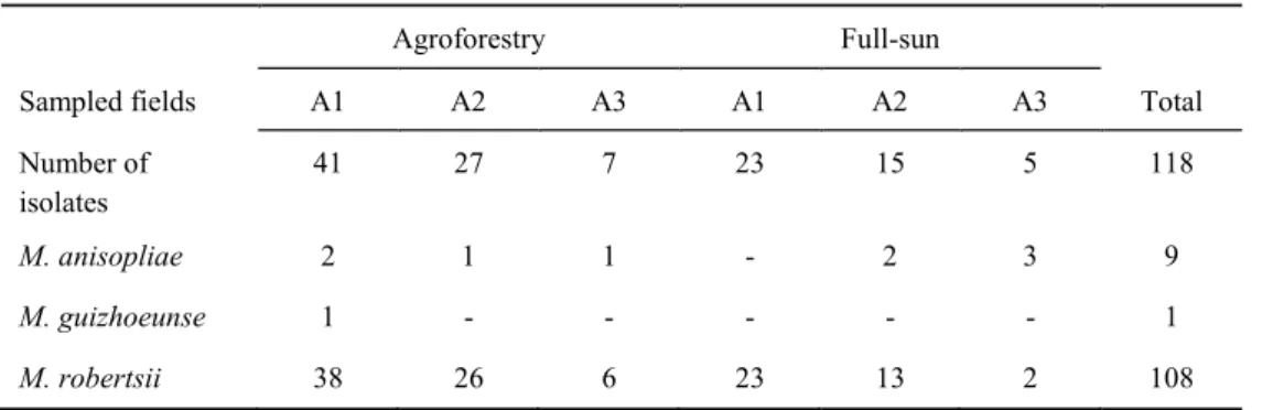
Metarhizium was the most frequently found genus we investigated its community composition and genetic variability in each production system. Understanding species distribution and pattern of plant association is the first step in the attempt to manage Metarhizium in a conservative biological control approach.
Figure 1. Comparison of the entomopathogenic fungi activity in soils under two coffee cultivation regimes:
agroforestry and full-sun. The differential “speed of kill” of the bait insects is taken as a measure of “ecosystem
services provided” by the entomopathogenic fungi, since the speed at which a fungus kill a bait is directly
correlated with the its virulence and potential of survive in that soil environment, and consequently it is directly
10 OVERVIEW
This thesis focuses on a means to establish a method to quantify Metarhizium colonization in plant roots, assess its genetic diversity in agroecosystems and also evaluate Metarhizium species diversity in the rhizosphere of coffee plants and non-crop plants present in the system.
In Chapter 1, we described a qPCR method to detect and quantify M. robertsii association in plant roots. The method showed specificity and reliable results through the utilization of SYBR green detection system. The method was validated through the quantification of a time course association of M. robertsii in bean plants from 7 to 35 days of association. Quantification through a cultivable method (CFU count) was used to compare the results. Laser scanning confocal microscopy was used to describe the association along the time course.
The three methods applied to quantify association displayed similar patterns. Association was higher in the first days of association followed by a decrease in the following days. The observed pattern could be associated with the massive growth of hyphae on the root surface following experimental inoculation. The method proposed is a valuable tool for investigation of M. robertsii colonization in experimental settings and detection in field plants.
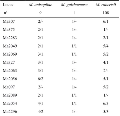
In Chapter 2, Metarhizium community and population diversity in coffee based agroforestry and full sun management were investigated. We hypothesized that the most diverse agroforestry system would harbor a more diverse Metarhizium community. A set of 118 isolates obtained from the both fields were characterized through molecular methods. Three species M. anisopliae, M. guizhouense and M. robertsii were recorded, and the last was the most abundant. Comparison of diversity indices between the fields in the area pairs revealed higher diversity in agroforestry system for two of the sampled areas and the overall
11
structuration according to the management systems. Three intraspecific clonal clades were detected in the M. robertsii. We suggested that the prevalence of M. robertsii could be due its preferential association with coffee plants.
12 REFERENCES
Behie S.W. & Bidochka M.J. (2014). Ubiquity of insect-derived nitrogen transfer to plants by endophytic insect-pathogenic fungi: an additional branch of the soil nitrogen cycle. Applied and Environmental Microbiology, 80, 1553-1560.
Behie S.W., Moreira C.M., Sementchoukova I., Barelli L., Zelisko P.M. & Bidochka M.J. (submitted). Trading insect nitrogen for photosynthate: Carbon translocation from a plant to an insect pathogenic, endophytic fungus. Science.
Behie S.W., Zelisko P.M. & Bidochka M.J. (2012). Endophytic insect-parasitic fungi translocate nitrogen directly from insects to plants. Science, 336, 1576-1577.
Bidochka M.J., Kamp A.M., Lavender T.M., Dekoning J. & De Croos J.N.A. (2001). Habitat association in two genetic groups of the insect-pathogenic fungus Metarhizium anisopliae: Uncovering cryptic species? Applied and Environmental Microbiology, 67, 1335-1342.
Bischoff J.F., Rehner S.A. & Humber R.A. (2009). A multilocus phylogeny of the Metarhizium anisopliae lineage. Mycologia, 101, 512-530.
Cardoso I.M., Guijt I., Franco F.S., Carvalho A.F. & Neto P.S.F. (2001). Continual learning for agroforestry system design: university, NGO and farmer partnership in Minas Gerais, Brazil. Agricultural Systems, 69, 235-257.
Enkerli J., Kölliker R., Keller S. & Widmer F. (2005). Isolation and characterization of microsatellite markers from the entomopathogenic fungus Metarhizium anisopliae. Molecular Ecology Notes, 5, 384-386.
Fang W. & St. Leger R.J. (2010). Mrt, a gene unique to fungi, encodes an oligosaccharide transporter and facilitates rhizosphere competency in Metarhizium robertsii. Plant Physiology, 154, 1549-1557.
Fegan M., Manners J.M., Maclean D.J., Irwin J.A.G., Samuels K.D.Z., Holdom D.G. & Li D.P. (1993). Random amplified polymorphic DNA markers reveal a high degree of genetic diversity in the entomopathogenic fungus Metarhizium anisopliae var. anisopliae. Microbiology, 139, 2075-2081.
Fisher J.J., Rehner S.A. & Bruck D.J. (2011). Diversity of rhizosphere associated entomopathogenic fungi of perennial herbs, shrubs and coniferous trees. Journal of Invertebrate Pathology, 106, 289-295.
Gao Q., Jin K., Ying S.-H., Zhang Y., Xiao G., Shang Y., Duan Z., Hu X., Xie X.-Q., Zhou G., Peng G., Luo Z., Huang W., Wang B., Fang W., Wang S., Zhong Y., Ma L.-J., St. Leger R.J., Zhao G.-P., Pei Y., Feng M.-G., Xia Y. & Wang C. (2011). Genome sequencing and comparative transcriptomics of the model entomopathogenic fungi Metarhizium anisopliae and M. acridum. PLoS Genet, 7, e1001264.
13
Hu G. & St Leger J. (2002). Field studies using a recombinant mycoinsecticide (Metarhizium anisopliae) reveal that it is rhizosphere competent. Applied and Environmental Microbiology, 68, 6383-6387.
Jha S., Bacon C.M., Philpott S.M., Ernesto Méndez V., Läderach P. & Rice R.A. (2014). Shade coffee: Update on a disappearing refuge for biodiversity. BioScience, 64, 416-428.
Kanzok S.M. & Jacobs-Lorena M. (2006). Entomopathogenic fungi as biological insecticides to control malaria. Trends in Parasitology, 22, 49-51.
Kepler R.M., Humber R.A., Bischoff J.F. & Rehner S.A. (2014). Clarification of generic and species boundaries for Metarhizium and related fungi through multigene phylogenetics. Mycologia, 106, 811-829.
Kepler R.M., Sung G.-H., Ban S., Nakagiri A., Chen M.-J., Huang B., Li Z. & Spatafora J.W. (2012). New teleomorph combinations in the entomopathogenic genus Metacordyceps. Mycologia, 104, 182-197.
Kepler R.M., Ugine T.A., Maul J.E., Cavigelli M.A. & Rehner S.A. (2015). Community composition and population genetics of insect pathogenic fungi in the genus Metarhizium from soils of a long-term agricultural research system. Environmental Microbiology, 17, 2791–2804.
Khan A., Hamayun M., Khan S., Kang S.-M., Shinwari Z., Kamran M., ur Rehman S., Kim J.-G. & Lee I.-J. (2012). Pure culture of Metarhizium anisopliae LHL07 reprograms soybean to higher growth and mitigates salt stress. World J Microbiol Biotechnol, 28, 1483-1494.
Leal S.C.M., Bertioli D.J., Butt T.M. & Peberdy J.F. (1994). Characterization of isolates of the entomopathogenic fungus Metarhizium anisopliae by RAPD-PCR. Mycological Research, 98, 1077-1081.
Li C., Huang B., Fan M., Lin Y. & Li Z. (2010). Metacordyceps guniujiangensis and its Metarhizium anamorph: a new pathogen on cicada nymphs. Mycotaxon, 111, 221-231.
Liao X., O’Brien T., Fang W. & St. Leger R. (2014). The plant beneficial effects of
Metarhizium species correlate with their association with roots. Appl Microbiol Biotechnol, 98, 7089-7096.
Lomer C.J., Bateman R.P., Johnson D.L., Langewald J. & Thomas M. (2001). Biological control of locusts and grasshoppers. Annual Review of Entomology, 46, 667-702. Lopes R.B., Souza D.A., Oliveira C.M. & Faria M. (2013). Genetic diversity and
pathogenicity of Metarhizium spp. associated with the white grub Phyllophaga capillata (Blanchard) (Coleoptera: Melolonthidae) in a soybean field. Neotropical Entomology, 42, 436-438.
Loureiro M.L. & Lotade J. (2005). Do fair trade and eco-labels in coffee wake up the consumer conscience? Ecological Economics, 53, 129-138.
14
Milner R.J. (1992). Selection and characterization of strains of Metarhizium anisopliae for control of soil insects in Australia. . In: Biological Control of Locusts and Grasshoppers. (eds. Lomer CJ & Prior C). CAB International Wallinford, pp. 200-207.
Moreira C.C., Celestino D., Guerra T.G., Cardoso i.M. & Elliot S.L. (submitted). Ecosystem services offered by entomopathogenic fungi: Agroforestry coffee soils are more insect-suppressive than Full-sun soils. Submitted to Journal of Invertebrate Pathology.
Moreira C.C. & Elliot S.L. (in prep.). Bait survival technique to estimate the potential for soil-borne insect-pathogenic fungi as cryptic providers of ecosystem services. In preparation.
Oulevey C., Widmer F., Kölliker R. & Enkerli J. (2009). An optimized microsatellite marker set for detection of Metarhizium anisopliae genotype diversity on field and regional scales. Mycological Research, 113, 1016-1024.
Pattemore J., Hane J., Williams A., Wilson B., Stodart B. & Ash G. (2014). The genome sequence of the biocontrol fungus Metarhizium anisopliae and comparative genomics of Metarhizium species. BMC Genomics, 15, 660.
Ratnadass A., Fernandes P., Avelino J. & Habib R. (2012). Plant species diversity for sustainable management of crop pests and diseases in agroecosystems: a review. Agron. Sustain. Dev., 32, 273-303.
Rezende J.M., Zanardo A.B.R., Lopes M.D., Delalibera I. & Rehner S.A. (2015). Phylogenetic diversity of Brazilian Metarhizium associated with sugarcane agriculture. Biocontrol, 60, 495-505.
Riba G., Bouvier-Fourcade I. & Caudal A. (1986). Isoenzymes polymorphism in Metarhizium anisopliae (Deuteromycotina: Hyphomycetes) entomogenous fungi. Mycopathologia, 96, 161-169.
Roberts D.W. & St. Leger R.J. (2004). Metarhizium spp., cosmopolitan insect-pathogenic fungi: Mycological Aspects. In: Advances in Applied Microbiology. Academic Press, pp. 1-70.
Rocha L.F.N., Inglis P.W., Humber R.A., Kipnis A. & Luz C. (2013). Occurrence of Metarhizium spp. in Central Brazilian soils. Journal of Basic Microbiology, 53, 251-259.
Roy H.E., Brodie E.L., Chandler D., Goettel M.S., Pell J.K., Wajnberg E. & Vega F.E. (2010). Deep space and hidden depths: understanding the evolution and ecology of fungal entomopathogens. In: The Ecology of Fungal Entomopathogens (eds. Roy H, Vega F, Chandler D, Goettel M, Pell J & Wajnberg E). Springer Netherlands, pp. 1-6.
Roy H.E., Steinkraus D.C., Eilenberg J., Hajek A.E. & Pell J.K. (2006). Bizarre interactions and end games: Entomopathogenic fungi and their arthropod hosts. Annual Review of Entomology, 51, 331-357.
15
Sasan R.K. & Bidochka M.J. (2013). Antagonism of the endophytic insect pathogenic fungus Metarhizium robertsii against the bean plant pathogen Fusarium solani f. sp. phaseoli. Canadian Journal of Plant Pathology, 35, 288-293.
Scholte E.-J., Ng'habi K., Kihonda J., Takken W., Paaijmans K., Abdulla S., Killeen G.F. & Knols B.G.J. (2005). An entomopathogenic fungus for control of adult african Malaria mosquitoes. Science, 308, 1641-1642.
Shah P.A. & Pell J.K. (2003). Entomopathogenic fungi as biological control agents. Appl Microbiol Biotechnol, 61, 413-423.
Sorokin N. (1883). Plant parasites of man and animals as causes of infectious diseases. In, pp. 268-291.
St. Leger R.J. (2008). Studies on adaptations of Metarhizium anisopliae to life in the soil. Journal of Invertebrate Pathology, 98, 271-276.
St. Leger R.J., May B., Allee L.L., Frank D.C., Staples R.C. & Roberts D.W. (1992). Genetic differences in allozymes and in formation of infection structures among isolates of the entomopathogenic fungus Metarhizium anisopliae. Journal of Invertebrate Pathology, 60, 89-101.
St. Leger R.J. & Screen S. (2001). Prospects for strain improvement of fungal pathogens of insects and weeds. In: Fungal biocontrol agents: progress, problems and potential (eds. Butt TM, Jackson C & Morgan N). CAB International Wallingford, UK, pp. 219-238.
Steinwender B.M., Enkerli J., Widmer F., Eilenberg J., Thorup-Kristensen K. & Meyling N.V. (2014). Molecular diversity of the entomopathogenic fungal Metarhizium community within an agroecosystem. Journal of Invertebrate Pathology, 123, 6-12. Sung G.-H., Hywel-Jones N.L., Sung J.-M., Luangsa-ard J.J., Shrestha B. & Spatafora J.W.
(2007). Phylogenetic classification of Cordyceps and the clavicipitaceous fungi. Studies in Mycology, 57, 5-59.
Tiago P.V., Souza H.M.d.L., Moysés J.B., Oliveira N.T.d. & Lima E.Á.d.L.A. (2011). Differential pathogenicity of Metarhizium anisopliae and the control of the sugarcane root spittlebug Mahanarva fimbriolata. Brazilian Archives of Biology and Technology, 54, 435-440.
Tigano-Milani M.S., Gomes A.C.M.M. & Sobral B.W.S. (1995). Genetic Variability among Brazilian Isolates of the Entomopathogenic Fungus Metarhizium anisopliae. Journal of Invertebrate Pathology, 65, 206-210.
Tscharntke T., Clough Y., Bhagwat S.A., Buchori D., Faust H., Hertel D., Hölscher D., Juhrbandt J., Kessler M., Perfecto I., Scherber C., Schroth G., Veldkamp E. & Wanger T.C. (2011). Multifunctional shade-tree management in tropical agroforestry landscapes – a review. Journal of Applied Ecology, 48, 619-629.
16
Wang C. & St Leger R.J. (2007). The MAD1 Adhesin of Metarhizium anisopliae links adhesion with blastospore production and virulence to insects, and the MAD2 adhesin enables attachment to plants. Eukaryotic Cell, 6, 808-816.
Wang S., O’Brien T.R., Pava-Ripoll M. & St. Leger R.J. (2011). Local adaptation of an introduced transgenic insect fungal pathogen due to new beneficial mutations. Proceedings of the National Academy of Sciences, 108, 20449-20454.
Wyrebek M. & Bidochka M.J. (2013). Variability in the insect and plant adhesins, Mad1and Mad2, within the fungal genus Metarhizium suggest plant adaptation as an evolutionary force. PLoS ONE, 8, e59357.
17
C
HAPTER
1
R
EAL TIMEPCR
QUANTIFICATION OF ENDOPHYTIC COLONIZATION OFM
ETARHIZIUM ROBERTSII AND THE DESCRIPTION OF ITS ASSOCIATION18 Abstract
19 Introduction
The insect-pathogenic fungus Metarhizium (Hypocreales: Clavicipitaceae) has been used extensively as a biological control agent (Shah & Pell 2003) and many of the biochemical and molecular factors involved in insect pathogenesis have been elucidated (Gao et al. 2011). However, there are many instances where Metarhizium has not performed optimally as a biological control agent in the field (Meyling & Eilenberg 2007). One of the main reasons for this may be that the ecological role of this fungus has generally been neglected (Vega et al. 2009). Regardless of the potential utility of the fungus as a biocontrol agent one cannot ignore its phylogenetic history as a relative of plant endophytes (Behie et al. 2013) and this must be considered in any biocontrol effort. The discovery of Metarhizium as a root endophyte (Sasan & Bidochka 2012) and its nutrient transfer to plants from insect cadavers (Behie et al. 2012) has highlighted its importance in the ecosystem. The endophytic capability of Metarhizium is now being evaluated alongside its use in biological control, and is potentially critical in developing novel and effective biological control strategies.
Endophytes play key roles in the ecosystem and influence health, evolution, and ecology of the host plant (Brundrett 2006). For example, Metarhizium was reported as plant growth promoter (Khan et al. 2012; Sasan & Bidochka 2012; Liao et al. 2014), plant pathogen antagonist, nitrogen transfer to a broad plant host range (Behie & Bidochka 2014b) and photosynthate compounds receiver from plant host (Behie et al. submitted). However, the mechanisms involved in endophytic associations have not been fully elucidated.
20
non-axenic conditions which could potentially lead to an inaccurate estimation of plant colonization (Porras-Alfaro & Bayman 2011). The use of green fluorescence protein (GFP)-transformed fungi and microscopic evaluation of plant tissues has proven useful in determining the presence of fungus in plant tissues (Maciá-Vicente et al. 2009), but it is not quantitatively relevant. Nevertheless, GFP-transformed fungus has the additional advantage of the description of the association spatially and the identification of fungal and plant special structures (Maciá-Vicente et al. 2009).
21 Material and Methods
Fungal and plant material
A Metarhizium robertsii (ARSEF 2575) transformant expressing green fluorescence protein (-GFP) was used for real time PCR calibration and quantification experiments, colony counting and for laser confocal microscopy. The construction of the GFP-expressing plasmids, transformation, and transgenic fungal lines have been previously described by Fang et al. (2006). The fungal isolate was maintained on PDA (39g l-1 of potato dextrose agar). For fungal DNA extraction, 100 µl of fungal conidia (107 per ml) were inoculated into 50 ml 0.2% (w/v) yeast extract, 1% peptone, 2% glucose broth (YPD) in flasks shaken at 280 rpm at 27°C for 5 days. Phaseolus vulgaris seeds (haricot bean, cultivar ‘Soldier’) were used as the host plant and seeds were obtained from OSC Seeds, Ontario, Canada.
Seed sterilization and fungal inoculation
Bean seeds were surface sterilized using chlorine gas method (Gamborg & Phillips 1995). Chlorine gas was produced by the combination of 100 ml of 5.25% hypochlorite (bleach) and 4 ml of 37% hydrochloric acid in a small beaker placed in glass dissector under the fume hood for 18 hr. Surface sterilized seeds were placed on water-agar (1.5 % agar) and kept at 25°C for a photoperiod of 16 h a day for 5 days in order to obtain seedlings. The seedling were inoculated with 100µl of ARSEF 2575-GFP conidial suspension (107 conidia/ml) in Triton X-100 (0.001%) and kept for a further 3 days in water agar. Control seedlings were inoculated with 100 µl of 0.01% sterile distilled solution of Triton X-100.
Planting, harvesting, CFU count and plant material processing
22
into six sections of 0.5 cm lengths. Six randomly chosen 0.5 cm pieces were placed into 5 ml distilled water and homogenized using a rotary homogenizer (Greiner Scientific). Samples (100 µl) of homogenate were spread, in duplicate, on to selective media, containing: PDA, 0.5 g cycloheximide, 0.2 g chloramphenicol, 0.5 g 65% dodine and 0.01 g crystal violet. The plates were kept at 27°C for 20 days and M. robertsii CFU’s were counted in five plant replicates.
The remaining root pieces were frozen at -80 °C for 12 hours and then freeze-dried for 24h. An aliquot of 2 mg of the lyophilized material was ground using a ball mill (Retsch, MM 300) and DNA was extracted from the powdered material.
DNA extractions
Genomic DNA was extracted from fungal isolate grown in YPD using the Wizard® Genomic DNA Purification Kit (Promega). The obtained DNA was used to evaluate primer sets for qPCR and also to construct standard calibration curves (DNA concentration versus qPCR CT values). DNA from uninoculated plants was also extracted using Plant DNeasy Mini Kit (Qiagen) as the negative control. DNA from lyophilized plant-ARSEF 2575-GFP association samples were extracted using a Soil Isolation DNA Kit (Norgen). This kit was finally selected after several attempts using various DNA extraction kits to produce DNA free of potential PCR inhibitors present in soil residues associated with plant roots. DNA quality verification and quantification were checked spectrophotometrically at 260 nm/280 nm (Implen NanophotometerTM Pearl). DNA from fungal, plant only and inoculated plants were stored until use at -20°C.
Design of PCR primers and specificity
23
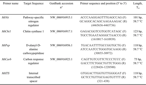
ng/µl), 12.5 µl Jump Start Taq Ready Mix (Sigma), 0.5 µM of forward and reverse primers and 8.5 µl PCR-grade water. The PCR conditions were; 2 min 98 °C, 40 cycles of 98 °C for 30 sec, 58.7 °C for 30 sec and 72 °C for 30 sec. The amplicons were visualized by ultraviolet fluorescence on 3% agarose gel stained with ethidium bromide. The MtNit primer pair provided the most consistent results in conventional PCR and was selected for the subsequent tests performed by qPCR.
Quantitative real time-PCR
The qPCR reactions contained SSO Fast Eva Green Ready mix (2x) (Bio-Rad), 0.3 µM of MtNit forward and reverse primers, 3 µl of template DNA and PCR-grade water to a final volume of 20 µl. Each reaction was performed in triplicate and controls were included in each reaction set. The qPCR reactions were performed using Real Time Detection Sytem MyIQ (BioRad Laboratories Ltda., Canada, Mississauga, Ont.) at: 2 min 98 °C, 40 cycles of 98 °C for 5 sec, 58.7 °C for 10 sec and 72 °C for 20s. The Ct cycle was calculated using the Optical System software, version 2.0 for MyIQ (BioRad). MtNit specificity was also checked by qPCR using template (ARSEF 2575-GFP), bean DNA and blanks without DNA to construct a dissociation curve. The qPCR products were also visualized by ultraviolet fluorescence on 3% agarose gel stained with ethidium bromide.
Standard curve using genomic DNA
24 Spike test
A spike test was conducted in order to verify the ability of the MtNit primers to recognize target DNA with an overwhelming concentration of non-target plant DNA. Fungal DNA concentrations ranging from 10 to 0.01ng were spiked with 20 ng of plant DNA and subjected to qPCR. Ct values were plotted versus the logarithmic value for each fungal DNA concentration.
Real time quantification of time course of colonization in roots
Plants from a time-course development of ARSEF 2575-GFP association and control plant (uninoculated) were harvested and processed as described above. Plant-ARSEF 2575-GFP association DNA was extracted also as described above, and used as templates for qPCR. The DNA from five plant replicates was used and three qPCR reactions were performed in each plant replicate. The result of the amount of fungal DNA in the root experimental sample was obtained as a Ct value. In order to obtain the amount of fungal DNA (ng) in the samples, the Ct values obtained in the given calibration curve were correlated with the amount of DNA used to construct the curve. Subsequently, the Ct value from experimental samples was interpolated in the calibration curve to estimate amounts of DNA (ng) in every experimental sample. To compare the mean amount of fungal colonization in the five plant replicates among the time points (7, 14, 21, 28, 35 days) a generalized linear model was constructed and an ANOVA with F test was performed using R (R Development Core 2008). Sampling was destructive, so separate plants were evaluated at each time point. For this reason, we did not use repeated measures in the analysis. The differences among the mean colonization (ng) at each time point were obtained through a simplification of the original model with amalgamation of time points, the models were then contrasted with the original model and the significance differences were obtained (Crawley 2007).
25
26 Results
Primer specificity
The fidelity of all the primers designed (Table 1) was tested by conventional PCR with M. robertsii ARSEF 2575-GFP (target) and bean (target) DNA templates (Fig. 1). Any non-specific homology was checked by Blast alignment of the primer target sequences from the Genbank. The primers were also tested using qPCR and the MtNit primer set was selected for its consistent performance based on Ct value and specificity visualized by the dissociation curve of the qPCR (Fig. 2A). The dissociation profile showed amplification in the presence of the ARSEF 2575-GFP template, but no amplification was observed using bean DNA or water (negative control); these results were also confirmed by agarose gel electrophoresis of the reaction product (Fig. 2B).
Table 1. Quantitative real-time polymerase chain reaction primers designed for the present study
Primer name Target Sequence GenBank accession n°
Primer sequence and position (5′ to 3′) Length, Tm
MtNit Pathway-specific nitrogen regulator
NW_006916915.1 ACCCAAGGAGTTTGAGCCAG (F) GCAGGCACAGCAAGAAAGAC (R)
(4465636-4465736)
101 bp, 58.7 °C
MtChi1 Chitin synthase 1 NW_006916917.1 GAGACGGTCGTGGTCATAGC (F) TGCCTGAATAGGGCTAACCG (R)
(1610817-1610939)
123 bp, 58.7 °C
MtPep D-alanyl-D-alanine carboxypeptidase
NW_006916956.1 TGACAATTTTGCCGGTGCTG (F) ATCCAATCCTGGGTGCAAGG (R)
(30855-30972)
118 bp, 58.7 °C
MtCarb Carbon response regulator
NW_006916923.1 CAGTTGTCGTTCTCCCTCCC (F) GACCTTCTGGCTGTTCTGGG (R)
(1228426-1228500)
75 bp, 58.7 °C
27
Figure 1. Specificity test of the five Metarhizium robertsii specific primer pairs by conventional polymerase chain
reaction amplification. Test for M. robertsii genomic DNA amplification from lanes 2-6. Test for plant (Phaseolus vulgaris) genomic DNA from lanes 7-11. Lane 1 is a 100-bp ladder (Norgen). Lanes 2 and 7: MtNit; lanes 3 and 8:
MtChi1, lanes 4 and 9: MtPep, lanes 5 and 10: MtCarb, lanes 6 and 11: MtITS.
Figure 2. Dissociation curve and electrophoresis gel of quantitative real-time polymerase chain reaction amplification using the MtNit primer. a) Specific amplification is seen by the amplification of the reaction containing M. robertsii genomic DNA as the template (-) . No amplification is seen with plant DNA (---) and negative control
(…). b) Specific amplification is also visualized in the electrophoresis. Lane 1 is a 100-bp ladder (Norgen).
Standard curve and Spike test
28
3A). The 101-bp M. robertsii ARSEF 2575- GFP fragment was amplified over a log concentration range showing a correlation coefficient of 0.99 and PCR efficiency of 102.65%.
In the spike test the concentration of plant genomic DNA used (20 ng), was 2000 times greater than the smallest amount (0.01ng) of fungal DNA. Figure 3B showns that the 101-bp MtNit target amplified was selectively amplified in the presence of contaminating plant DNA with a high correlation coefficient.
29
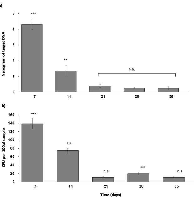
Real time quantification of time course of M. robertsii colonization in roots and CFU count
Real time PCR with the primer MtNit template DNA from root experimental samples shows production of templates in inoculated plants (Fig. 4A). Uninoculated plants did not yield substantial production of templates. Colonization at seven days was much higher than at all the other time points (4.30 ng± 0.30 S.E.M.; P<0.001). At fourteen days, reduced colonization was detected (1.33±0.38; P<0.001). At the other time points, colonization was detected only at low levels and no significant differences were detected.
30
Figure 4. Time course quantification (7, 14, 21, 28 and 35 days) of endophytic colonization of Metarhizium robertsii ARSEF 2575-GFP in bean roots. (a) Time course quantification obtained by qPCR. (b) Time course quantification obtained from CFU counts. *** P <0.001, ** P <0.01, n.s. – not significant.
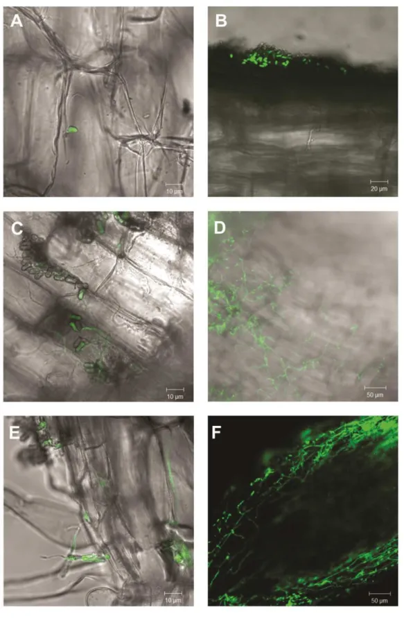
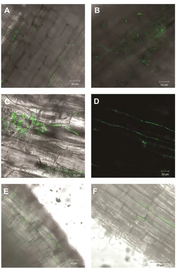
Laser Confocal Scanning Microscopy of the plant- ARSEF 2575-GFP association
31
32
33
34 Discussion
The recent discovery that Metarhizium can form intimate and apparently mutualistic associations with plants could have great agricultural potential to plant protection from insect pests, pathogens and plant nutrition (Fang & St. Leger 2010; Behie et al. 2012; Sasan & Bidochka 2012, 2013; Behie & Bidochka 2014b; Behie et al. submitted). At present, though, studies of the association between roots and insect pathogenic fungi are at a very initial stage; much still needs to be explored regarding molecular and biochemical mechanisms, interaction dynamics and consequences of the association for both partners. One of the first steps for this is to establish methods to assess, quantify and describe the development of the association. Here we established a qPCR based method to detect and quantify M. robertsii in plant root tissues. We also described the time course colonization of bean roots using qPCR, a culturing based method and GFP detection with laser scanning confocal microscopy. Although it could be considered that this is a very specific tool, it is becoming ever more apparent that plant root associations with M robertsii and congeneric fungi are the norm rather than isolated instances (Behie & Bidochka 2014b; Behie et al. 2015), with potentially important implications for plant biology and ecology in general.
35
The MtNit primer was sufficiently sensitive to detect up to 1 pg of M. robertsii purified DNA. The linear range of amplification obtained in the standard curve provided a reliable degree of confidence in quantification of experimental samples. Also, the degree of specificity seen under spike test conditions in a fixed large amount of non-specific DNA is a strong indication of the consistency and sensitivity of the qPCR diagnostic. The methodology provided here can be extremely useful for further investigation of the M. robertsii-plant association. Association detection and quantification can be included as a response variable in experimental set-ups investigating the effects of M. robertsii in plants such as: protection against biotic and abiotic stress, production of toxic compounds, activation of host defense mechanisms and others, unveiling molecular and biochemical paths of the interaction and others.
The use of isolates with knockouts of specific genes is one of the most informative approaches to unravelling the roles of those genes (Fang et al. 2006); such mutants have proven to be useful to investigate the role of specific genes in plant-fungus associations (Fang & St. Leger 2010; Wang et al. 2011; Liao et al. 2014). The comparison of the amount of association of the wild type and mutants can be a relevant response and explain much of the role of the surveyed genes; the method presented here can be a very valuable tool to understand the role of specific genes involved in nutrient transfer between Metarhizium and plants.
36
used in order to reach a detectable amount of the fungal DNA, high water content in roots can also reduce the amount of plant material processed for DNA extraction and consequently the amount of fungal DNA in the sample. The utilization the fine-ground freeze dried material for DNA extraction optimized the recovery of fungal DNA due the removal of water from sample and increasing the contact surface of the DNA isolation substances present in the kit. The small particles of the plant-fungus material also increased the recovery of fungal DNA in the sample.
Besides the differences in the scale, qPCR detection and CFU counting seems to follow the same pattern along the time course development of the association. Usually cultivation methods relying on CFU counts tend to be challenging under non-axenic conditions (Porras-Alfaro & Bayman 2011) but here this problem was solved through the utilization of selective media. Cultivating methods can also over- or underestimate colonization when accounting the amount of colonization through the percent of fragments colonized. In this case, a root fragment completely colonized by the endophyte and a fragment containing only a small propagule are scored as positive (Maciá-Vicente et al. 2009). Unless a great number of fragments are used, the method is frequently saturated. Here the homogenate method resulted in a more uniform sampling and consequently in a more accurate translation of the colonization pattern.
37
Leger 2010), while externally growing fungal mycelium is essential for increasing nutrient scavenging (Behie & Bidochka 2014a) communication between plants (Badri et al. 2009), nutrient recycle and exchange (Morgan et al. 2005) and influence the controlo f soil pests (Keyser et al. 2014). Based on that, external mycelial growth was kept in the analysis; however, if the interest is solely in the fungus growing inside the roots alternative methods for surface sterilization without sodium hypochlorite can be tested.
The great amount of colonization at the seventh day achieved by the both quantification methods is clearly influenced by the inoculum applied to the roots initially. The observation of the colonization pattern obtained by the GFP-tagged M. robertsii supported this observation. A great amount of germinating spores and the formation of an extensive external mycelial network is observed in the first days. Penetrating hyphae are also observed and at 7 days post inoculation and it is already possible to see growth in the intercellular spaces in the epidermal layer. Penetration seems to occur through the formation of appressorium-like structures. Regions with degraded hyphae showing loss of fluorescence were frequently observed, which may indicate hyphal death by the action of the host defense mechanisms. Further studies can elucidate what plant defense mechanisms are triggered by M. robertsii colonization and what factors maintain the long term association.
38
39 References
Alkan N., Gadkar V., Coburn J., Yarden O. & Kapulnik Y. (2004). Quantification of the arbuscular mycorrhizal fungus Glomus intraradices in host tissue using real-time polymerase chain reaction. New Phytologist, 161, 877-885.
Badri D.V., Weir T.L., van der Lelie D. & Vivanco J.M. (2009). Rhizosphere chemical dialogues: plant–microbe interactions. Current Opinion in Biotechnology, 20, 642-650. Behie S.W. & Bidochka M.J. (2014a). Nutrient transfer in plant–fungal symbioses. Trends in
Plant Science, 19, 734-740.
Behie S.W. & Bidochka M.J. (2014b). Ubiquity of insect-derived nitrogen transfer to plants by endophytic insect-pathogenic fungi: an additional branch of the soil nitrogen cycle. Applied and Environmental Microbiology, 80, 1553-1560.
Behie S.W., Jones S.J. & Bidochka M.J. (2015). Plant tissue localization of the endophytic insect pathogenic fungi Metarhizium and Beauveria. Fungal Ecology, 13, 112-119. Behie S.W., Moreira C.M., Sementchoukova I., Barelli L., Zelisko P.M. & Bidochka M.J.
(submitted). Trading insect nitrogen for photosynthate: Carbon translocation from a plant to an insect pathogenic, endophytic fungus. Science.
Behie S.W., Padilla-Guerrero I.E. & Bidochka M.J. (2013). Nutrient transfer to plants by phylogenetically diverse fungi suggests convergent evolutionary strategies in rhizospheric symbionts. Communicative & Integrative Biology, 6, e22321.
Behie S.W., Zelisko P.M. & Bidochka M.J. (2012). Endophytic insect-parasitic fungi translocate nitrogen directly from insects to plants. Science, 336, 1576-1577.
Bell A.S., Blanford S., Jenkins N., Thomas M.B. & Read A.F. (2009). Real-time quantitative PCR for analysis of candidate fungal biopesticides against malaria: Technique validation and first applications. Journal of Invertebrate Pathology, 100, 160-168. Brundrett M. (2006). Understanding the Roles of Multifunctional Mycorrhizal and Endophytic
Fungi. In: Microbial Root Endophytes (eds. Schulz BE, Boyle CC & Sieber T). Springer Berlin Heidelberg, pp. 281-298.
Crawley M.J. (2007). The R Book. Wiley Publishing.
Fang W., Pei Y. & Bidochka M.J. (2006). Transformation of Metarhizium anisopliae mediated by Agrobacterium tumefaciens. Canadian Journal of Microbiology, 52, 623-626. Fang W. & St. Leger R.J. (2010). Mrt, a gene unique to fungi, encodes an oligosaccharide
transporter and facilitates rhizosphere competency in Metarhizium robertsii. Plant Physiology, 154, 1549-1557.
Gamborg O. & Phillips G. (1995). Sterile Techniques. In: Plant Cell, Tissue and Organ Culture (eds. Gamborg O & Phillips G). Springer Berlin Heidelberg, pp. 35-42.
40
Hallmann J., Berg G. & Schulz B. (2006). Isolation Procedures for Endophytic Microorganisms. In: Microbial Root Endophytes (eds. Schulz BE, Boyle CC & Sieber T). Springer Berlin Heidelberg, pp. 299-319.
Jacobs S., Zechmann B., Molitor A., Trujillo M., Petutschnig E., Lipka V., Kogel K.-H. & Schäfer P. (2011). Broad-spectrum suppression of innate immunity is required for colonization of Arabidopsis Roots by the fungus Piriformospora indica. Plant Physiology, 156, 726-740.
Keyser C.A., Thorup-Kristensen K. & Meyling N.V. (2014). Metarhizium seed treatment mediates fungal dispersal via roots and induces infections in insects. Fungal Ecology, 11, 122-131.
Khan A., Hamayun M., Khan S., Kang S.-M., Shinwari Z., Kamran M., ur Rehman S., Kim J.-G. & Lee I.-J. (2012). Pure culture of Metarhizium anisopliae LHL07 reprograms soybean to higher growth and mitigates salt stress. World J Microbiol Biotechnol, 28, 1483-1494.
Lahrmann U., Ding Y., Banhara A., Rath M., Hajirezaei M.R., Döhlemann S., von Wirén N., Parniske M. & Zuccaro A. (2013). Host-related metabolic cues affect colonization strategies of a root endophyte. Proceedings of the National Academy of Sciences, 110, 13965-13970.
Landa B.B., López-Díaz C., Jiménez-Fernández D., Montes-Borrego M., Muñoz-Ledesma F.J., Ortiz-Urquiza A. & Quesada-Moraga E. (2013). In-planta detection and monitorization of endophytic colonization by a Beauveria bassiana strain using a new-developed nested and quantitative PCR-based assay and confocal laser scanning microscopy. Journal of Invertebrate Pathology, 114, 128-138.
Liao X., O’Brien T., Fang W. & St. Leger R. (2014). The plant beneficial effects of
Metarhizium species correlate with their association with roots. Appl Microbiol Biotechnol, 98, 7089-7096.
Lovett B. & St. Leger R.J. (2014). Stress is the rule rather than the exception for Metarhizium. Curr Genet, 1-9.
Maciá-Vicente J.G., Jansson H.-B., Talbot N.J. & Lopez-Llorca L.V. (2009). Real-time PCR quantification and live-cell imaging of endophytic colonization of barley (Hordeum vulgare) roots by Fusarium equiseti and Pochonia chlamydosporia. New Phytologist, 182, 213-228.
Malvick D.K. & Grunden E. (2005). Isolation of fungal DNA from plant tissues and removal of DNA amplification inhibitors. Molecular Ecology Notes, 5, 958-960.
Meyling N.V. & Eilenberg J. (2007). Ecology of the entomopathogenic fungi Beauveria bassiana and Metarhizium anisopliae in temperate agroecosystems: Potential for conservation biological control. Biological Control, 43, 145-155.
Morgan J.A.W., Bending G.D. & White P.J. (2005). Biological costs and benefits to plant– microbe interactions in the rhizosphere. Journal of Experimental Botany, 56, 1729-1739.
41
Phe M.H., Dossot M. & Block J.C. (2004). Chlorination effect on the fluorescence of nucleic acid staining dyes. Water Research, 38, 3729-3737.
Porras-Alfaro A. & Bayman P. (2011). Hidden Fungi, Emergent Properties: Endophytes and Microbiomes. Annu. Rev. Phytopathol., 49, 291-315.
R Development Core T. (2008). R: A Language and Environment for Statistical Computing. In. R Foundation for Statistical Computing Vienna, Austria.
Sasan R.K. & Bidochka M.J. (2012). The insect-pathogenic fungus Metarhizium robertsii (Clavicipitaceae) is also an endophyte that stimulates plant root development. American Journal of Botany, 99, 101-107.
Sasan R.K. & Bidochka M.J. (2013). Antagonism of the endophytic insect pathogenic fungus Metarhizium robertsii against the bean plant pathogen Fusarium solani f. sp. phaseoli. Canadian Journal of Plant Pathology, 35, 288-293.
Schneider S., Rehner S.A., Widmer F. & Enkerli J. (2011). A PCR-based tool for cultivation-independent detection and quantification of Metarhizium clade 1. Journal of Invertebrate Pathology, 108, 106-114.
Schulz B. & Boyle C. (2005). The endophytic continuum. Mycological Research, 109, 661-686. Shah P.A. & Pell J.K. (2003). Entomopathogenic fungi as biological control agents. Appl
Microbiol Biotechnol, 61, 413-423.
Tellenbach C., Grünig C.R. & Sieber T.N. (2010). Suitability of Quantitative Real-Time PCR To Estimate the Biomass of Fungal Root Endophytes. Applied and Environmental Microbiology, 76, 5764-5772.
Thornbury D. & Farman M. (2000). Reusing nylon-membrane for radioactive hybridizations. Bio Techniques, 29, 1250-1254.
Vega F.E., Goettel M.S., Blackwell M., Chandler D., Jackson M.A., Keller S., Koike M., Maniania N.K., Monzon A., Ownley B.H., Pell J.K., Rangel D.E.N. & Roy H.E. (2009). Fungal entomopathogens: new insights on their ecology. Fungal Ecology, 2, 149-159.
Wang S., O’Brien T.R., Pava-Ripoll M. & St. Leger R.J. (2011). Local adaptation of an introduced transgenic insect fungal pathogen due to new beneficial mutations. Proceedings of the National Academy of Sciences, 108, 20449-20454.
42
C
HAPTER
2
M
ETARHIZIUM
ASSOCIATED WITH AGROFORESTRY AND FULL
SUN COFFEE SYSTEMS
:
COMMUNITY CHARACTERIZATION AND
43
Abstract
Metarhizium
species are insect pathogens and plant symbionts very abundant in
soils worldwide. They also have the potential to provide important ecosystem
services in agricultural settings as reduction of insect populations, improved plant
nutrient uptake and protection from plant pathogens.
The use of more diverse and
sustainable land use practices such as tree
-
based intercropping can have the
potential to reduce the negative impact of agricultural practices on beneficial
microorganisms communities, however the effects of those practices
in
Metarhizium
species diversity and distribution is unknown.
In this paper we
compare the effects of agroforestry and full sun coffee cultivation systems
on
Metarhizium
community diversity and population structure.
One hundred and
eighteen isolates from three areas, containing each an agroforestry and full sun
field were included in this study. The isolates were characterized through
sequencing of the final portion of the translation enlongation factor 5’TEF and
SSR markers. Phylogeny of 5’TEF region reveled three species,
with
M.
robertsii
predominating. Comparison of diversity indices between the
44 Introduction
The substantial and irreversible loss of biodiversity and the consequent deterioration of ecosystem services are consequences of the growing amount of land employed to agriculture and agricultural intensification (Matson et al. 1997; Tilman et al. 2002). Paradoxically, the biodiversity loss can affect ecosystem functioning and the delivery of services and consequently decreasing soil fertility and agricultural production (Tilman et al. 2002; Foley et al. 2005). The role of biodiversity in the provision of ecosystem services is widely recognized nowadays (Cardinale et al. 2012). However the importance of organisms that provide support (e.g., soil formation and fertility) and regulation (e.g., pest control and crop pollination) services are generally neglected (Bommarco et al. 2013).
45
Metarhizium as a provider of ecosystem services. One of the breakthrough findings in Metarhizium ecology is its ability to transfer nutrients from insect cadavers to plants (Behie et al. 2012), representing an additional branch in the nitrogen cycle. This ability seems to be widespread within the genus and across plant taxa (Behie & Bidochka 2014). Also, Metarhizium species had already been reported as plant growth promoters and plant pathogen antagonists (Sasan & Bidochka 2012). Thereby, these fungi provide supporting ecosystem services, providing nutrition to plants, and regulation ecosystem servives, controlling insect pest and plant pathogens.
Faced with the above findings, we predict that in environments with greater plant abundance and variety, and consequently more complex root systems, there could be more opportunities to enter in association with plants, due to the increased availability of plant hosts. Once in association with the plant root system, the fungi will be more widely distributed within the soil systems (Keyser et al. 2014), increasing the possibility of encountering insect hosts and ultimately facilitating the provision of ecosystem services by these organisms. Also, the distribution of Metarhizium species can be correlated with specific plant taxa (Fisher et al. 2011; Wyrebek et al. 2011). These characteristics highlight the need to consider specific crops of interest, plant diversity and management systems in diversity surveys.