P . b . b . 0 2 Z 0 3 1 1 0 5 M , V e r l a g s p o s t a m t : 3 0 0 2 P u r k e r s d o r f , E r s c h e i n u n g s o r t : 3 0 0 3 G a b l i t z
Indexed in EMBASE/Excerpta Medica/Scopus
www.kup.at/kardiologie
Member of the ESC-Editors’ Club
Member of the
Offizielles Organ des
Österreichischen Herzfonds
Homepage:
www.kup.at/kardiologie
Online-Datenbank mit
Autoren- und Stichwortsuche
Increased Platelet Activation by
PTFE-Covered Coronary Stent Grafts:
A Flow Cytometric Analysis in a
Pulsed Floating Model of
Recirculating Human Plasma
Beythien Ch, Brockmann MA
Gutensohn K, Nienaber CA
Journal für Kardiologie - Austrian
Journal of Cardiology 2004; 11
Jetzt in 1 Minute
Früh-erkennung der PAVK: boso
ABI-system 100
PAVK – Die unterschätzte Krankheit
Die periphere arterielle Verschlusskrank-heit (PAVK) ist weitaus gefährlicher und verbreiteter als vielfach angenommen. Die getABI-Studie [1] zeigt, dass 20 % der > 60-Jährigen eine PAVK-Prävalenz aufweisen. Die PAVK wird oft zu spät diagnostiziert. Das liegt vor allem da-ran, dass die Betroffenen lange Zeit be-schwerdefrei sind und eine entsprechen-de Untersuchung daher meist erst in akuten Verdachtsfällen erfolgt. Mit dem Knöchel-Arm-Index („ankle- brachial in dex“ [ABI]) ist die Diagnose einer PAVK durchführbar. Der Knöchel-Arm-Index (ABI) ist ein wesentlicher Marker zur Vorhersage von Herzinfarkt, Schlag-anfall und Mortalität.
PAVK-Früherkennung mit dem boso ABI-system 100: Ein Gewinn für alle. Eine präzise und schnelle, vaskulär orientierte Erst untersuchung.
Der entscheidende Wert für die Dia-gnose der PAVK ist der Knöchel-Arm-Index („ankle-brachial index“ [ABI]). Das boso ABI-system 100 ermittelt die-sen Wert zeitgleich und oszillometrisch an allen 4 Extremitäten. Die eigentliche Messung dauert dabei nur ca. 1 Minu-te. Ein ABI-Wert < 0,9 weist im
Ver-gleich mit dem Angiogramm als Gold-standard mit einer Sensitivität von bis zu 95 % auf eine PAVK hin und schließt umgekehrt die Erkrankung mit nahezu 100 % Spezifität bei gesunden Perso-nen aus.
Das boso ABI-system 100 wurde wei-terentwickelt und ist jetzt optional mit der Messung der Pulswellenge-schwindigkeit ausgestattet.
Optional ist das boso ABI-system 100 ab sofort auch mit der Möglichkeit zur Messung der
Pulswellengeschwindig-keit (ba) verfügbar. Mit der Messung der Pulswellengeschwindigkeit („pulse wave velocity“ [PWV]) kann eine arteri-elle Gefäßsteifigkeit diagnostiziert wer-den. Die Steifigkeit der arteriellen Ge-fäße nimmt mit einer fortschreitenden Arteriosklerose zu, was sich durch eine Erhöhung der Pulswellengeschwindig-keit darstellt. PWV und ABI-Wert er-möglichen eine noch fundiertere Risi-kostratifizierung von kardiovaskulären Ereignissen.
Literatur: 1. http://www.getabi.de
Weitere Informationen:
Boso GmbH und Co. KG Dr. Rudolf Mad
A-1200 Wien
322 J KARDIOL 2004; 11 (7–8)
Increased Platelet Activation by PTFE-Covered Coronary Stent Grafts
Received: September 25th, 2003; revision received: October 28th, 2003; accepted:
April 18th, 2004.
From the University Hospital Rostock, Department of Cardiology [C.B., C.N.], Rostock, Germany; University Hospital Hamburg-Eppendorf, Department of Transfusion Medi-cine and Transplantation Immunology [M.A.B., K.G.], Hamburg, Germany
Correspondence to: Christian Beythien, PD Dr. med. habil., University Hospital Rostock, Department of Cardiology, Ernst-Heydemann-Straße 6, D-18055 Rostock, Germany; E-Mail: [email protected]
Abstract: Various designs are under investigation to resolve the problem of acute thrombosis and resteno-sis after stent implantation in coronary arteries. The interaction of stent surface with both platelets and the coagulation system has been shown to play a major role in this process. To investigate effects of PTFE (Teflon) coating stents of the same design with and without PTFE cover (n = 12 for each condition) were placed in an in vitro model of recirculating human platelet rich plasma. Samples were drawn every two min until stent thrombosis to analyze platelet activa-tion by flow cytometry using monoclonal antibodies against activation dependent epitopes CD62p (p-selectin) and CD63 (GP53) expressed as „mean chan-nel fluorescence intensity“ (MCFI). Additionally, time until appearance of macroscopic visible platelet aggre-gates, and time until stent thrombosis were measured. Flow cytometric analyses revealed a maximum expres-sion for CD62p (MCFI 37.2 ± 5.8) after 8 min, and after 10 min for CD63 (MCFI 40.0 ± 5.0) with PTFE-coated stents, meanwhile with bare stents maximum expres-sion of both epitopes (MCFI 38.1 ± 8.4) was reached after 12 min; coated vs. bare stents p = 0.05.
PTFE-Increased Platelet Activation by PTFE-Covered
Coronary Stent Grafts: A Flow Cytometric Analysis in a
Pulsed Floating Model of Recirculating Human Plasma
C. Beythien, M. A. Brockmann, K. Gutensohn, C. A. Nienaber
coating induced platelet aggregates after 5.7 ± 1.7 min, whereas uncoated stents after 6.8 ± 2.7 min; p = 0.04. Likewise, time until stent thrombosis was shorter in systems with PTFE-covered stents compared to uncoated stents; 11.1 ± 1.4 min versus 12.6 ± 2.2 min; p = 0.035. Thus, PTFE-coating increases in vitro thrombo-genicity by activating platelets and coagulation.
Kurzfassung: Erhöhte Plättchenaktivierung durch PTFE-ummantelte Stents. Durchflußzytometrische Analyse in einem In-vitro-Modell mit rezirku-lierendem plättchenreichem Plasma. Verschiede-ne StentversioVerschiede-nen werden zur Zeit untersucht, um das Problem der akuten Thrombose und Restenose nach Implantation in Koronararterien zu lösen. Eine Interak-tion zwischen der Stentoberfläche, Thrombozyten und dem Gerinnungssystem hat sich als wichtiger Faktor dabei erwiesen. Um Effekte einer PTFE-Ummantelung (Teflon) zu erforschen, wurden jeweils 12 baugleiche Stents mit und ohne Ummantelung in einem In-vitro-Modell mit rezirkulierendem humanem plättchen-reichem Plasma untersucht. Um die
Plättchenaktivie-rung zu quantifizieren, wurden bis zur Stentthrombose alle zwei Minuten Proben entnommen und mittels Durchflußzytometrie die Aktivierungsmarker CD62p (p-Selektin) und CD63 (GP53) bestimmt. Die Darstel-lung erfolgte als „mean channel fluorescence intensity“ (MCFI). Außerdem wurde die Zeit bis zum Auftreten makroskopisch sichtbarer Plättchenaggre-gate und bis zur Stentthrombose gemessen. Die durchflußzytometrischen Analysen bei den PTFE-um-mantelten Stents ergaben eine maximale Expression des CD62p nach 8 min (MCFI 37,2 ± 5,8) und von CD63 nach 10 min (MCFI 40,0 ± 5,0), während bei den unbeschichteten Stents die maximale Expression erst nach 12 min (MCFI 38,1 ± 8,4) erreicht war; beschich-tete vs. unbeschichbeschich-tete Stents p = 0,05. Die PTFE-Um-mantelung induzierte eine Plättchenaggregatbildung nach 5,7 ± 1,7 min, während diese bei unbeschich-teten Stents erst nach 6,8 ± 2,7 min auftrat; p = 0,04. Entsprechend war die Zeit bis zur Stentthrombose ver-kürzt; 11,1 ± 1,4 versus 1,4 versus 12,6 ± 2,2 min (p = 0,035). In vitro führt also eine PTFE-Ummantelung zu einer gesteigerten Aktivierung von Plättchen und Gerinnungssystem. J Kardiol 2004; 11: 322–5.
Introduction
To resolve the problem of acute stent-thrombosis and restenosis after implantation in coronary arteries, different de-signs and coatings are currently under investigation to im-prove the interaction of foreign surface with platelets and the coagulation system. The Jostent® coronary stent graft (JOMED/ SITOmed) with expandable polytetrafluoro-ethylene (PTFE, Teflon) graft material is a new stent design previously used in the treatment of aortocoronary vein graft lesions,and coro-nary aneurysms [1–4]. To investigate the PTFE covered stents and their interaction with platelets and the coagulation sys-tem, we utilized an in vitro model of recirculating human platelet rich plasma to monitor (1) stent induced expression of activation dependent glycoproteins on platelet surface, (2) the time until the occurrence of macroscopic visual platelet aggre-gates, and (3) the time until stent thrombosis. The results were compared to the characteristics of the identical stainless steel (316L) stent without PTFE cover.
Methods
To quantify stent induced platelet activation the expression of glycoproteins were measured in an previously described in
vitro model [5–7]. Experiments were conducted on blood from 12 obviously healthy, non-smoking male volunteers (age 29 ± 5 years) without any medication during and/or 14 days prior to blood collection. Forty ml of blood was collected via 16G needles from large antecubital veins in plastic syringes filled with sodium citrate (1:10) without the use of a tourni-quet. Platelet rich plasma (PRP) was prepared by centrifuga-tion of the whole blood at 300 g for 10 min at room tempera-ture (20 ± 2 °C). After removal of the PRP, the remaining plasma was centrifuged at 2500 g for 10 minutes at room tem-perature to prepare platelet-poor plasma (PPP). Using a Coul-ter CounCoul-ter STKR® (Beckman-Coulter, Fullerton, CA, USA) the platelet count within PRP was measured, and the PRP was diluted with PPP to a final concentration of 250 plt/nl.
Commercialized PTFE covered and uncoated stainless steel stents with a length of 12 mm in expanded condition were mounted on a conventional polyethylene percutaneous transluminal balloon angioplasty catheter (Boston Scientific, Watertown, MA, USA), and implanted into silicon tubings with 12 atm for 30 s to a final diameter of 4 mm to simulate in-human situations. Then PRP was carefully filled into the tub-ing systems. After recalcification to physiological concentra-tions PRP was rotated by a roller pump (Ismatec, Zurich, Switzerland) with a flow rate of 8 ml/min, and a flow velocity of 2 cm/s. The temperature of the in vitro tubing systems was
324 J KARDIOL 2004; 11 (7–8)
kept stable at 37 °C by a water bath. In three parallel silicon tubings one system was equipped with a PTFE covered stent, one with uncovered stent, and one without stent (control).
Aliquots of 100 µl PRP were removed before the circula-tion was started and after 2, 4, 6, 8, 10, 12, and 14 min. The samples were immediately fixed with 0.15 M phosphate-buff-ered saline (PBS; Gibco BRL, Eggenstein, Germany) con-taining glyoxal 0.2 % w/v (Merck, Darmstadt, Germany), and paraformaldehyde 0.4 % w/v (Serva, Heidelberg, Germany). Stabilization was performed by dilution 1:10 with PBS con-taining 0.2 % w/v Glycine (Serva, Heidelberg, Germany), labeled directly with monoclonal antibodies, and then stored at +4 °C in the dark [8]. Monoclonal antibodies CD41a, CD62p, and CD63 (Immunotech, Hamburg, Germany) were used. Flow cytometry analysis was performed within 2 hrs on a FACScan® cytometer (Becton Dickinson, Mountain View, USA). A life gate was set around CD41a positive cells; only those cells expressing this platelet specific membrane protein were included, and 20,000 events were analyzed. Results were expressed as „mean channel fluorescence intensity“ (MCFI). Antibody positive cells were defined as cells with fluorescence higher than isotype control.
In addition, time until stent thrombosis, and macroscopic visible platelet aggregates was measured.
Statistical analysis
Data comparison over the course of flow cytometric analysis were performed using the Friedman test. Statistical compari-sons were performed with „Student’s t-Test“ for paired data; p values of 0.05 or less were considered to be statistically sig-nificant. All values indicated are mean ± standard error of mean.
Results
Flow cytometric analyses
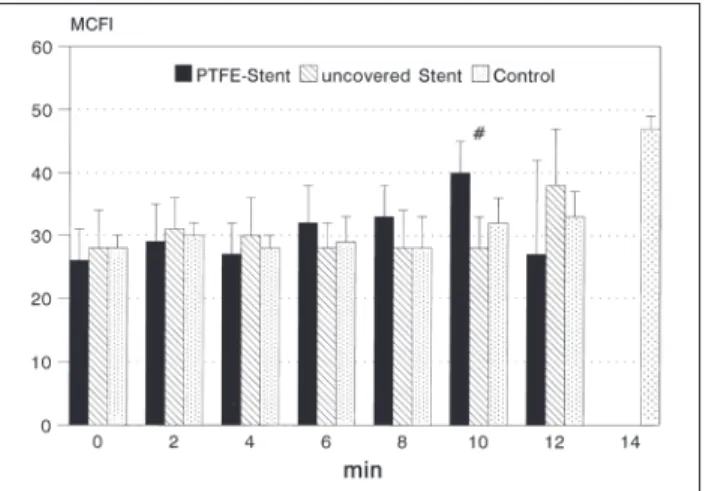
After starting of circulation the expression of monoclonal anti-bodies increased with a maximum after 8 min for CD62p, and after 10 min for CD63 in the tubing with PTFE covered stents. In the circulation with uncovered stents the maximum expression appeared after 12 min for all measured antibodies (p = 0.05, Figs. 1, 2). In the control tubings the expression of CD62p and CD63 was increasing up to the end of measurement.
In vitro
circulating modelFirst macroscopic visual platelet aggregates appeared after 5.6 ± 1.6 min in systems with PTFE-covered stents, whereas
Figure 1: Bar chart of the changes of the expression of the platelet antigen CD62p (P-selectin) over 14 min course of circulation of platelet-rich-plasma in the in vitro model. Values are means ± SD; # p = 0.05 vs. uncovered stents.
Figure 2: Bar chart of the changes of the expression of the platelet epitope CD63 (GP 53) over 14 min course of circulation of platelet-rich-plasma in the in vitro model. Values are means ± SD; # p = 0.05 vs. uncovered stents.
Figure 3: Bar graph of the time until the occurrence of platelet aggregates after ini-tiation of circulation of platelet-rich-plasma in the in vitro model. Values are means ± SD; # p = 0.04 vs. PTFE stents, p < 0.0005 vs. control system without stent.
Figure 4: Bar graph of the time until stent thrombosis, and thrombus development in the tubing system of the in vitro model respectively. Values are means ± SD;
J KARDIOL 2004; 11 (7–8) Increased Platelet Activation by PTFE-Covered Coronary Stent Grafts
325 first aggregates in systems with uncoated stents where
ob-served after 6.7 ± 2.7 min; p = 0.04 (Fig. 3). The time until stent thrombosis within the in vitro system containing PTFE-covered stents was 11.0 ± 1.3 min, whereas systems contain-ing uncoated stents occluded after 12.5 ± 2.1 min; p = 0.035 (Fig. 4).
Discussion
PTFE-covered stents have the advantage of reducing peri-interventional distal thrombotic embolization [9], and at least the theoretical benefit of reducing restenosis by blocking plaque protrusion, attenuating diffusion of cytokines, and re-ducing transmigration of inflammatory cells. However, these advantages only apply for the center part of the stent, whereas the uncovered distal and proximal parts of the stent lead to focal stent edge renarrowing, what influences the overall restenosis rate [10]. Initial case reports and small series dem-onstrated promising results [11, 12]. Other authors reported about restenosis and acute thrombotic occlusions after clopidogrel was abandoned [13]. Clinical data from a non-randomized study comparing covered and uncovered stents failed to demonstrate significant differences, they could only reveal trends or showed no differences [2]. Colombo and co-workers found in the RECOVERS trial an increased rate of myocardial infarctions both in hospital and in follow up period, whereas restenosis rate was comparable [14]. Proliferation from both the edges and small ruptures of the stent membrane during implantation are potential explanations for the lack of beneficial impact [15]. PTFE-coated guide-wire demonstrate a thrombus formation rate from 25 % to 69 % dependent on their design [16]. Thus, available data are still controversial.
There are other in vitro models to test thrombogenicity of coronary stents that investigate coagulation factors, platelet beta-thromboglobulin, 111Indium labeled platelet
accumula-tion, fibrinogen adsorpaccumula-tion, and platelet adhesion. However, comparable in vitro data and test models are unfortunately not existing. The present model does deliberately not apply whole blood. To study the interaction of platelets with e. g. leuko-cytes, monocytes or other blood corpuscles was not the target. Platelets and platelet activation are inherently not simple to detect by the technique of flow cytometry, standard deviations are substantial anyway. Thus, only platelet rich plasma was used to minimize additional artifacts induced by the other blood corpuscles. Our data revealed a time dependent activa-tion of both platelets and the coagulaactiva-tion system. After detect-ing a maximum of epitopic antigen expression on platelets the number of measurable antigens decreased demonstrating a consumption of available platelets by aggregation, adhesion and thrombus formation in the tubing system. Thus, after 10 min in the tubings with PTFE-stents more measurements were not feasible. P-selectin and glycoprotein 53 are expressed in conditions of activated platelets, and seem to play a key role and appear to be most closely associated with an increase in thrombotic risk [17]. With PTFE-covered stents the expres-sion of these glycoproteins was significantly higher as an evi-dence of additional platelet activation in contrast to the uncov-ered stents. In addition, thrombus formation was induced even by a higher degree by the teflon layer. In a recently published animal model, thrombus formation within the stent-graft in-terface was shown to promote neointimal development [18].
Other authors demonstrated a retarded neointimal hyperplasia only at the midportion of the devices, but did not prevent neointimal pannus ingrowth at the proximal and distal ends [14]. Thus, the question, whether an increased activation of platelets and the coagulation system even may enhance a neointimal covering of the stent can not be answered at this moment.
However, in vitro studies are not necessarily reflecting clinical conditions especially the additional administration of clopidogrel and aspirin as currently recommended. They can only evaluate one fragment of a puzzle. This is a noteworthy limitation of this in vitro model. Increased activation of both platelets and coagulation system may or may not be of advan-tage in conjunction with stent-grafts. Although this in vitro model offers the opportunity to investigate in vitro and ex vivo impact of inhibitors of platelet aggregation and the coagula-tion cascade the purpose of this present study was exclusively to explore the genuine influence of PTFE on activation of platelets and coagulation. Additional investigations are needed to further evaluate the biological interactions and im-plications of PTFE-covered stents.
Acknowledgements
We would like to thank the company JOMED GmbH (Rangendingen, Germany) for supplying the investigated stents.
References
1. Baldus S, Köster R, Elsner M, Walter DH, Arnold R, Auch-Schwelk W, Berger J, Rau M, Meinertz T, Zeiher AM, Hamm CW. Treatment of aortocoronary vein graft lesions with mem-brane-covered stents. A multicenter surveil-lance trial. Circulation 2000; 102: 2024–7. 2. Briguori C, De Gregorio J, Nishida T, Adamian M, Albiero R, Tucci G, Di Mario C, Colombo A. Polytetrafluoroethylene-covered stent for the treatment of narrowings in aorticocoronary saphenous vein grafts. Am J Cardiol 2000; 86: 343–6.
3. Von Birgelen C, Haude M, Liu F, Ge J, Gorge G, Welge D, Wieneke H, Baumgart D, Opherk D, Erbel R. Treatment of a coronary pseudoaneurysm by stent-graft implantation. DMW 1998; 123: 418–22.
4. Di Mario C, Inglese L, Colombo A. Treat-ment of a coronary aneurysm with a new polytetrafluoroethylene-coated stent: a case report. Catheter Cardiovasc Interv 1999; 46: 463–5.
5. Beythien C, Terres W, Hamm CW. In vitro model to test the thrombogenicity of coronary stents. Thromb Res 1994; 79: 581–90. 6. Gutensohn K, Beythien C, Bau J, Meinertz T, Kuehnl P. Flow cytometric analysis of coronary stent-induced alteration of platelet antigens in an in vitro model. Thromb Res 1997; 86: 49–56.
7. Brockmann MA, Gutensohn K, Bau J, Kuehnl P, Meinertz T, Nienaber C, Beythien C. Influence of heparin coating of coronary stents and ex vivo efficacy of different doses of ace-tylsalicylic acid and ticlopidine in a pulsed floating model of recirculating human plasma. Platelets 2002; 13: 443–9.
8. Ruf A, Patscheke H. Flow cytometric detec-tion of activated platelets: comparison of de-termining shape change, fibrinogen binding, and P-selectin expression. Sem Thromb Hemostasis 1995; 21: 146–51. 9. Lefkovits J, Holmes RD, Califf RM, Safian RD, Pieper K, Keeler G, Topol EJ. Predictors and sequelae of distal embolization during saphenous vein graft intervention from the CAVEAT-II trail. Circulation 1995; 92: 734–40.
10. Lukito G, Vandergoten P, Jaspers L, Dendale P, Benit E. Six months clinical, angio-graphic, and IVUS follow-up after PTFE graft stent implantation in native coronary arteries. Acta Cardiol 2000; 55: 255–60.
11. Elsner M, Auch-Schwelk W, Britten M, Walter DH, Schachinger V, Zeiher AM. Coro-nary stent grafts covered by polytetrafluoro-ethylene membrane. Am J Cardiol 1999; 84: 335–8.
12. von Birgelen C, Haude M, Herrmann J, Altmann C, Klinkhart W, Welge D, Wieneke H, Baumgart D, Sack S, Erbel R. Early clinical ex-perience with the implantation of a novel syn-thetic coronary stent graft. Cathet Cardiovasc Intervent 1999; 47: 496–503.
13. Campbell PG, Hall JA, Harcombe AA, de Belder MA. The Jomed Covered Stent Graft for coronary artery aneurysms and acute perfora-tion: a successful device which needs careful deployment and may not reduce restenosis. J Invasive Cardiol 2000; 12: 272–6. 14. Stankovic G, Colombo A, Presbitero P, van den Branden F, Inglese L, Cernigliaro C, Niccoli L, Bartorelli AL, Rubartelli P, Reifart N, Heyndrickx GR, Saunamäki K, Morice MC, Sgura FA, Di Mario C. The Randomized Evalua-tion of polytetrafluoroethylene COVERed stent in Saphenous vein grafts (RECOVERS) Trial. Cir-culation 2003; 108: 37–42.
Die neue Rubrik im Journal für Kardiologie:
Clinical Shortcuts
In dieser Rubrik werden Flow-Charts der Kardiologie kurz und bündig vorgestellt
Zuletzt erschienen:
Interventionelle kathetergestützte
Diagnostik der Synkope
Aortenklappenimplantation (TAVI)
J Kardiol 2015; 22 (5–6): 132–4.
J Kardiol 2014; 21 (11–12): 334–7.
Einsatz einer perioperativen Blockertherapie
Kardiologische Rehabilitation nach
zur Reduktion von Morbidität und Mortalität
akutem Koronarsyndrom (ACS)
J Kardiol 2015; 22 (1–2): 38–40.
J Kardiol 2015; 22 (9–10): 232–5.
Besuchen Sie unsere Rubrik
P
聺
Medizintechnik-Produkte
boso ABI-system 100 Boso GmbH & Co KG IntelliSpace Cardiovascular
Philips Austria GmbH, Healthcare
BioMonitor 2
BIOTRONIK Vertriebs-GmbH CT TAVI Planning mit
syngo.CT Cardiac Function-Valve Pilot Siemens AG Österreich
STA R Max
Stago Österreich GmbH