TCS1, a Microtubule-Binding Protein,
Interacts with KCBP/ZWICHEL to Regulate
Trichome Cell Shape in
Arabidopsis thaliana
Liangliang Chen1,2☯, Yuancheng Peng1,3☯, Juan Tian4, Xiaohong Wang5, Zhaosheng Kong4, Tonglin Mao5, Ming Yuan5, Yunhai Li1*
1State Key Laboratory of Plant Cell and Chromosome Engineering, CAS Center for Excellence in Molecular Plant Sciences, Institute of Genetics and Developmental Biology, Chinese Academy of Sciences, China, 2University of Chinese Academy of Sciences, China,3School of Life Science, Anhui Agricultural
University, China,4State Key Laboratory of Plant Genomics, Institute of Microbiology, Chinese Academy of Sciences, China,5State Key Laboratory of Plant Physiology and Biochemistry, Department of Plant Sciences, College of Biological Sciences, China Agricultural University, China
☯These authors contributed equally to this work.
Abstract
How cell shape is controlled is a fundamental question in developmental biology, but the genetic and molecular mechanisms that determine cell shape are largely unknown. Arabi-dopsis trichomes have been used as a good model system to investigate cell shape at the single-cell level. Here we describe thetrichome cell shape 1(tcs1) mutants with the reduced trichome branch number in Arabidopsis.TCS1encodes a coiled-coil domain-con-taining protein. Pharmacological analyses and observations of microtubule dynamics show that TCS1 influences the stability of microtubules. Biochemical analyses and live-cell imag-ing indicate that TCS1 binds to microtubules and promotes the assembly of microtubules. Further results reveal that TCS1 physically associates with KCBP/ZWICHEL, a microtubule motor involved in the regulation of trichome branch number. Genetic analyses indicate that kcbp/zwiis epistatic totcs1with respect to trichome branch number. Thus, our findings define a novel genetic and molecular mechanism by which TCS1 interacts with KCBP to regulate trichome cell shape by influencing the stability of microtubules.
Author Summary
The particular shape of plant cells is not only crucial for their biological functions but also affects the overall shape of organs. How cell shape is controlled is a fundamental question in developmental biology, and the study of plant cell shape regulation is an interesting part of plant biology. Arabidopsis trichomes have been used as a good model system to investi-gate cell shape at the single-cell level. In this study, we use Arabidopsis trichomes as a model to identify thetrichome cell shape 1(tcs1) mutants with the reduced trichome branch number.TCS1encodes a microtubule binding protein, which is required for the a11111
OPEN ACCESS
Citation:Chen L, Peng Y, Tian J, Wang X, Kong Z, Mao T, et al. (2016) TCS1, a Microtubule-Binding Protein, Interacts with KCBP/ZWICHEL to Regulate Trichome Cell Shape inArabidopsis thaliana. PLoS Genet 12(10): e1006266. doi:10.1371/journal. pgen.1006266
Editor:David G Oppenheimer, University of Florida, UNITED STATES
Received:February 16, 2016
Accepted:July 28, 2016
Published:October 21, 2016
Copyright:©2016 Chen et al. This is an open access article distributed under the terms of the
Creative Commons Attribution License, which permits unrestricted use, distribution, and reproduction in any medium, provided the original author and source are credited.
Data Availability Statement:All relevant data are within the paper and its Supporting Information files.
Funding:This work was supported by the National Natural Science Foundation of China(http://www. nsfc.gov.cn) (Grant 31425004) to YL. The funders had no role in study design, data collection and analysis, decision to publish, or preparation of the manuscript.
stability of microtubules. We further find that TCS1 physically interacts with a microtu-bule motor involved in the regulation of trichome branch number. TCS1 acts genetically with this microtubule motor to control trichome branch number. Thus, our findings pro-vide important insights into how the microtubule cytoskeleton determines cell shape.
Introduction
The particular shape of plant cells not only relates to their functions but also influences the overall shape of organs. Arabidopsis trichomes are well established as a system for studying cell shape at the single-cell level [1–3]. Arabidopsis trichomes differentiate from single epidermal cells, which stop proliferating and begin endoreduplication cycle or endocycle. After three or four endoreduplication cycles, trichome cells have two successive branching events and mor-phological changes, and then form mature trichomes [1]. Trichomes on Arabidopsis leaves are regularly spaced and exhibit a distinctive shape with a stalk and three or four branches. The cytoskeletons appear to be important for establishing and maintaining the branching pattern of trichomes [4–6]. It is generally accepted that mutations in genes involved in the regulation of actin cytoskeleton often cause distorted trichomes, while the disruption of genes regulating the microtubule cytoskeleton usually influences the number of trichome branches [4,5,7–12]. However, the genetic and molecular mechanisms by which the cytoskeletons determine tri-chome cell shape remain largely unknown in plants.
In trichomes, microtubules, a major component of the plant cytoskeletons, not only regulate anisotropic cell expansion but also control cell branching. Several factors that regulate trichome branch number by influencing the microtubule cytoskeleton have been described in Arabidop-sis. Arabidopsis TUBULIN FOLDING COFACTOR (TCF) C and TCFA have been suggested to be required for microtubule biogenesis, and their loss-of-function mutants show the reduced trichome branch number and shape as well as multiple growth defects [13,14], suggesting that the formation of new microtubules is likely to be important for the formation of new branches. KINESIN-13A has the microtubule-depolymerizing activityin vitroandin vivo, and kinesin-13amutants produce trichomes with more branches [15]. Kinesin-like calmodulin-binding protein (KCBP/ZWICHEL) is involved in the regulation of microtubule stability and trichome morphogenesis in plants [4,16]. Trichomes onkcbp/zwichel(zwi) leaves have a short stalk and only one or two branches compared with wild-type trichomes with three or four branches [16]. KCBP-interacting Ca2+binding protein (KIC) represses the activity of KCBP in response to Ca2+and regulates trichome branching [17]. Plants overexpressingKICproduce trichomes with reduced branch number [17]. KCBP also physically interacts with ANGUSTIFOLIA (AN) in yeast cells, which is involved in the regulation of the microtubule cytoskeleton [18]. Tri-chomes onanleaves have one or two branches, indicating AN is required for normal trichome branching [18,19]. KCBP has been suggested to function with suppressors ofzwi(SUZ) in a complex to control the number of trichome branches, but theSUZgenes remain to be cloned in Arabidopsis [20]. KCBP has also been recently reported to interact with both microtubules and F-actin to affect trichome branch initiation and elongation, respectively [21]. These studies imply that KCBP acts as an important node linking cytoskeletons with trichome cell shape.
To further understand the genetic and molecular mechanisms of cell shape control, we char-acterizetcs1mutants, which form trichomes with the reduced branch number. Mutations in
to control the number of trichome branches. Thus, our findings reveal a novel genetic and molecular mechanism of TCS1 and KCBP in trichome cell shape control.
Results
The
tcs1
mutants exhibit the reduced number of trichome branches
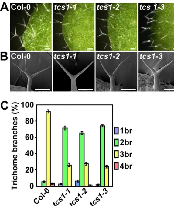
We isolated thetrichome cell shape 1(tcs1) mutants in a screen of publicly available T-DNA mutant collections ofArabidopsis thaliana. Thetcs1-1,tcs1-2andtcs1-3trichomes had the reduced branch number compared with wild-type trichomes (Fig 1). By contrast, thetcs1
Fig 1.tcs1mutants show the reduced number of trichome branches.(A) Light microscope images of Col-0, tcs1-1,tcs1-2andtcs1-3trichomes. Bars = 100μm. (B) Scanning electron microscope images of Col-0,tcs1-1, tcs1-2andtcs1-3trichomes. Bars = 100μm. (C) Trichome branch (br) distribution of Col-0,tcs1-1,tcs1-2and tcs1-3first pair of leaves at 15 days after germination (DAG). Values are given as mean±SE.
mutants did not show any obvious defects in plant growth. Progeny of crosses of the three lines indicated that they are allelic. We further measured the number of trichome branches using the first pair of leaves. In wild-type leaves, trichomes normally had two branching points with three branches (92%), although trichomes with four branches were occasionally found (Fig 1C). By contrast, about 70% and 25% of trichomes ontcs1leaves had two and three branches, respectively (Fig 1C). The tips oftcs1trichome branches were sharp, as those observed in wild-type trichome branches (Fig 1B). Thus, these results show thatTCS1influences the number of trichome branches in Arabidopsis.
Trichomes of
tcs1
are hypersensitive to the microtubule-disrupting drug
oryzalin and the microtubule-stabilizing drug paclitaxel
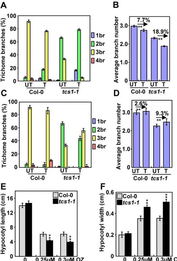
In Arabidopsis, the reduced branch number of trichomes is often correlated with a decrease in the level of endoreduplication or the destabilization of microtubules [18,22]. We firstly investigated whetherTCS1could affect endoreduplication in trichome cells. As the nuclear size is often associ-ated with the ploidy level, we measured the nuclear size of Col-0 andtcs1-1trichomes. The average nuclear size oftcs1-1trichomes was similar to that of wild-type trichomes (S1A and S1B Fig). The ploidy levels intcs1-1leaves were comparable with those in wild-type leaves (S1 Fig). These results suggest thatTCS1may not regulate endoreduplication. We then asked whetherTCS1could influ-ence the microtubule cytoskeleton. The microtubule-disrupting drug oryzalin has been shown to destabilize microtubules, leading to a decrease in the number of trichome branches in Arabidopsis [23]. We therefore treated 4-day-old seedlings of Col-0 andtcs1-1with 20 μM oryzalin for 2 hours. After a 10-day recovery on ½ MS medium, we examined the branch number of Col-0 and
tcs1-1trichomes. As shown inFig 2A and 2B, the oryzalin treatment caused a 7.7% decrease in the average number of Col-0 trichome branches, while the oryzalin treatment resulted in an 18.9% reduction in the average number oftcs1-1trichome branches. The microtubule-stabilizing drug paclitaxel (taxol) has been reported to stabilize microtubules [23]. We asked whether taxol could rescue the trichome branch phenotype oftcs1. Four-day-old seedlings of Col-0 andtcs1-1were treated with 20 μM taxol for 2 hours. After a 10-day recovery on ½ MS medium, we examined the branch number of Col-0 andtcs1-1trichomes. In our growth condition, the taxol treatment caused a 2.6% increase in the average number of Col-0 trichome branches, while the taxol treat-ment resulted in a 9.3% increase in the average number oftcs1-1trichome branches (Fig 2C and 2D), suggesting that taxol partially rescues the phenotype oftcs1-1trichome branches.
As the microtubule is crucial for hypocotyl elongation [24], we asked whether TCS1 affects hypocotyl growth. As shown inFig 2E and 2F, the average length and width of dark-grown
tcs1-1hypocotyls was comparable with that of dark-grown Col-0 hypocotyls. We then treated dark-grown Col-0 andtcs1-1seedlings with oryzalin and measured their hypocotyl length and width. After oryzalin treatment, hypocotyls oftcs1-1were significantly shorter and wider than those of the wild type (Fig 2E and 2F). Epidermal cells intcs1-1hypocotyls were short and wide in comparison with those in wild-type hypocotyls (S2 Fig). These results show that hypocotyls oftcs1-1are hypersensitive to oryzalin treatment than wild-type hypocotyls.
Disruption of
TCS1
influences the stability of microtubules
microtubule arrays disappeared faster intcs1-1trichome cells than those in wild-type trichome cells. We counted the number of cortical microtubules in the trichome branch junction. Micro-tubules were similar in density before oryzalin treatment. However, more cortical microMicro-tubules were disrupted intcs1-1than in the wild type after drug treatment. These results indicate that
TCS1influences the stability of microtubules in trichomes. Similarly, we observed that micro-tubule arrays disappeared relatively faster in epidermal cells oftcs1-1cotyledons than those in epidermal cells of wild-type cotyledons after oryzalin treatment (S3 Fig).
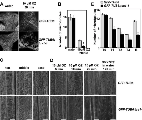
Astcs1hypocotyls were hypersensitive to the microtubule-disrupting drug oryzalin, we investigated whetherTCS1is required for the stability of microtubules in hypocotyl cells. Corti-cal microtubule arrays in epidermal cells ofGFP-TUB6;tcs1hypocotyls were comparable with those ofGFP-TUB6hypocotyls (Fig 3C and 3E). We then applied the microtubule-disrupting drug oryzalin to epidermal cells of etiolatedGFP-TUB6andGFP-TUB6;tcs1-1hypocotyls. Cor-tical microtubule arrays disappeared relatively faster in epidermal cells oftcs1-1hypocotyls than those in epidermal cells of wild-type hypocotyls (Fig 3D and 3E). When oryzalin was washed off after the treatment, the recovery of cortical microtubules in epidermal cells oftcs1-1
hypocotyls was slower than that in epidermal cells of wild-type hypocotyls (Fig 3D and 3E). Taken together, these results indicate thatTCS1influences the stability of microtubules.
TCS1
encodes a coiled-coil domain-containing protein
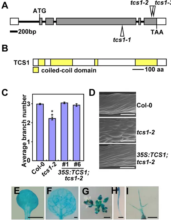
Thetcs1-1,tcs1-2andtcs1-3mutants were identified from the T-DNA insertions in the fourth exon and the sixth exon of the geneAt1g19835, respectively (Fig 4A). T-DNA insertions were confirmed using T-DNA specific and flanking primers (S4A–S4C Fig). We further investigated the expression level ofAt1g19385intcs1-1,tcs1-2andtcs1-3mutant seedlings. As shown inS4D Fig, the full length transcript ofAt1g19835was not detected intcs1mutants, suggesting thattcs1
mutants are loss-of-function alleles. A plasmid containing wild-typeAt1g19835cDNA driven by a 35S promoter was introduced into thetcs1-2mutant. Transgenic plant exhibited complementa-tion oftcs1-2phenotypes (Fig 4C and 4D). In addition, transformation oftcs1-1with TCS1-GFP fusion protein under the control of theTCS1promoter (pTCS1:TCS1-GFP) restored a wild-type phenotype (S4E Fig). Therefore, these results indicate thatAt1g19835is theTCS1gene.
TCS1encodes a 982-amino-acid protein that contains four coiled-coil domains, which belongs to a family of long coiled-coil protein that consists of 7 members in Arabidopsis [25] (Fig 4B;S5 Fig). Although the family members have been named as filament-like plant proteins (AtFPP), their biochemical and biological functions are totally unknown in Arabidopsis [25]. By performing a BLAST search in the databases, we identified TCS1 homologs inBrassica rapa,Gossypium raimondii,Sorghum bicolor,Zea mays, andOryza sativa, but we did not find convincing homologs from animals and yeasts (S5 Fig), suggesting that TCS1 and its homologs might have evolved to control cell morphogenesis in plants.
To determine the expression pattern ofTCS1, RNA from roots, flowers, seedlings and leaves were investigated by RT-PCR analysis.TCS1mRNA was detected in all plant organs tested (S6 Fig). Tissue-specific expression pattern ofTCS1was examined using histochemical assay of
tcs1-1treated with (T) or without (UT) 20μM paclitaxel for 2 hours. The branch number of Col-0 andtcs1-1 trichomes was examined after a 10-day recovery on½MS medium. (D) The average number of Col-0 andtcs1-1 trichome branches treated with (T) or without (UT) 20μM paclitaxel for 2 hours. The branch number of Col-0 and tcs1-1trichomes was examined after a 10-day recovery on½MS medium. (E) The average hypocotyl length of Col-0 andtcs1-1seedlings grown in½MS containing 0μM, 0.25μM and 0.3μM oryzalin (OZ) for 15 days in dark. (F) The average hypocotyl width of Col-0 andtcs1-1seedlings grown in½MS containing 0μM, 0.25μM and 0.3μM oryzalin (OZ) for 15 days in dark. Values (A-F) are given as mean±SE.**P<0.01 compared with the wild type (Student’sttest).
GUS activity of transgenic plants containing theTCS1promoter:GUSfusion (pTCS1:GUS). GUS activity was detected in cotyledons, leaves, inflorescences and developing etiolated hypo-cotyls (Fig 4E–4H). GUS activity was also observed in trichomes (Fig 4I), consistent with the role ofTCS1in trichome morphogenesis.
TCS1 binds to microtubules and promotes microtubule assembly
Astcs1affects the stability of microtubules, we asked whether TCS1 could directly bind to the microtubules. A cosedimentation assay was used to analyze the binding of TCS1 to
taxol-Fig 3. Disruption ofTCS1influences the stability of microtubules in trichomes.(A) Cortical microtubules in trichome cells ofGFP-TUB6andGFP-TUB6;tcs1-1treated with water or 10μM oryzalin (OZ) for 20 minutes. Bars = 20μm. (B) The microtubule density at the branch junction ofGFP-TUB6andGFP-TUB6;tcs1-1trichome cells treated with water or 10μM oryzalin for 20 minutes. ImageJ software was employed to quantify the numbers of cortical microtubules. A line of fixed length (10μm) perpendicular to the orientation of the most cortical microtubules at the trichome branch junction was drawn, and the number of cortical microtubules across the line was counted. At least 10 cells from each treatment were used, and four lines of fixed length were drawn for each cell. Values are given as mean±SE.**P<0.01 compared with the wild type after oryzalin treatment (Student’st test). (C) Cortical microtubules in epidermal cells ofGFP-TUB6andGFP-TUB6;tcs1-1etiolated hypocotyls. GFP-TUB6andGFP-TUB6;tcs1-1seedlings were grown in dark for 72 hours. Epidermal cells in the top, middle and basal regions ofGFP-TUB6andGFP-TUB6;tcs1-1hypocotyls were observed by confocal microscopy. Bars = 10μm. (D) Cortical microtubules in epidermal cells in the middle region ofGFP-TUB6and GFP-TUB6;tcs1-1etiolated hypocotyls treated with 10μM oryzalin (OZ) for 5, 10 and 20 minutes (min). Oryzalin was then washed off, and cortical microtubules were imaged after 2 hours. Bars = 10μm. (E) Quantification of cortical microtubules in hypocotyl epidermal cells ofGFP-TUB6andGFP-TUB6;tcs1-1seedlings treated with 10μM oryzalin for 0 min (T0), 5 min (T1), 10 min (T2) and 20 min (T3), respectively (n>30 cells for each sample). The R represents that oryzalin was then washed off for 2 hours. The y axis represents the number of cortical microtubules across a fixed line (20μm) vertical to the orientation of most cortical microtubules in the cell. Values are given as mean±SE.
**P<0.01 compared withGFP-TUB6(Student’sttest).
Fig 4. Identification of theTCS1gene.(A) TheTCS1gene structure. The start codon (ATG) and the stop codon (TAA) are indicated. Closed boxes indicate the coding sequences, open boxes show the 5’ and 3’ untranslated regions, and the line between boxes indicates the intron. The T-DNA insertion sites (tcs1-1,tcs1-2andtcs1-3) in TCS1are shown. (B) The predicted TCS1 protein contains four coiled-coil domains. aa, amino acids. (C) The average number of trichome branches in Col-0,tcs1-2,35S:TCS1;tcs1-2#1and35S:TCS1;tcs1-2#6leaves. Values are given as mean±SE.**P<0.01 compared with Col-0 (Student’sttest). (D) Scanning electron
microscope images of epidermal cells in Col-0,tcs1-2and35S:TCS1;tcs1-2hypocotyls grown in½MS containing 0.3μM oryzalin for 15 days in dark. Bars = 200μm. (E-I)TCS1expression activity was monitored bypTCS1:GUS transgene expression. Histochemical analysis of GUS activity in a cotyledon (E), a leaf (F), an inflorescence (G), a 4-day-old dark-grown seedling (H) and a trichome (I). Bars = 1 mm in (E-H) and 0.1 mm in (I).
stabilized microtubules. TCS1 was expressed as a maltose binding protein (MBP) fusion pro-tein (MBP-TCS1) inE.coli. As shown inFig 5AandS7A Fig, MBP-TCS1 was cosedimented
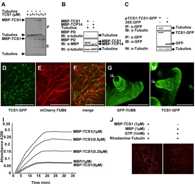
Fig 5. TCS1 binds to microtubules and promotes microtubule assembly.(A) MBP-TCS1 fusion protein was cosedimented with paclitaxel-stabilized microtubules prepolymerized from 8μM tubulins. After high-speed
centrifugation, the amount of MBP-TCS1 in pellets (P) increased when higher concentrations of MBP-TCS1 proteins were added. MBP-TCS1 mostly appeared in the supernatant (S) in the absence of tubulins. (B) TCS1 directly interacts with tubulinsin vitro. Tubulins were pulled down (PD) by MBP-TCS1 immobilized on amylose resin and analyzed by immunoblotting (IB) using an anti-tubulin antibody. MBP-TCP14 was used as a negative control.(C) TCS1 interacts with tubulins in Arabidopsis. Total proteins frompTCS1:TCS1-GFPand35S:GFPtransgenic plants were immunoprecipitated (IP) with GFP-Trap-A, and the immunoblot (IB) was detected with anti-GFP and anti-tubulin antibodies, respectively. Tubulins were detected in the immunoprecipitated GFP-TCS1 complex but not in the control (GFP). (D-F) TCS1 colocalizes with cortical microtubules (MTs) in Arabidopsis epidermal pavement cells. TCS1-GFP (D), mCherry-TUB6 (E) and merged (F) images are shown. TCS1-GFP localizes along cortical microtubules (mCherry-TUB6) in a punctate pattern. Scale bars = 5μm. (G and H) GFP fluorescence of GFP-TUB6 (G) and TCS1-GFP (H) in elongating trichome branches. The white arrows indicate the three dimension (3-D) reconstruction of the microtubule-depleted zone at the extreme apex of elongating trichome branches. Bars = 20μm.(I) MBP-TCS1 promotes the assembly of microtubules. The time course of tubulin polymerization from 20μM tubulins in the presence of different concentrations of MBP-TCS1 was detected turbidimetrically by absorbance at 350 nm. 1μM MBP was used as a negative control. (J) Confocal microscopy analysis showed that TCS1 promotes tubulin assemblyin vitro. Rhodamine labeled tubulins (20μM) were incubated with 1μM MBP-TCS1 and 1μM MBP at 37˚C for 30 min, respectively. Microtubules were visualized under confocal microscopy.
with the microtubules. The binding of TCS1 to microtubules was saturated at a stoichiometry of about 0.38 M MBP-TCS1 per mole of tubulin dimers (S7C Fig). The binding of the positive control (AUGMIN subunit 8, AUG8) to microtubules was saturated at a stoichiometry of about 0.22 M His-AUG8 per mole of tubulin dimers in our experimental conditions (S7B and S7C Fig) [26]. We then asked whether TCS1 could directly interact with tubulins. To test this, we conducted pull-down experiments. As shown inFig 5B, MBP-TCS1 bound to tubulins, while the negative control (MBP-TCP14) did not interact with tubulins. Thus, these results indicate that TCS1 physically interacts with tubulinsin vitro.
We further performed co-immunoprecipitation analyses to detect the interaction of TCS1 with tubulins in Arabidopsis. Total proteins frompTCS1:TCS1-GFPor35S:GFPplants were isolated and incubated with GFP-Trap-A agarose beads to immunoprecipitate TCS1-GFP and GFP. The anti-GFP and anti-tubulin antibodies were used to examine immunoprecipitated proteins, respectively. As shown inFig 5C, tubulins were found in the immunoprecipitated TCS1-GFP complex but not in the negative control (GFP), indicating that TCS1 physically associates with tubulins in Arabidopsis.
To further investigate whether TCS1 localizes to cortical microtubules, we conducted live-cell imaging using a functional TCS1-GFP fusion under the control ofTCS1promoter. As shown inFig 5D–5F, TCS1-GFP localizes to puncta along cortical microtubules (mCherry-TUB6) in pavement cells, indicating that TCS1 binds to the microtubules. We then investigated the co-localization of TCS1-GFP and microtubules in developing trichomes. We have previ-ously showed that it is difficult to observe the signal of mCherry labeled-microtubules in tri-chomes [21]. We therefore usedpTCS1:TCS1-GFPandGFP-TUB6-expressing lines to compare TCS1 with microtubules. InGFP-TUB6trichomes, transverse microtubule arrays formed rings encircling the elongating branches, without the signal at the extreme apex (Fig 5G) [21]. Similarly, we observed that TCS1-GFP was present in elongating trichome branches, but leave a TCS1-depleted zone at the extreme apex (Fig 5H). These results indicate that TCS1 and microtubules exhibit similar organization patterns in trichomes, further suggesting that TCS1 is a microtubule-binding protein.
As TCS1 directly interacts with microtubules, we asked whether TCS1 could affect microtu-bule assembly. We therefore added various concentrations of MBP-TCS1 (0, 0.25, 0.5 and 1 μM) and 1 μM MBP to a 20 μM tubulin solution, and tubulin polymerization was investi-gated by measuring turbidity. As shown inFig 5I, the presence of MBP-TCS1 increased turbid-ity, indicating that MBP-TCS1 increases microtubule mass. The assembly rate of tubulins was increased in a dosage-dependent manner with the addition of MBP-TCS1. To confirm this result, we observed the assembly of rhodamine-labeled tubulins incubated with MBP and MBP-TCS1 under confocal microscopy. As shown inFig 5J, the assembly of microtubules was detected in the presence of MBP-TCS1 rather than MBP. Taken together, these results indicate that TCS1 promotes microtubule assembly.
TCS1 physically interacts with KCBP/ZWI
in vitro
and
in vivo
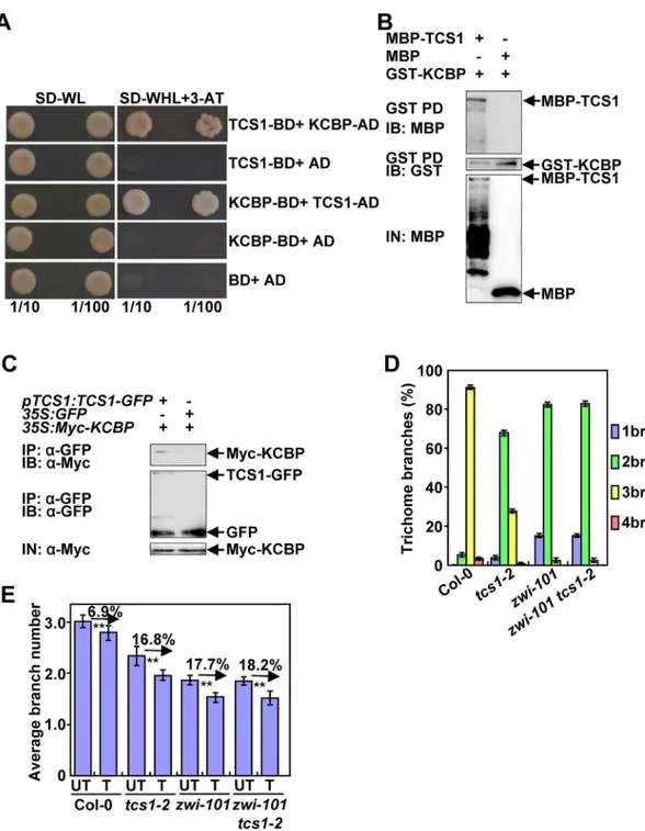
pull-Fig 6. TCS1 physically and genetically interacts with KCBP to control the number of trichome branches. (A) TCS1 interacts with KCBP in yeast cells. (B) TCS1 physically interacts with KCBPin vitro. MBP-TCS1 was pulled down (PD) by GST-KCBP immobilized on Glutathione Sepharose 4B and analyzed by immunoblotting (IB) using an anti-MBP antibody. MBP was used as a negative control. (C) TCS1 interacts with KCBPin vivo. Total proteins frompTCS1:TCS1-GFP;35SMyc-KCBPand35S:GFP; 35SMyc-KCBPplants were immunoprecipitated with GFP-Trap-A (IP), and the immunoblots (IB) were probed with anti-GFP and anti-Myc antibodies, respectively. Myc-KCBP was detected in the immunoprecipitated TCS1-GFP complex. (D) Trichome branch (br) distribution of Col-0,tcs1-2,zwi-101andzwi-101 tcs1-2first pair of leaves at 15 days after germination (DAG). Values are given as mean±SE. (E) The average number of Col-0,tcs1-2,zwi-101,zwi-101 tcs1-2trichome branches treated with (T) or without (UT) 20μM oryzalin for 2 hours. The branch number of Col-0,tcs1-2,zwi-101,zwi-101 tcs1-2 trichomes was examined after a 10-day recovery on½MS medium.
down experiments. TCS1 was expressed as a maltose binding protein (MBP) fusion protein (MBP-TCS1), while KCBP was expressed as a glutathione S-transferase (GST) fusion protein (GST-KCBP). As shown inFig 6B, MBP-TCS1 bound to GST-KCBP, while the negative con-trol (MBP) did not bind to GST-KCBP. This result indicates that TCS1 physically interacts with KCBPin vitro.
We further performed co-immunoprecipitation analysis to investigate the association of TCS1 with KCBP in Arabidopsis. We generated35S:Myc-KCBPtransgenic plants. We crossed thepTCS1:TCS1-GFPand35S:GFPtransgenic lines with35S:Myc-KCBPtransgenic plants to generatepTCS1:TCS1-GFP;35S:Myc-KCBPand35S:GFP;35S:Myc-KCBPplants, respectively. Total proteins were isolated and incubated with GFP-Trap-A agarose beads to immunoprecipi-tate TCS1-GFP and GFP. The anti-GFP and anti-Myc antibodies were used to detect immuno-precipitated proteins, respectively. Myc-KCBP was found in the immunoimmuno-precipitated
TCS1-GFP complex but not in the negative control (GFP) (Fig 6C), indicating that TCS1 phys-ically associates with KCBP in Arabidopsis.
TCS1
genetically interacts with
KCBP
to control the number of trichome
branches
As TCS1 physically interacts with KCBP, andtcs1mutants showed similar trichome branching phenotypes tokcbp/zwimutants, we sought to establish genetic relationships betweenTCS1
andKCBPin the regulation of trichome branch number. We obtained thezwi-101mutant (SALK_017886) harboring the T-DNA insertion in theKCBP/ZWIgene (S8 Fig). The full length mRNA ofKCBPcould not be detected inzwi-101, suggesting thatzwi-101is a loss-of-function allele. Thezwi-101trichomes exhibited the reduced number of branches (Fig 6D), consistent with previous results [16]. We then generated azwi-101 tcs1-2double mutant and investigated its trichome branch number. As shown inFig 6DandS9 Fig, the branch number ofzwi-101 tcs1-2double mutant trichomes was comparable to that ofzwi-101single mutant trichomes, suggesting thatzwi-101is epistatic totcs1-2with respect to the number of trichome branches. Considering that both TCS1 and KCBP affect the stability of microtubules, we asked whether genetic interactions betweenTCS1andKCBPin trichome branch number are related to microtubule stability. We therefore treated 4-day-old seedlings of Col-0,zwi-101,tcs1-2and
zwi-101 tcs1-2with 20 μM oryzalin for 2 hours. After a 10-day recovery on ½ MS medium, the number of Col-0,zwi-101,tcs1-2andzwi-101 tcs1-2trichome branches was investigated. After oryzalin treatment, the number ofzwi-101 tcs1-2trichome branches was similar to that of zwi-101trichome branches (Fig 6E). The oryzalin treatment caused a similar decrease in the aver-age number ofzwi-101 tcs1-2andzwi-101trichome branches. These results suggest thatTCS1
acts genetically withKCBPto regulate the number of trichome branches by influencing the sta-bility of microtubules.
KCBP was reported to physically interact with AN in yeast cells [18]. Theanmutants showed the reduced branches of trichomes in leaves [18,19]. We asked whetherTCS1andAN
Discussion
A fundamental question in developmental biology is how cell shape is controlled. In plants, cell shape is crucial not only for the function of the individual cell, but also for its role in organ shape and size control. However, the genetic and molecular mechanisms that determine cell shape remain largely unknown in plants. In this study, we report that theTCS1gene, which encodes a microtubule binding protein with long coiled-coil domains, is required for trichome cell shape in Arabidopsis. TCS1 directly binds to microtubules and promotes microtubule assembly. TCS1 physically and genetically interacts with KCBP/ZWICHEL to regulate the number of trichome branches by influencing microtubule stability. Thus, our findings reveal a novel genetic and molecular mechanism of TCS1 and KCBP in trichome cell shape control.
TCS1 regulates trichome cell shape by influencing the stability of
microtubules
Thetcs1trichomes showed the reduced branch number (Fig 1), althoughtcs1plants appear to be similar to wild-type plants. Trichome branching is a complicated process, which is regulated by a number of factors. In Arabidopsis, DNA replication (endoreduplication) in trichome cells influences the number of trichome branches [22,27,28]. However, mutations inTCS1did not affect ploidy levels in leaves and nuclear size in trichome cells (S1 Fig). Thus, it is unlikely that TCS1 regulates trichome branch number by influencing DNA replication events. After endore-duplication, a cytoskeleton-dependent polarization event happens during trichome morpho-genesis [23], resulting in a total of three to four branches in the mature trichome on leaves. Molecular-genetic and pharmacological studies have established that microtubules are essential for trichome branching in Arabidopsis [23,29]. Interestingly, the trichomes oftcs1were hyper-sensitive to the microtubule-disrupting drug oryzalin in comparison with those of the wild type (Fig 2A and 2B). Similarly,tcs1hypocotyls were more sensitive to oryzalin than wild-type hypocotyls (Fig 2E, 2F, andS2 Fig). By contrast, the microtubule-stabilizing drug taxol treat-ment partially rescued the branch number oftcs1trichomes (Fig 2C and 2D). These results suggest thatTCS1may affect the stability of microtubules, which are crucial for trichome cell morphogenesis. Consistent with this notion, we observed that microtubules intcs1cells disap-peared faster than those in wild-type cells when treated with oryzalin (Fig 3andS3 Fig). Thus, these results support that mutations inTCS1influence the stability of microtubules, resulting in the altered trichome cell shape in Arabidopsis.
TCS1 is a microtubule-binding protein and promotes microtubule
assembly
TheTCS1gene encodes a coiled-coil domain-containing protein, which belongs to a family of long coiled-coil protein that consists of 7 members in Arabidopsis [25]. However, the biological functions of the TCS1 family members are totally unknown in Arabidopsis [25]. Therefore, TCS1 is a novel regulator of trichome cell shape in Arabidopsis. Sequence analyses show that TCS1 homologs are plant-specific proteins (S5 Fig), suggesting that TCS1 and its homologs might have evolved to regulate cell shape in plants. Expression ofTCS1was detected in all tested tissues (S6 Fig), although the only visible phenotype intcs1mutants was found in tri-chomes. It is possible that TCS1 might function redundantly with other proteins to influence cell growth in other tissues or cell types.
that TCS1 directly bound to the microtubule in Arabidopsis cells (Fig 5D–5F). In addition, TCS1-GFP and GFP-TUB6 showed similar organization patterns in elongating trichome branches (Fig 5G and 5H). These results support that TCS1 is a microtubule binding protein. It is possible that TCS1 directly binds to microtubules during trichome development and stabi-lizes microtubules, thereby influencing the formation of trichome branches in Arabidopsis. Biochemical analyses showed that TCS1 promotes microtubule assembly, consistent with the function of TCS1 in stabilizing microtubules (Fig 5I and 5J). The microtubule assembly has been known to influence trichome branch number. For example, mutations in the TUBULIN FOLDING COFACTOR (TCF) C and TCFA result in the unbranched trichome phenotype [13,14]. These mutants were proposed to affect the making of assembly componentα/βtubulin dimmers and possibly decrease the assembly of new microtubules. Mutations inKINESIN-13A, which promotes microtubule depolymerization, resulted in the increased number of trichome branches [15,32]. Thus, it is possible that TCS1 promotes microtubule assembly and increases the stability of microtubules, thereby influencing trichome branch number in Arabidopsis.
A possible genetic and molecular mechanism of TCS1 in regulating the
number of trichome branches
KCBP, a microtubule motor, regulates cell division and trichome cell shape in Arabidopsis [16,17]. Trichomes onzwileaves had one or two branches with blunt tips. It has been suggested that KCBP participates in the trichome morphogenesis by regulating the local reorientation and stability of microtubules [30,31]. Similarly, TCS1 regulates trichome branch number by influencing the stability of microtubules. Thetcs1mutants showed similar trichome branch number phenotype tokcbp/zwimutants, suggesting that TCS1 could genetically interact with KCBP to control the branch number of trichomes. Consistent with this idea, our genetic analy-ses show thatzwiis epistatic totcs1with respect to trichome branch number. Further results demonstrated that TCS1 physically interacted with KCBPin vitroandin vivo(Fig 6A–6C). As both TCS1 and KCBP influence the stability of microtubules, it is possible that TCS1 functions with KCBP to control trichome branch number by affecting the dynamics and stability of microtubules in Arabidopsis. Supporting this notion,zwi-101 tcs1-2andzwi-101trichomes showed a similar level of hypersensitivity to oryzalin (Fig 6E). A recent study have shown that KCBP interacts with both microtubules and actin cytoskeleton to regulate trichome branching and elongation in Arabidopsis [21]. Thezwitrichomes had the reduced number of branches, shortened stalks and stunted branches [16]. The reduced number of branches inzwitrichomes is likely caused by defects in microtubules. By contrast, the transverse cortical F-actin cap at the trichome branch apex has been proposed to regulate polarized branch elongation and tip sharpening [21].tcs1mutants only affected the trichome branch number and had normal stalks and trichome branch tips (Fig 1A and 1B), suggesting that KCBP and TCS1 may have the over-lapped function in the regulation of microtubule cytoskeleton rather than actin cytoskeleton.
with KCBP, it is possible that TCS1 and other members in the KCBP complex may have genetic interactions in trichome branching. Supporting this notion, we found an epistatic interaction betweenANandTCS1with respect to the number of trichome branches, although TCS1 does not physically interact with AN (S12 Fig). It will be a worthwhile challenge to build up the genetic and molecular interactions between TCS1 and other members of the KCBP complex in the future. Taken together, our findings reveal a novel genetic and molecular mechanism by which TCS1 interacts with KCBP to control trichome cell shape by influencing the stability of microtubules.
Materials and Methods
Plant materials and growth conditions
Thetcs1-1(SAIL_403_D02),tcs1-2(SALK_040648),tcs1-3(SALK_078664),zwi-101
(SALK_017886), andan1-101(SALK_026489) mutants were obtained from the Nottingham Arabidopsis Stock Centre (NASC). The T-DNA insertions were verified by PCR and sequenc-ing ussequenc-ing the primers described inS1 Table. Arabidopsis seeds were sterilized with 100% iso-propanol for 2 min and 10% NaClO (v/v) for 10 min and then washed six times with sterile water. Arabidopsis seeds were dispersed on ½ Murashige and Skoog (MS) medium containing 0.9% agar and 1% glucose and then stored at 4°C for 3 days in the darkness. Plants were grown at 22°C under long-day conditions (a 16-h-light /8-h-dark cycle). To observe etiolated hypo-cotyls, we grow plants in dark at 22°C.
Construction and plant transformation
A PCR-based Gateway system was used to generate35S:TCS1,pTCS1:TCS1-GFPandpTCS1:
GUSconstructs. TheTCS1CDS was amplified using the primers CDS-LP and TCS1-CDS-RP (S1 Table). PCR product was subcloned into thepCR8/GW/TOPO TAcloning vector (Invitrogen) using TOPO enzyme. TheTCS1CDS was then subcloned into the Gateway binary vectorpMDC32to generate the35S:TCS1construct. TheTCS1genomic sequence containing a 2012-bp promoter sequence and 3298bp gene was amplified using the primers gTCS1-GFP-LP and gTCS1-GFP-RP. PCR products were firstly cloned into thepCR8/GW/TOPO TAcloning vector (Invitrogen) using TOPO enzyme. TheTCS1genomic sequence was then subcloned into thepMDC107vector to generate the constructpTCS1:TCS1-GFP. The 2164bp promoter sequence ofTCS1was amplified using the primers TCS1pro-LP and TCS1pro-RP. PCR prod-ucts were cloned into thepCR8/GW/TOPO TAcloning vector (Invitrogen) using TOPO enzyme. TheTCS1promoter was then subcloned into thepMDC164vector to generate the transformation plasmidpTCS1:GUS. The plasmids35S:TCS1,pTCS1:TCS1-GFPandpTCS1:
GUSwere transferred intotcs1-2or Col-0 plants using Agrobacterium GV3101, and medium with hygromycin (30μg/mL) was used to select transgenic plants.
Morphological and cellular analysis
Trichome branches on the first pair of Col-0 andtcs1-1leaves were counted at 15 days after germination (DAG). Leaves and etiolated hypocotyls of wild-type andtcs1-1mutants were fixed in a solution (formalin, acetic acid, ethanol and H2O in a ratio of 1: 0.5: 4.75: 3.75) for 24 hours, dehydrated with a graded ethanol series and dried at critical point in liquid CO2. Sam-ples were coated with gold and then observed in an S-4160 Field Emission Scanning Electron Microscope (SEM) (Hitachi).
and hypocotyl epidermal cells of the wild type (Col-0) andtcs1-1for specific times. The micro-tubule-stabilizing drug taxol (paclitaxel, Sigma-Aldrich) was used to treat trichomes of the wild type andtcs1-1.
To quantify the numbers of cortical microtubules in trichome and hypocotyl cells, the Ima-geJ software was employed. A line of fixed length (10 μm or 20 μm) perpendicular to the orien-tation of the most cortical microtubules was drawn, and the number of cortical microtubules across the line was counted. At least 10 cells from each treatment were used, and four lines of fixed length were drawn for each cell. The average number of cortical microtubules before and after treatments was calculated. The Student’s test was used to analyze the significance of the difference.
GUS staining
Samples (pTCS1:GUS) were putted into a GUS staining solution [0.1% Nonidet P-40, 1 mM 5-bromo-4-chloro-3-indolyl-b-D-glucuronic acid, 10 mM EDTA, 100 mM Na3PO4buffer, and 3 mM each K3Fe(CN)6/K4Fe(CN)6] and incubated at room temperature for 6 hours. After GUS staining, 70% ethanol was used to remove chlorophyll.
Confocal microscopy observation
GFP fluorescence in cells of trichomes and hypocotyls was detected using a Zeiss LSM710 META confocal microscope. GFP was observed using wave lengths of 510 to 530 nm. To study the co-localization of TCS1 and microtubules, we crossedpTCS1:TCS1-GFPtransgenic plants withmCherry-TUB6expressing plants. Seeds were germinated on ½ Murashige and Skoog (MS) medium supplemented with 0.9% agar and 1% glucose. Leaves of 6-day-old mCherry-TUB6;pTCS1:TCS1-GFPseedlings were observed under a spinning disk confocal microscope equipped with lasers for GFP and mCherry (Intelligent Design).
RNA isolation and semiquantitative RT-PCR analysis
Leaves, stems, cotyledons and roots from 12-day-old seedlings were collected to isolate total RNAs using an RNeasy Plant Mini kit (TIANGEN). Reverse transcription (RT)-PCR was per-formed using Superscript III reverse transcriptase (Invitrogen).ACTIN2mRNA was an inter-nal control. The specific primers used for RT-PCR are shown inS1 Table.
Yeast two-hybrid assays
The coding sequence ofTCS1was cloned intoNotI andSalI sites of the bait vectorpDBleu
(Invitrogen) and the prey vectorpEXP-AD502(Invitrogen) to generateTCS1-BDand
TCS1-ADconstructs, respectively. The specific primers forTCS1-BDandTCS1-ADwere TCS1-Y2H-SalI-LP and TCS1-Y2H-NotI-RP (S1 Table). The coding sequence ofKCBPwas cloned intoNotI andSalI sites of the bait vectorpDBleu(Invitrogen) and the prey vector pEX-P-AD502(Invitrogen) to generateKCBP-BDandKCBP-ADconstructs. The specific primers forKCBP-BDandKCBP-ADconstructs were KCBP-Y2H-SalI-LP and KCBP-Y2H-NotI-RP (S1 Table). The prey and bait plasmids were co-transformed into the yeast strain PT69-4A to investigate their interactions.
In vitro
protein-protein interaction
The coding sequence ofTCS1was cloned into the vectorpMAL-C2to generate theMBP-TCS1
pGEX-4T-1(Amersham-Pharmacia) to generate theGST-KCBPconstruct. The specific primers for theGST-KCBPconstructs were GST-KCBP-LP and GST-KCBP-RP (S1 Table). The coding sequence ofANwas subcloned into the vectorpET-NTto generate theAN-Hisconstruct. The specific primers for theAN-Hisconstruct were AN-His-LP and AN-His-RP (S1 Table). The
MBP-TCS1,GST-KCBPandAN-Hisplasmids were transformed and expressed inE.coli
Rosetta (DE3).
To investigate protein-protein interaction, we performed the pull-down assay. Bacterial lysates containing ~15 μg of MBP-TCS1 or MBP-TCP14 fusion proteins were mixed with ~20 μg of tubulins. Bacterial lysates containing ~15 μg of MBP-TCS1 or MBP proteins were mixed with `~20 μg of AN-His fusion proteins. Amylose resin (30 μL; New England Biolabs) was added into each combined solution with gently rocking at 4°C for 1 h. Bacterial lysate con-taining ~20 μg of GST-KCBP was mixed with lysate concon-taining ~15 μg of MBP-TCS1 or MBP. Glutathione Sepharose 4B (30 μL;GE Healthcare) was added into each combined solution with gently rocking at 4°C for 1 h. The TGH buffer (1× Complete Protease Inhibitor cocktail [Roche], 150 mM NaCl, 10% glycerol, 1.5 mM MgCl2, 50 mM HEPES, pH 7.5, 1 mM phenyl-methylsulfonyl fluoride, 1 mM EGTA, pH 8.0, and 1% Triton X-100) was used to wash beads for five times. The isolated proteins were then analyzed by 10% SDS-polyacrylamide gels and determined by immunoblot analysis with MBP, tubulin, GST, and His anti-bodies (Abmart), respectively.
Assays of microtubule cosedimentation and assembly
For the microtubule cosedimentation assay, different concentrations of MBP-TCS1 were added to paclitaxel-stabilized microtubules in the PEMT buffer (1 mM MgCl2, 1 mM EGTA, 100 mM PIPES, and 20 μM taxol, pH 6.9). After incubation at 25°C for 30 min, the samples were centrifuged at 100,000g at 25°C for 30 min to separate supernatants and pellets. They were then analyzed by 10% SDS-PAGE and determined by staining the gels with Coomassie Brilliant Blue R 250.
For the microtubule polymerization assay, different concentrations of MBP-TCS1 were added to 20 μM tubulin solution in the PEM buffer (1 mM MgCl2, 1 mM EGTA, 1 mM GTP, and 100 mM PIPES, pH6.9). The polymerization was investigated turbidimetrically by absor-bance at 350 nm with a 0.4-cm path quartz cell at 37°C in a DU-640 spectrophotometer (Beck-man Coulter, Fullerton, CA). 1 μM MBP was used as a negative control. The data was recorded from time 0 to 35min, when the turbidity in all samples did not increase any more.
For the observation of microtubule assembly, 1μM MBP-TCS1, 20μM Rhodamine labeled tubulins and 1mM GTP were incubated at 37°C for 30 min. The microtubule polymerization was then stopped using 1% glutaraldehyde. Spinning-Disc Confocal Microscopy Imaging was performed on an Olympus IX81 inverted microscope equipped with a Yokogawa spinning-disc confocal head (Yokogawa Electric) and an Andor iXon charge-coupled device camera (Andor Technology). 1μM MBP was used as a negative control. Images were captured using Andor iQ software, version 1.1 (Andor Technology), and processed using ImageJ software.
Co-immunoprecipitation
isolated with the extraction buffer (1× Complete protease inhibitor cocktail, 50 mM Tris/HCl, pH 7.5, 2% Triton X-100, 1 mM EDTA, 150 mM NaCl, 20 mg/mL MG132, and 20% glycerol) and mixed with GFP-Trap-A (Chromotek) for 1 h at 4°C. Beads were washed four times with the wash buffer (1×Complete protease inhibitor cocktail, 50mMTris/HCl, pH7.5, 150mM NaCl, and 0.1% Triton X-100). The immunoprecipitates were analyzed by 10% SDS-polyacryl-amide gel and determined by immunoblot analysis with anti-GFP (Abmart) and anti-Myc (Abmart) antibodies, respectively.
Accession numbers
Arabidopsis Genome Initiative locus identifiers for the genes mentioned in this article are as follows: AT1G19835 (TCS1), AT5G65930 (KCBP), and AT1G01510 (AN).
Supporting Information
S1 Fig.TCS1does not affect endoreduplication.(A) The size of nuclei in wild-type Col-0 and
tcs1-1trichomes. The nuclei were stained by DAPI. (B) The average area of nuclei in Col-0 and
tcs1-1trichomes. (C) Nuclear DNA ploidy distribution of cells in Col-0 andtcs1-1first pair of leaves measured at 13 days after germination (DAG). Values (B and C) are given as mean ± SE. Bars = 20 μm in (A).
(PDF)
S2 Fig. Hypocotyl cells oftcs1-1are hypersensitive to the microtubule-disrupting drug ory-zalin.(A) Scanning electron microscope images of Col-0 andtcs1-1cells in the top and middle regions of etiolated hypocotyls grown in ½ MS for 15 days in dark. Bars = 200 μm. (B) Scan-ning electron microscope images of Col-0 andtcs1-1cells in the top and middle regions of etio-lated hypocotyls grown in ½ MS containing 0.3 μM oryzalin for 15 days in dark.
Bars = 200 μm. (C) The average length of epidermal cells in the middle regions of Col-0 and
tcs1-1hypocotyls treated with oryzalin. Col-0 andtcs1-1seedlings were grown in ½ MS con-taining 0, 0.25 μM and 0.3 μM oryzalin (OZ) for 15 days in dark. (D) The average width of epi-dermal cells in the middle regions of Col-0 andtcs1-1hypocotyls treated with oryzalin. Col-0 andtcs1-1seedlings were grown in ½ MS containing 0, 0.25 μM and 0.3 μM oryzalin (OZ) for 15 days in dark. Values (C and D) are given as mean ± SE.P<0.01 compared with the wild type (Student’sttest).
(PDF)
S3 Fig. Microtubules in epidermal cells oftcs1-1cotyledons are hypersensitive to the micro-tubule-disrupting drug oryzalin.Cortical microtubules in epidermal cells ofGFP-TUB6and
GFP-TUB6;tcs1-1cotyledon veins treated with 5 μM oryzalin for 10 minutes. Bars = 20 μm. (PDF)
S4 Fig. Identification of theTCS1gene.(A) PCR identification of the T-DNA insertion in
tcs1-1with T-DNA specific primers (LB1) and flanking primers (LP and RP). (B) PCR identifi-cation of the T-DNA insertion intcs1-2with T-DNA specific primers (LBa1) and flanking primers (LP and RP). (C) PCR identification of the T-DNA insertion intcs1-3with T-DNA specific primers (LBa1) and flanking primers (LP and RP). (D) RT-PCR analysis ofTCS1
expression in Col-0,tcs1-1,tcs1-2andtcs1-3seedlings. RT-PCR was performed on first-strand cDNA prepared from 2-week-old seedlings. cDNA was standardized by reference to an
ACTIN2standard. (E) The average trichome branch number of Col-0,tcs1-1,pTCS1:
(Student’sttest). (PDF)
S5 Fig. Phylogenetic tree of TCS1 and its homologs in different species.The phylogenetic tree was constructed using the neighbor-joining method of the MEGA6 program (http://www. megasoftware.net/mega.html). Values at nodes represent percentages of 1000 bootstrap repli-cates. The scale bar at the bottom represents the genetic distance.
(PDF)
S6 Fig. Expression of theTCS1gene.RT-PCR analysis ofTCS1expression in roots, flowers, 10-day-old seedlings, rosette leaves and cauline leaves.
(PDF)
S7 Fig. Quantification of the binding affinity of TCS1 and AUG8 with microtubules.(A) MBP-TCS1 fusion protein was cosedimented with paclitaxel-stabilized microtubules (5 μM). After high-speed centrifugation, the amount of MBP-TCS1 in pellets increased when higher concentrations of MBP-TCS1 proteins were added before reaching saturation. (B) His-AUG8 fusion protein was cosedimented with paclitaxel-stabilized microtubules (5 μM). After high-speed centrifugation, the amount of His-AUG8 in pellets increased when higher concentra-tions of His-AUG8 proteins were added before reaching saturation. (C) Quantification of the binding affinity of TCS1 with microtubules shown in (A) compared with that of AUG8 with microtubules shown in (B). The binding of TCS1 and AUG8 to microtubules was saturated at a stoichiometry of about 0.38 M MBP-TCS1 and 0.22 M His-AUG8 per mole of tubulin dimers, respectively.
(PDF)
S8 Fig. Identification of thezwi-101mutant.(A) The insertion of T-DNA inzwi-101 (SALK_017886) is shown. (B and C) PCR identification of the T-DNA insertion inzwi-101
with T-DNA specific primers (LBa1) and flanking primers (LP and RP). (D) Expression levels ofKCBPin Col-0 andzwi-101seedlings as determined by RT-PCR.
(PDF)
S9 Fig.zwi-101is epistatic totcs1-2with respect to trichome branch number.(A) The aver-age number of Col-0,tcs1-2,zwi-101andzwi-101 tcs1-2trichome branches of the first pair of leaves at 15 days after germination (DAG). (B) Scanning electron microscope images of Col-0,
tcs1-2,zwi-101andzwi-101 tcs1-2trichome branches of first pair of leaves at 15 days after ger-mination (DAG). Values (A) are given as mean ± SE.P<0.01 compared with the respective controls (Student’sttest). Bars = 100 μm.
(PDF)
S10 Fig. Identification of thean-101mutant.(A) The insertion of T-DNA inan-101 (SALK_026489) is shown. (B and C) PCR identification of the T-DNA insertion inan-101
with T-DNA specific primers (LBa1) and flanking primers (LP and RP). (D) Expression levels ofANin Col-0 andan-101seedlings as determined by RT-PCR.
(PDF)
S11 Fig.an-101is epistatic totcs1-2with respect to trichome branch number.(A) The aver-age number of Col-0,tcs1-2,an-101andan-101 tcs1-2trichome branches of the first pair of leaves at 15 days after germination (DAG). (B) Scanning electron microscope images of Col-0,
tcs1-2,an-101andan-101 tcs1-2trichome branches of the first pair of leaves at 15 days after germination (DAG). Values (A) are given as mean ± SE. Bars = 100 μm.
S12 Fig. TCS1 does not physically interact with AN.AN-His proteins were pulled down (PD) by MBP-TCS1 immobilized on amylose resin and analyzed by immunoblotting (IB) using an anti-His antibody. MBP was used as a negative control.
(PDF)
S1 Table. List of primers used in this study (PDF)
Acknowledgments
We thank Dr. Ying Fu for theHis-AUG8construct and helpful discussions and the Arabidopsis Stock center NASC fortcs1,zwiandanmutants.
Author Contributions
Conceived and designed the experiments:LC YP YL.
Performed the experiments:LC YP JT XW.
Analyzed the data:LC YP JT ZK TM MY YL.
Contributed reagents/materials/analysis tools:ZK TM MY.
Wrote the paper:LC YL.
References
1. Marks MD (1997) MOLECULAR GENETIC ANALYSIS OF TRICHOME DEVELOPMENT IN ARABI-DOPSIS. Annu Rev Plant Physiol Plant Mol Biol 48: 137–163. doi:10.1146/annurev.arplant.48.1.137
PMID:15012260
2. Hulskamp M, Schnittger A, Folkers U (1999) Pattern Formation and Cell Differentiation: Trichomes in Arabidopsis as a Genetic Model System. Int Rev Cytol 186: 147–178. PMID:9770299
3. Oppenheimer DG (1998) Genetics of plant cell shape. Curr Opin Plant Biol 1: 520–524. PMID:
10066632
4. Reddy AS, Day IS (2000) The role of the cytoskeleton and a molecular motor in trichome morphogene-sis. Trends Plant Sci 5: 503–505. PMID:11120459
5. Schellmann S, Hulskamp M (2005) Epidermal differentiation: trichomes in Arabidopsis as a model sys-tem. Int J Dev Biol 49: 579–584. doi:10.1387/ijdb.051983ssPMID:16096966
6. Ivakov A, Persson S (2013) Plant cell shape: modulators and measurements. Front Plant Sci 4: 439. doi:10.3389/fpls.2013.00439PMID:24312104
7. Mathur J, Mathur N, Kirik V, Kernebeck B, Srinivas BP, et al. (2003) Arabidopsis CROOKED encodes for the smallest subunit of the ARP2/3 complex and controls cell shape by region specific fine F-actin formation. Development 130: 3137–3146. PMID:12783786
8. Deeks MJ, Kaloriti D, Davies B, Malho R, Hussey PJ (2004) Arabidopsis NAP1 is essential for Arp2/3-dependent trichome morphogenesis. Curr Biol 14: 1410–1414. doi:10.1016/j.cub.2004.06.065PMID:
15296761
9. El-Assal Sel D, Le J, Basu D, Mallery EL, Szymanski DB (2004) Arabidopsis GNARLED encodes a NAP125 homolog that positively regulates ARP2/3. Curr Biol 14: 1405–1409. doi:10.1016/j.cub.2004. 06.062PMID:15296760
10. El-Din El-Assal S, Le J, Basu D, Mallery EL, Szymanski DB (2004) DISTORTED2 encodes an ARPC2 subunit of the putative Arabidopsis ARP2/3 complex. Plant J 38: 526–538. doi:10.1111/j.1365-313X. 2004.02065.xPMID:15086808
11. Hulskamp M (2004) Plant trichomes: a model for cell differentiation. Nat Rev Mol Cell Biol 5: 471–480. doi:10.1038/nrm1404PMID:15173826
13. Kirik V, Grini PE, Mathur J, Klinkhammer I, Adler K, et al. (2002) The Arabidopsis TUBULIN-FOLDING COFACTOR A gene is involved in the control of the alpha/beta-tubulin monomer balance. Plant Cell 14: 2265–2276. doi:10.1105/tpc.003020PMID:12215519
14. Kirik V, Mathur J, Grini PE, Klinkhammer I, Adler K, et al. (2002) Functional analysis of the tubulin-fold-ing cofactor C in Arabidopsis thaliana. Curr Biol 12: 1519–1523. PMID:12225668
15. Lu L, Lee YR, Pan R, Maloof JN, Liu B (2005) An internal motor kinesin is associated with the Golgi apparatus and plays a role in trichome morphogenesis in Arabidopsis. Mol Biol Cell 16: 811–823. doi:
10.1091/mbc.E04-05-0400PMID:15574882
16. Oppenheimer DG, Pollock MA, Vacik J, Szymanski DB, Ericson B, et al. (1997) Essential role of a kine-sin-like protein in Arabidopsis trichome morphogenesis. Proc Natl Acad Sci U S A 94: 6261–6266. PMID:9177205
17. Reddy VS, Day IS, Thomas T, Reddy ASN (2004) KIC, a Novel Ca2+ Binding Protein with One EF-Hand Motif, Interacts with a Microtubule Motor Protein and Regulates Trichome Morphogenesis. Plant Cell 16: 185–200. doi:10.1105/tpc.016600PMID:14688294
18. Kim GT, Shoda K, Tsuge T, Cho KH, Uchimiya H, et al. (2002) The ANGUSTIFOLIA gene of Arabidop-sis, a plant CtBP gene, regulates leaf-cell expansion, the arrangement of cortical microtubules in leaf cells and expression of a gene involved in cell-wall formation. EMBO J 21: 1267–1279. doi:10.1093/ emboj/21.6.1267PMID:11889033
19. Folkers U, Kirik V, Schobinger U, Falk S, Krishnakumar S, et al. (2002) The cell morphogenesis gene ANGUSTIFOLIA encodes a CtBP/BARS-like protein and is involved in the control of the microtubule cytoskeleton. EMBO J 21: 1280–1288. doi:10.1093/emboj/21.6.1280PMID:11889034
20. Krishnakumar S, Oppenheimer DG (1999) Extragenic suppressors of the arabidopsis zwi-3 mutation identify new genes that function in trichome branch formation and pollen tube growth. Development 126: 3079–3088. PMID:10375500
21. Tian J, Han L, Feng Z, Wang G, Liu W, et al. (2015) Orchestration of microtubules and the actin cyto-skeleton in trichome cell shape determination by a plant-unique kinesin. Elife 4: e09351.
22. Heyman J, Van den Daele H, De Wit K, Boudolf V, Berckmans B, et al. (2011) Arabidopsis ULTRAVIO-LET-B-INSENSITIVE4 maintains cell division activity by temporal inhibition of the anaphase-promoting complex/cyclosome. Plant Cell 23: 4394–4410. doi:10.1105/tpc.111.091793PMID:22167059
23. Mathur J, Chua N-H (2000) Microtubule Stabilization Leads to Growth Reorientation in Arabidopsis Tri-chomes. Plant Cell 12: 465–477. PMID:10760237
24. Li J, Wang X, Qin T, Zhang Y, Liu X, et al. (2011) MDP25, A Novel Calcium Regulatory Protein, Medi-ates Hypocotyl Cell Elongation by Destabilizing Cortical Microtubules in Arabidopsis. Plant Cell 23: 4411–4427. doi:10.1105/tpc.111.092684PMID:22209764
25. Gindullis F, Rose A, Patel S, Meier I (2002) Four signature motifs define the first class of structurally related large coiled-coil proteins in plants. BMC Genomics 3: 9. doi:10.1186/1471-2164-3-9PMID:
11972898
26. Cao L, Wang L, Zheng M, Cao H, Ding L, et al. (2013) Arabidopsis AUGMIN subunit8 is a microtubule plus-end binding protein that promotes microtubule reorientation in hypocotyls. Plant Cell 25: 2187– 2201. doi:10.1105/tpc.113.113472PMID:23735294
27. Schnittger A, Schobinger U, Bouyer D, Weinl C, Stierhof YD, et al. (2002) Ectopic D-type cyclin expres-sion induces not only DNA replication but also cell diviexpres-sion in Arabidopsis trichomes. Proc Natl Acad Sci U S A 99: 6410–6415. doi:10.1073/pnas.092657299PMID:11983922
28. Schnittger A, Weinl C, Bouyer D, Schobinger U, Hulskamp M (2003) Misexpression of the cyclin-dependent kinase inhibitor ICK1/KRP1 in single-celled Arabidopsis trichomes reduces endoreduplica-tion and cell size and induces cell death. Plant Cell 15: 303–315. doi:10.1105/tpc.008342PMID:
12566574
29. Szymanski DB (2009) Plant cells taking shape: new insights into cytoplasmic control. Curr Opin Plant Biol 12: 735–744. doi:10.1016/j.pbi.2009.10.005PMID:19914858
30. Reddy ASN, Narasimhulu SB, Safadi F, Golovkin M (1996) A plant kinesin heavy chain-like protein is a calmodulin-binding protein. Plant J 10: 9–21. PMID:8758976
31. Reddy ASN, Safadi F, Narasimhulu SB, Golovkin M, Hu X (1996) A Novel Plant Calmodulin-binding Protein with a Kinesin Heavy Chain Motor Domain. J Biol Chem 271: 7052–7060. PMID:8636137
32. Oda Y, Fukuda H (2013) Rho of Plant GTPase Signaling Regulates the Behavior of Arabidopsis Kine-sin-13A to Establish Secondary Cell Wall Patterns. Plant Cell 25: 4439–4450. doi:10.1105/tpc.113. 117853PMID:24280391