No Evidence for AID/MBD4-Coupled DNA
Demethylation in Zebrafish Embryos
Nobuyoshi Shimoda1*., Kentaro Hirose2., Reiya Kaneto2¤
, Toshiaki Izawa3, Hayato Yokoi4, Naohiro Hashimoto1, Yutaka Kikuchi2
1.Department of Regenerative Medicine, National Institute for Longevity Sciences, National Center for Geriatrics and Gerontology, 35 Gengo, Morioka, Obu, Aichi 474-8522, Japan,2.Department of Biological Science, Graduate School of Science, Hiroshima University, Kagamiyama 1-3-1, Higashi-Hiroshima, Hiroshima 739-8526, Japan,3.Max Planck Institute of Biochemistry, Am Klopferspitz 18, 82152 Martinsried, Germany,4.Graduate School of Agricultural Science, Tohoku University, 1-1 Tsutsumi-Dori Amamiya-Machi, Aoba-Ku, Sendai 981-8555, Japan
.These authors contributed equally to this work.
¤ Current address: Nagasaki City Office, Nagasaki 850-0031, Japan
Abstract
The mechanisms responsible for active DNA demethylation remain elusive in Metazoa. A previous study that utilized zebrafish embryos provided a potent mechanism for active demethylation in which three proteins, AID, MBD4, and GADD45 are involved. We recently found age-dependent DNA hypomethylation in zebrafish, and it prompted us to examine if AID and MBD4 could be involved in the phenomenon. Unexpectedly, however, we found that most of the findings in the previous study were not reproducible. First, the injection of a methylated DNA fragment into zebrafish eggs did not affect either the methylation of genomic DNA, injected methylated DNA itself, or several loci tested or the expression level of aid, which has been shown to play a role in demethylation. Second, aberrant
methylation was not observed at certain CpG islands following the injection of antisense morpholino oligonucleotides against aid and mbd4. Furthermore, we demonstrated that zebrafish MBD4 cDNA lacked a coding region for the methyl-CpG binding domain, which was assumed to be necessary for guidance to target regions. Taken together, we concluded that there is currently no evidence to support the proposed roles of AID and MBD4 in active demethylation in zebrafish embryos.
OPEN ACCESS
Citation:Shimoda N, Hirose K, Kaneto R, Izawa T, Yokoi H, et al. (2014) No Evidence for AID/MBD4-Coupled DNA Demethylation in Zebrafish Embryos. PLoS ONE 9(12): e114816. doi:10.1371/ journal.pone.0114816
Editor:Dajun Deng, Peking University Cancer Hospital and Institute, China
Received:March 24, 2014
Accepted:October 28, 2014
Published:December 23, 2014
Copyright:ß2014 Shimoda et al. This is an open-access article distributed under the terms of theCreative Commons Attribution License, which permits unrestricted use, distribution, and repro-duction in any medium, provided the original author and source are credited.
Data Availability:The authors confirm that all data underlying the findings are fully available without restriction. The mbd4 cDNA sequence is available from the DDBJ database (accession number: AB918737).
Funding:This study was supported by a Grant-in-Aid for Scientific Research from the MEXT (KAKENHI 23616002;http://www.mext.go.jp/) to YK.
Introduction
We recently reported age-dependent decreases in 5-methylcytosine (5mC) in the zebrafish genome, and found that these were observed earlier than initially expected [1]. A slight decrease in global methylation was noted when late embryos (48 hours post fertilization; 48 hpf) were compared with early embryos (4 hpf, blastula stage), and we identified the CpG island shore as the preferred region for
age-related hypomethylation by bisulfite sequencing [1]. Although the
mechan-isms responsible for hypomethylation remain unknown, a failure in the
maintenance of a methylation pattern or the enzymatic removal of 5mC at CpG island shores during aging has been implicated.
Collas first reported that anin vitro-methylated plasmid injected into fertilized
zebrafish eggs was gradually demethylated starting from 1,2 hpf until 12 hpf,
which indicated the presence of demethylation activity in zebrafish embryos [2]. Demethylation was found to be replication-independent, thereby suggesting an enzymatic reaction that converted 5mC into cytosine (C) in zebrafish embryos. Rai et al. also injected methylated DNA fragments (M-DNA) into zebrafish eggs and demonstrated that exogenous DNA elicited demethylation of not only
M-DNA itself, but also the genomic M-DNA of M-M-DNA-injected zebrafish embryos [3].
Although the biological significance of demethylation elicited by this exogenous DNA was not described in their study, by utilizing this artificial demethylation system, Rai et al. tested for candidate enzymes, the function of which may explain the possible scenario for demethylation: conversion of 5mC to thymine (T) via deamination, followed by the removal of the mismatching T, and subsequent
replacement with C through base excision repair [3]. The candidates in the
deamination processes that were examined included all three members of the activation-induced deaminase (AID)/Apolipoprotein B RNA-editing catalytic component (APOBEC) deaminase family in zebrafish; AID, Apobec2a, and
Apobec2b [4,5]. Although zebrafish AID was recently shown to deaminate
methylated cytidines in vitro [6], whether Apobec2a and Apobec2b exhibit
deaminase activity has yet to be determined. In this context, it is important to note that mouse Apobec2 did not exhibit deaminase activity, and this is in contrast to its paralogs, Apobec1 and some Apobec3 genes, which have been
shown to deaminate particular RNAs and DNAs, respectively [7,8]. Another
candidate enzyme tested was a likely ortholog of human G: T-mismatch glycosylase, methyl-binding domain protein 4 (MBD4), which removes T mispairing with G in vitro [9,10]. MBD4 is also known to play a rolein vivo by
suppressing mutations at CpG sites in mammalian genomes [11,12]; however, a
defect in DNA methylation was not observed in mbd4knockout mice, which are
viable and fertile [11]. The non-enzymatic factor Growth arrest and
DNA-damage-inducible protein 45 alpha (Gadd45a), which was shown to be involved in
the global DNA demethylation of human culture cells [13], was also examined,
while contradictory results were obtained using the same cells or Gadd45a2/2
mice [14,15]. Rai et al. demonstrated that the knockdown of AID or MBD4
heterogeneous combination of genes, zebrafish aid and humanMBD4 elicited DNA demethylation in zebrafish embryos, which was promoted by the additional expression ofgadd45a[3]. Based on these findings, together with other supporting data, they proposed a mechanism for demethylation that is exerted by the concerted action of the three products: the conversion of 5mC to T via deamination by AID, followed by thymine base excision repair by MBD4, can occur in zebrafish embryos, and is facilitated by Gadd45a, which may serve as a scaffold to physically and/or functionally couple AID and MBD4. To the best of our knowledge, this was the first evidence for the involvement of T-G mismatches in vertebrate DNA in global demethylation and provided the rationale for the three factors to be investigated for active demethylation in mammalian cells; however, questions were raised on their model [16]. Following this study, Rai et al. suggested that the proposed demethylation machinery may be involved in regulating intestinal cell fating [17].
We speculated whether the active demethylation system described by Rai et al.
[3] in zebrafish embryos may be involved in the age-related hypomethylation of
CpG island shores. To investigate this possibility further, we examined the reproducibility of several experiments in their previous study [3], and
demonstrated that most of their findings were not reproducible. This finding, together with the absence of actively-demethylated loci in the genome of zebrafish embryos [18,19], cast doubt on the occurrence of active demethylation during the early development of zebrafish.
Results
No discernible hypomethylation of genomic DNA or
methylated-DNA (M-methylated-DNA) itself was observed following the injection of M-methylated-DNA
into zebrafish fertilized eggs
Rai et al. reported that the injection of 200 pg of in vitro-methylated DNA
(M-DNA) in fertilized zebrafish eggs caused not only the significant demethylation of M-DNA itself, but also demethylation of 20%–40% of the bulk genome, both of which peaked at 13 hpf, which corresponded to the early somite stage of zebrafish development [3]. This artificially-induced demethylation system was utilized
throughout the study to identify candidate genes, such as aid andmbd4, in the
demethylation activity observed in zebrafish embryos. Therefore, M-DNA-dependent demethylation represented a fundamental experiment to confirm the reproducibility of their experimental results.
To determine whether the demethylation system was reproducible, we prepared the same DNA fragment (736 bp) as that used by Rai et al. [3] (Fig. 1A), and methylated itin vitrousing HpaII methylase (Fig. 1B). Methylation was confirmed at four CCGG sites in the fragment by its resistance to HpaII, which cleaved the
tetranucleotide when C was not methylated (Fig. 1B). We injected 200 pg of
running undigested genomic DNA on an agarose gel (Fig. 1C). The purity of genomic DNA was verified by digestion with MspI, which is a methylation-insensitive isoschizomer of HpaII (Fig. 1D). To prepare a reference of a globally demethylated genome, we suppressed the activity of DNA methyltransferase 1 (Dnmt1) using an injection of antisense morpholino oligonucleotides (MO) against the dnmt1 gene [20,21] into zebrafish eggs, and then extracted genome
DNA at 48 hpf. The knockdown of dnmt1 resulted in approximately 20%
hypomethylation at the several loci examined by bisulfite sequencing [22], which should be similar to the moderately demethylated genome in M-DNA-injected
embryos [3]. The digestion of the genome fromdnmt1 MO-injected embryos by
HpaII or HpyCH4IV, another methylation-sensitive enzyme, was more sensitive to the methylation-sensitive enzymes than the control genome, which indicated
that the methylation level in dnmt1MO-injected embryos was lower than that in
the control embryos (white arrows inFig. 1E and 1F). In contrast, we observed no significant differences in digestion patterns between the genomes from M-DNA-injected and control embryos at any of the developmental stages examined (Fig. 1E and 1F), even though a M-DNA injection was shown to cause genome-wide demethylation that was detectable with HpaII digestion, followed by electrophoresis on agarose gels [3].
Rai et al. also demonstrated that M-DNA injected into embryos clearly lost its methylation at 13 hpf [3], which was in accordance with the findings reported by
Collas [2]. To confirm this, we recovered injected M-DNA at 13 hpf from
embryos and examined its methylation using bisulfite sequencing. In contrast to the findings of Collas [2] and Rai et al. [3], we demonstrated that injected
M-DNA remained highly methylated at any time point, including 13 hpf (Fig. 2A).
For these experiments we used the zebrafish line, named AB/Tu¨, which was derived from a cross of AB and Tu¨bingen (Tu¨) lines. On the other hand, Rai et al used Tu¨ line. To examine if the discrepancies we observed above could be attributed to the difference in zebrafish lines, we performed the same experiments with Tu¨ line but were still unable to reproduce the results that Rai et al. described (S1 Fig.). In accordance with these results, we observed no demethylation in any of the randomly-chosen seven sequences, which included three types of repetitive sequences (two LINEs; KenoDr1 and LINE-1, two SINEs; DANA and SINE3-1a,
and 5S rRNA) and two unique sequences (runx1and fli1a), in M-DNA-injected
embryos at 13 hpf (Fig. 2B). On the other hand, as we previously reported [22], a reduction was observed in methylation levels in all of the selected sequences from
dnmt1 MO-injected embryos (Fig. 2B). Thus, we concluded that the injection of M-DNA into zebrafish eggs had no discernible effect on the methylation level of
(M), anddnmt1MO-injected (MO) embryos at the indicated time points (hours post fertilization; hpf) were run on an agarose gel. (D, E, F) The same genomic DNAs as those used and shown in (C) were digested with MspI (D), HpaII (E), or HpyCH4IV (F), and run on an agarose gel. The molecular weight markers of DNA loaded on the first lanes of gels, shown as MW, were 100 bp (B) or 1 kb ladders (C, D, E, and F). Note that smearing down of high-molecular DNA, thereby reducing the methylation level, was discernible only in the genomic DNA fromdnmt1MO-injected embryos (white arrows in E and F).
genomic DNA from M-DNA-injected zebrafish embryos or the injected M-DNA itself. These results demonstrated that the first and fundamental experiments of Rai et al. [3] were not reproducible.
Fig. 2. No demethylation at M-DNA or randomly chosen loci was detected by the M-DNA-injection into eggs.(A) Bisulfite sequencing revealed no significant changes in the methylation levels of injected M-DNA recovered from 10 embryos at any of the indicated time points. (B) Bisulfite sequencing analysis showed that the conserved regions of the five repetitive sequences (KenoDr1, DANA, SINE3-1a, LINE-1, and 5S rRNA) or confined regions of two single genes (runx1andfli1a) were highly methylated in M-DNA-injected embryos at 13 hpf (M-DNA) as in controls (WT), whereas moderate
hypomethylation was observed in the equivalent regions indnmt1MO-injected embryos (dnmt1MO). Black and white circles are methylated and unmethylated cytosines, respectively. Crosses denote the positions at which CpG was absent due to polymorphisms. Values represent the ratio of the numbers of methylated cytosines in the total number of CpG dinucleotides in the regions surveyed.
Injection of M-DNA into zebrafish eggs does not increase the
expression level of
aid
and
apobec2a
Rai et al. also reported that the injection of M-DNA into zebrafish eggs increased the expression levels of all three annotated members of the AID/Apobec
deaminase family, i.e. AID, Apobec2a, and Apobec2b [3]. Based on this finding,
these genes were selected as candidates that could play roles in artificially induced demethylation, as described above. However, we wanted to confirm whether the expression of these deaminase genes was upregulated in an M-DNA-dependent manner because the M-DNA injection did not cause demethylation in zebrafish embryos. We examined the expression of the deaminase genes by quantitative real time PCR (qPCR), and found no significant increases in the expression of either
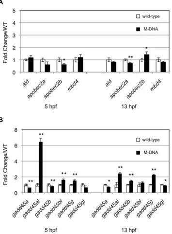
aid or apobec2a at 13 hpf (Fig. 3A). In contrast, although the expression of
apobec2b was upregulated (Fig. 3A), the upregulation level of this gene
Fig. 3. M-DNA did not induce the expression ofaidorapobec2a.(A) The expression levels of AID/Apobec family genes at the indicated time points were quantified by qPCR, and these expression levels in wild-type embryos (white rectangles) were compared with those in M-DNA-injected embryos (black rectangles). (B) The expression levels of Gadd45-family genes between wild-type and M-DNA-injected embryos were compared using qPCR. Gene expression was normalized againstrpl13a(ribosomal protein L13agene) and data represent the mean¡standard error of the mean (s.e.m.) from three independent experiments; **P,0.01 and *P,0.05.
(approximately 1.5-fold change/WT) was lower than that reported previously [3]
(approximately 5-fold change/WT), and no upregulation of apobec2b was
observed when Tu¨ line was used (S2A Fig.). Our results indicated thataid would not be selected as a candidate gene that could be involved in artificially induced demethylation. In addition, Rai et al. showed that four of the six Gadd45 family genes examined, namely gadd45a,gadd45a like(gadd45al),gadd45b, andgadd45g, were all markedly upregulated either at 5 hpf, 13 hpf, or at both time points by
the M-DNA injection, and gadd45a was chosen for further analyses and was
shown to enhance demethylation [3]. We examined the expression of all six
gadd45genes, and showed that none of them, except forgadd45al,were markedly
upregulated by M-DNA (Fig. 3Band S2B Fig.). Rather than being increased, the
expression level of gadd45a in M-DNA-injected embryos was slightly lower than
that in control embryos at 13 hpf (Fig. 3B).
Zebrafish MBD4 lacked a methyl-CpG binding domain (MBD)
In most vertebrates, MBD4 consists of two well-conserved and functional domains, an N-terminal methyl-CpG binding domain (MBD) and C-terminal DNA glycosylase domain, which are separated by a poorly conserved spacer region
[9]. Rai et al. showed that the co-expression of zebrafish AID/Apobec and human
MBD4 in zebrafish embryos led to the widespread DNA demethylation of the genome, including the retrotransposable elements, KenoDr1 and LINE-1, which are typically highly and constitutively methylated [3]. They suggested that the overexpression of human MBD4 with a MBD and a DNA glycosylase domain may account for the extended demethylation of the genome. Rai et al. used the human
mbd4gene instead of the zebrafishmbd4in the co-expression experiment, and this
may have been because the 59end of zebrafish mbd4cDNA had not been
determined at that time [3]. Therefore, in their previous study, it was assumed that zebrafish MBD4 had a MBD, similar to human MBD4, and zebrafish MBD4 was regarded as being interchangeable with human MBD4 for demethylation. MBD4 in other fish species such as Coelacanth, Fugu, medaka, cave fish, Tilapia,
Stickleback, and cod all have a MBD at the N-termini (http://www.ensembl.org/
info/about/species.html). However, using 59RACE, we found that the complete
cDNA structure of the sole zebrafish homologue of humanmbd4only coded for a
DNA glycosylase domain and lacked a MBD domain (DDBJ accession number: AB918737), as were the rare cases for Cionaand chicken [23,24] (http://www. ensembl.org/info/about/species.html) (Fig. 4A, see alsoS3 Fig.). An in-frame stop codon and eleven repetitive sequences were detected upstream of the first ATG of the open reading frame; therefore, there may be no additional coding sequences
upstream of the first ATG (S3 Fig.). The most recent zebrafish genome-build
(Zv9) remains incomplete at the mbd4locus, whereas some contigs of
Illumina-generated sequences in the Sanger database (http://www.sanger.ac.uk/resources/
the MBD of mouse MBD4 as a query, only MBD1, MBD3, and MeCP2 (methyl-cytosine binding protein 2) were retrieved.
In addition to the lack of a MBD-coding sequence, zebrafishmbd4was unique
in that alternative splicing produced a minor isoform that lacked exon 6 (150 bp, 50 amino acids), a middle region of the conserved DNA glycosylase domain Fig. 4. Zebrafish MBD4 lacks a MBD andmbd4MO did not interfere splicing of transcripts.(A) Schematic structures of MBD4 proteins in the chordata are indicated. The predominant form of MBD4 had MBD and a glycosylase domain near the N- and C- termini, respectively, similar to human MBD4. As rare cases in chordate, such as Chicken and Ciona, two zebrafish MBD4 proteins generated via alternative splicing lacked MBD. The shorter isoform lacked thirty amino acids, which corresponded to exon 6 (marked in grey). (B) The schematic structure of the zebrafishmbd4gene. Boxes show exons, and black and white boxes show translated and untranslated regions, respectively. Alternative splice events are indicated as diagonal lines. Five splicing variants of the 59UTR and a shorter isoform that skipped exon 6 (grey box) were obtained by RT-PCR. The approximate positions of MO and primer pairs designed by Rai et al. [3] to detect aberrant splicing by MO are indicated by a small bar and arrows, respectively. The lengths of introns that may have been included in the RT-PCR product are shown. (C) With the primer set shown in (B), a region ofmbd4cDNA was amplified from wild-type (WT) embryos in which the indicated amounts ofmbd4MO were injected. The same banding pattern was observed irrespective of the MO injection. In addition to the expected band (II), three faint, but distinct bands (I, III, and IV) appeared both in wild-type and MO-injected embryos. The bottom band (I) was derived from an alternative splicing variant that skipped exon 6. (D) PCR amplification of band I and II in a single tube reproduced the same banding pattern on agarose gel as that in (C). The possible structures of bands III and IV are shown on the right of the gel.
(Fig. 4, see also S3 and S4A Figs.). It is currently not known whether the two forms of zebrafish MBD4, especially the smaller one, exhibit DNA glycosylase activity or function in combination or independently. Given the lack of MBD in zebrafish MBD4, together with the presence of two forms of the protein in zebrafish embryos, it would be premature to suggest that the functions of zebrafish MBD4 proteins could be deduced from the results obtained with the overexpression of full-length human MBD4 in zebrafish embryos.
mbd4
MO was non-functional, whereas
aid
MO blocked its own
transcription
To knockdown the activities of MBD4 and AID, Rai et al. designed MOs against
the boundaries between the first exon and first intron of thembd4 oraidgene to
inhibit the correct splicing of their transcripts (Figs. 4B and5A), and confirmed their efficacy by detecting their unspliced transcripts [3]. To confirm these findings, we injected two different amounts of AID and MBD4 MOs, 4pg and 5ng,
the former of which was used in their previous study [3]. Since 4pg of MO was
Fig. 5.aidMO suppressed the expression of theaidgene.(A) The genomic structure of theaidgene was drawn, as in Fig. 4 (B). (B) qPCR analyses of theaidtranscripts from ScraidMO andaidMO-injected AB/Tu¨ embryos were conducted by the primer set shown as arrowheads in (A). ScraidMO is a negative control for
aidMO that has a scrambled sequence foraidMO. (C) A MO-induced reduction in the expression ofaidwas confirmed with Tu¨ line by qPCR with a primer set that differed from the primer set used in (B), and was shown as arrows in (A). Gene expression was normalized againstrpl13a, and data represent the mean¡standard error of the mean (s.e.m.) from three independent experiments. (D) cDNAs derived from wild-type (WT) and MO-injected embryos were used for 59RACE ofEF1a1. The products were checked as in (B).
approximately three orders of magnitude lower than the amount of MO
commonly used [5,25,26], we also used 1,000 times more of the former, 5ng of
MO. We then attempted to detect missplicing of the mbd4 or aid transcript in
mbd4oraidMO-injected embryos, respectively, with the same primer sets used in
the previous study (Figs. 4B and 5A). When PCR amplified a region of mbd4
cDNA that spanned exon 6 and 7, we encountered two unanticipated results. First, in addition to a band of the expected size of 340 bp (fragment II inFig. 4C), three
faint DNA fragments were generated from control embryos (Fig. 4Cand
S4A Fig.), in contrast to the one amplified fragment from control embryos in the
previous study [3]. We cloned and sequenced all four fragments, and found that
they may have been derived from two alternatively spliced mRNA: the bottom
band of 190 bp (fragment I inFig. 4C) was from cDNA that skipped exon 6 of the
mbd4gene, the splicing variant, as described above. The second bottom band was
from cDNA of 340 bp, which retained exon 6 (fragment II in Fig. 4C). The two
other larger fragments, depicted as III and IV in Fig. 4C, appeared to be a
heteroduplex of fragments I and II (Fig. 4D) because the sequences of cloned
fragments III and IV were identical to either the sequence of fragment I or II. When we re-amplified fragments I and II in a single tube, four DNA bands again appeared on the agarose gel (Fig. 4D). Therefore, it was likely that the looped-out exon 6 region of heteroduplex DNA could be an obstacle for migration in agarose and the degree of delayed migration may further differ between the two forms of the heteroduplex, depending on the strand that is looped-out.
Second, the same levels of the four PCR products (fragments I, II, III, and IV)
were amplified from mbd4MO-injected embryos (Fig. 4C), which indicated that
the mbd4 MO designed by Rai et al. [3] did not cause the missplicing of mbd4
transcripts at the position indicated in the previous study at either 4 pg or 5ng. We doubted whether the position of PCR amplification that spanned exon 6 and 7 would be adequate to detect missplicing because it was located apart from the MO position (exon 1/intron 1 boundary). Targeting a splice junction of the first exon in pre-mRNA by MO generally triggers the complete or partial inclusion of intron 1 [26,27,28]. Therefore, we PCR amplified a region that spanned introns 1 and 2 of the mbd4gene, and still could not detect any aberrant splicing variants (S4B Fig.). These results indicated thatmbd4 MO did not exert splicing blocker activity at the region specified in the previous study or at the exon 1/intron 1 boundary. Thus, whether zebrafish MBD4 is involved in demethylation has yet to be confirmed.
aid MO was also designed to anneal the first exon and first intron boundary
(Fig. 5A) and reportedly caused missplicing [3]. However, we demonstrated by
qPCR analyses that the injection of aidMO suppressed the expression ofaideven
at a very low amount (4 pg) in AB/Tu¨ strain (Fig. 5B). The primer set used was
designed for the amplification of exons 4 and 5 of aid (Fig. 5A); therefore, it is
unlikely that intron 1 (870 bp), which may be left unspliced byaidMO, hampered
PCR. In accordance with this result, qPCR analyses with a different set of primers
that spanned intron 2 revealed a marked decrease in the amount of aid
elongation factor 1a1 (EF1a1) could be obtained not only from wild-type, but also
from aid MO-injected AB/Tu¨ embryos, which showed that our inability to
amplify aid cDNA was not due to the degradation of mRNA extracted fromaid
MO-injected embryos (Fig. 5D). Since the binding site ofaidMO was close to the 59end of theaidtranscript (50,60 bp downstream of the transcription start site),
aid MO may prevent the progression of transcription. Although aid MO did not
induce aberrant splicing as reported,aidMO may have suppressed the function of
the AID protein in zebrafish embryos.
No gain in methylation at the CpG islands of
neurod2
,
sox1a
, or
atoh1a
by
aid
or
mbd4
MO
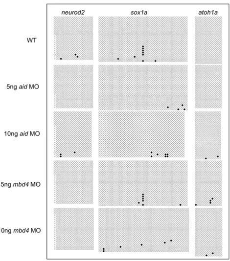
Based on the finding that the knockdown of AID, Gadd45a, or MBD4 by MO caused the loss of neuronal gene expression at 24 hpf, Rai et al. compared the Fig. 6. NeitheraidMO normbd4MO elicited methylation at the CpG islands of neuronal genes.Bisulfite sequence analysis was used to examine the methylation of the CpG islands in the three genes indicated. Black and white circles are methylated and unmethylated cytosines, respectively. Crosses denote the positions at which CpG was absent due to polymorphisms.
methylation status of transcription factor genes involved in neurogenesis at the
CpG islands in aid or mbd4 injected embryos with those in control
MO-injected embryos at 80% epiboly [3]. Using methylated DNA
immunoprecipita-tion (MeDIP) alone or in combinaimmunoprecipita-tion with bisulfite sequence analyses, Rai et al. reported that a pronounced increase in CpG methylation at the CpG islands of
neurod2,sox1a,andatoh1agenes following the injection ofaidMO or mbd4MO [3]. To confirm the reproducibility of these findings, we repeated the experiment
three times by two different operators; one operator used 5 ng and 10 ng of aid
andmbd4MO (Fig. 6) and the other used four different amounts ofaidandmbd4
MO (4 pg, 10 pg, 5 ng, 10 ng) for AB/Tu¨ (S5 Fig.) and 5 ng ofaidandmbd4MO
for Tu¨ (S6 Fig.). However, bisulfite sequence analysis in each case revealed no increases in methylation at the CpG islands at any amounts of the two MOs. The
absence of an increase in methylation bymbd4MO was consistent withmbd4MO
being non-functional. In contrast, we consider it unlikely that AID is involved in
maintaining the hypomethylated status of these CpG islands because aid MO
severely attenuated aid transcription, as described above.
Discussion
Rai et al. previously reported demethylation activity in zebrafish embryos that was
catalyzed by the concerted actions of AID, MBD4, and Gadd45 [3]. They later
demonstrated that demethylation activity regulated intestinal cell fating [17]. To the best of our knowledge, no other studies have confirmed these findings since their publication; therefore, we repeated the experiments performed in the former study. Rai et al. showed that the injection of M-DNA into zebrafish embryos caused the demethylation of both genomic DNA and the injected DNA fragment itself [3], a unique response whose biological significance remains unclear. This finding was the rationale for the presence of demethylation activity in zebrafish embryos, and the phenomenon was utilized as an assay system to uncover the
involvement of three proteins, AID, MBD4, and Gadd45, in demethylation [3].
Therefore, we first examined the M-DNA injection into zebrafish eggs, but were unable to confirm the phenomenon even though the same M-DNA fragment was used. To detect the M-DNA-dependent global demethylation of the zebrafish genome, we adopted one of the two procedures used in their previous study: the digestion of genomic DNA with the methylation-sensitive enzyme HpaII, together with HpyCH4IV, an enzyme that has been used to sensitively detect age-related hypomethylation in the zebrafish genome [1]. Qualitative analysis with restriction enzymes was sufficiently sensitive to detect the small decrease that occurred in
global methylation in dnmt1 MO-injected embryos. In addition, we employed
bisulfite sequencing to detect DNA methylation changes at specific loci. We, however, did not detect demethylation of M-DNA or several genomic loci, both of which were examined in our study.
apobec2a, and apobec2b, was clearly induced by the introduction of M-DNA into
zebrafish embryos, and that these genes were involved in demethylation [3].
However, we did not observe any significant increases in the expression ofaid or
apobec2a genes, except for apobec2b. We cannot explain these discrepancies between our results and the findings reported by Rai et al [3]. Our inability to
detect any signs of the demethylation or induction of aid or apobec2a raises
serious concerns about the reality of M-DNA-related phenomena described in their previous study [3].
The presence of active demethylation in early development of the zebrafish remains controversial. Recent analyses of the whole-genome bisulfite sequence in zebrafish embryos until the germ ring stage (5.7 hpf) revealed that no loci were subjected to active demethylation [18,19]. While these studies identified distinct loci in the maternal genome in which demethylation proceeds between 1-cell and 16/32-cell stages, demethylation is considered to be passive based on the gradual and apparently replication-dependent dilution process [18,19]. Alternatively, as suggested by Rai et al., CpG islands, which were generally hypomethylated regardless of gene expression [29], may be under surveillance of active
demethylation, which removes aberrant methylation from CpG islands [3].
However, some neurogenesis-related CpG islands shown to be methylated by aid
MO in the previous study remained hypomethylated regardless of the suppression
of aid transcription. We once again cannot account for this discrepancy, and it
remains to be determined whether CpG islands in zebrafish are guarded by
demethylation activity against de novo methylation.
Rai et al. showed thatmbd4 MO also caused thede novomethylation of some
neurogenesis-related CpG islands, similar toaid MO [3]. However, we could not
verify these findings, even when the amount of MOs used (10 ng) was 2500 times
higher than those in the previous study (4 pg). Therefore, mbd4 MO, which was
used in the previous study, may not have been appropriate for testing the involvement of zebrafish MBD4 in demethylation because aberrant splicing was
not induced by mbd4MO. Further analyses are clearly required to determine
whether zebrafish MBD4 is related to demethylation.
Rai et al. suggested that the demethylation complex consisting of AID, MBD4,
and Gadd45 could be recruited to 5mC by MBD in MBD4 [3]. In contrast to the
assumption by Rai et al. [3], we found that the zebrafish MBD4 protein lacked a
MBD. This result, together with the finding that AID or Gadd45 had no DNA binding motif, makes it difficult to explain how the proposed demethylation complex consisting of AID, MBD4, and Gadd45 could be recruited to 5mC. Thus, we did not examine the overexpression of zebrafish AID or zebrafish MBD4 in zebrafish eggs. Although Rai et al. reported that the forced co-expression of
zebrafish AID and human MBD4 elicited demethylation [3], the possibility that
the heterogeneous combination of the two proteins exerted artificial demethyla-tion activity in zebrafish embryos cannot be ruled out because human MBD4 possesses a MBD.
A recent study revealed that zebrafish AID was unique among all orthologs
knockdown of aidin our experiments did not induce global methylation changes in the zebrafish genome at 5 or 13 hpf, which suggested that AID-dependent active demethylation in zebrafish may be loci-specific if it occurs at embryonic stages. The requirement of AID for DNA demethylation has not yet reached consensus, even in mouse systems; it has been supported by the findings of studies
performed using primordial germ cells [30] and pluripotent ES/iPS cells
[31,32,33], but not in those using activated B cells [34] in which there is adequate
amounts of AID to mutate a number of non-Ig loci [35].
Given that AID is involved in active demethylation, another unresolved issue was the substrate of AID for demethylation. Recent findings on the TET DNA dioxygenase, which oxidized 5mC to 5-hydroxymethylcytosine (5hmC), suggest
that 5hmC, but not 5mC could be a substrate of AID for demethylation [36];
5hmC is deaminated by AID/APOBECs to produce 5-hydroxymethyluracil (5hmU), which can then be excised by 5hmU glycosylases and finally replaced with an unmethylated cytosine by base excision repair. The zebrafish genome encodes all three members of the TET family proteins, each of which converts
5mC to 5hmC [25]. However, 5hmU was not detected in mouse neuronal or
HEK293 cells [36,37], and recentin vitrofindings showed that the deamination of
5hmC by AID was unlikely to occur [38,39]. These studies challenge the
plausibility of the proposed pathway that invoked 5hmC deamination in DNA demethylation.
We consider two points to be critical in order to confirm the reproducibility of the findings reported by Rai et al [3]: the demethylation of both zebrafish genomic DNA and M-DNA itself by the injection of M-DNA into zebrafish eggs, and the
aberrant methylation at certain CpG islands due to aid MO or mbd4 MO. We
were unable to duplicate either of these points, and the reasons for the
discrepancies between our results and the findings reported by Rai et al. [3] are unknown. It is important to note that the procedures used for the experiments described above were straightforward and well established, and the materials used were all purchased from manufacturers, except the zebrafish embryos, which makes it unlikely that we overlooked changes that should have been induced by M-DNA or MOs.
Taken together, we obtained the following results that were contradictory to previous studies: 1) demethylation was not detected following the injection of a
methylated DNA fragment, 2) zebrafish MBD4 lacked a MBD, 3) mbd4MO was
Conclusions
Our results raise serious concerns regarding the previously proposed model involving the concerted action of AID, MBD4 and GADD45 in active DNA demethylation in zebrafish.
Materials and Methods
Ethics statement
All animal experiments were conducted in strict accordance with relevant national and international guidelines: ‘Act on Welfare and Management of Animals’ (Ministry of Environment of Japan) and all steps were taken to minimize animal discomfort. Zebrafish embryos were euthanized by overdose with Tricaine. Ethics was approved from the Hiroshima University Animal Research Committee (Permit Number: F13-1).
Zebrafish care
Wild-type (AB/Tu¨ and Tu¨bingen lines) zebrafish were maintained at 28.5
˚
C in a14 hr light: 10 hr dark cycle in our fish facility.
Preparation of methylated DNA (M-DNA) and genomic DNA
To prepare M-DNA, a 736 bp-DNA fragment was PCR amplified from the
luciferase gene in pGL3-SV40 (Promega), as described previously [3]. The PCR
fragment was purified with QIA quick mini-spin columns (Qiagen) and
methylatedin vitrowith HpaII methylase (New England Biolabs) according to the
manufacturer’s instructions. The resultant M-DNA was purified with phenol/ chloroform/isoamyl alcohol followed by precipitation with ethanol. Prior to the injection, M-DNA was diluted in Danieau solution [58 mM NaCl, 0.7 mM KCl,
0.4 mM MgSO4, 0.6 mM Ca(NO3)2, 5.0 mM HEPES, pH7.6] [40] and it was
injected at 200 pg per embryo. To prepare genomic DNA, zebrafish embryos were harvested at the designated time points and DNA was extracted using Proteinase
K and SDS [40]. M-DNA and genomic DNA were dissolved in 10 mM Tris-HCl
(pH 8.0)/0.1 mM EDTA, and M-DNA and genomic DNA concentrations were measured on a NanoDrop 2000 spectrophotometer (NanoDrop Technologies Inc., ThermoFisher Scientific).
Digestion of M-DNA and genomic DNA by restriction enzymes
Fifty nanograms of M-DNA was digested with either MspI (5 units) or HpaII (5
units) for 2 hr at 37
˚
C. One hundred nanograms of genomic DNA was digestedcut genomic DNA were separated on a 1.2% agarose gel with a 1-kb DNA ladder marker. Gels were stained with ethidium bromide (0.5 mg/ml) and then destained in water. DNA-size markers and restriction enzymes were purchased from New
England Biolabs, except for the 100-bp ladder maker in Fig. 5B (Invitrogen).
Morpholino (MO) Injections
The MO sequences used were identical to those used by Rai et al. [3]. MOs
obtained from Gene-tools LLC Ltd. (Philomath, OR) were dissolved in water, and the concentrations of dissolved MOs were determined by the procedure shown on
the home page of Gene-tools (http://www.gene-tools.com/) using a NanoDrop
2000 spectrophotometer. Prior to the injection, MOs were diluted in Danieau solution [40]. M-DNA or MO was injected into zebrafish eggs at the one-cell stage and ten embryos were then used to extract genomic DNA.
Bisulfite sequence analyses
In the methylation analysis for zebrafish genes and M-DNA, 200 ng of genomic DNA was processed using the EZ DNA Methylation-Gold Kit (Zymo Research, Orange, CA, USA) to convert unmethylated cytosines into uracils. Injected M-DNA was recovered from ten embryos with genomic M-DNA at different time points. The primer pairs used to amplifyneurod2,sox1a, andathoh1aCpG islands
were the same as those used in the previous study [3]. We used the MethPrimer
program to choose the sequences of the other primer pairs for bisulfite sequencing
[41]. Primer sequences and PCR conditions are shown in S1 Table. PCR
fragments were cloned into the pGEM-T Easy vector (Promega, Madison, WI,
USA) and were used to transform the Escherichia coli DH5alpha strain. Plasmids
isolated from 24 and 12 transformants for zebrafish genes and M-DNA, respectively, were sequenced with the DYEnamic ET Terminator Cycle Sequencing kit (GE Healthcare, Piscataway, NJ, USA) on an ABI3100 genetic analyzer (Applied Biosystems, Foster City, CA, USA). QUMA was used to analyze sequence data and drawing figures [42].
Preparation of total RNA and first strand cDNA synthesis
To extract total RNA from embryos, pools of 20 live embryos were homogenized
in 500 ml TRIzol Reagent (Invitrogen, Carlsbad, CA, USA) using a Kontes Pellet
Pestle in companion tubes (Thermo Fisher Scientific, Waltham, MA, USA). Extracted RNA was then purified by an RNeasy Mini kit (QIAGEN, Hilden, Germany). The quantity of total RNA was measured on a NanoDrop 2000 spectrophotometer. Five hundred nanograms of total RNA was mixed with an RNA loading buffer containing 0.4 mg/ml ethidium bromide, and electrophor-esed on a 1.0% formaldehyde-containing agarose gel to check the quality of RNA. One mg of total RNA from embryos at 4 hpf, 13 hpf, and 28 hpf fish was
cDNA Reverse Transcription kit (Applied Biosystems, Foster City, CA, USA). In all cases, a reverse-transcriptase negative control was used to test genomic DNA contamination.
Quantitative real-time RT-PCR (qPCR)
Standard reactions for qPCRs were prepared as follows: 10 ml of SsoFast EvaGreen
Supermix (BioRad, Hercules, CA, USA), 0.25 ml of the forward and reverse
primers (20 mM each), 0.67 ml of the template, and 8.83ml of water. Templates
were 1:20 diluted cDNA samples, and in the case of negative controls, cDNAs were replaced by water. All real time assays were performed in triplicate using a LightCycler (Roche Diagnostics, Mannheim, Germany). After denaturing samples at 95
˚
C for 30 s, forty amplification cycles were performed, with each cycle consisting of 95˚
C for 5 s followed by 59˚
C for 20 s. Primer information is shown on S1 Table. P-valueswere calculated using an unpaired t test.RT-PCR and 5
9
RACE
Using first strand cDNA as a template, aid andmbd4 were PCR amplified either
with a set of primers designed by Rai et al. [3] or with primers designed in this
study. 59RACE was performed using a SMART RACE cDNA Amplification Kit
(Clontech) following the manufacturer’s instructions. PCR products were run on
a 2% agarose gel, except for the RT-PCR product ofmbd4(exon1,exon3), which
was separated on a 10,20% gradient polyacrylamide gel (Wako, Japan).
Supporting Information
S1 Fig. Absence of genome demethylation following the injection of a methylated DNA fragment into zebrafish eggs of Tu¨ line. (A) Undigested
genomic DNA of control (W), M-DNA-injected (M), and dnmt1 MO-injected
(MO) embryos at the indicated time points (hours post fertilization; hpf) were run on an agarose gel. (B, C, D) The same genomic DNAs as those used and shown in (A) were digested with MspI (B), HpaII (C), or HpyCH4IV (D), and run on an agarose gel. The molecular weight markers of DNA loaded on the first lanes of gels, shown as MW, were 1 kb ladders (A, B, C, and D). Note that smearing down of high-molecular DNA, thereby reducing the methylation level, was
discernible only in the genomic DNA from dnmt1 MO-injected embryos (white
arrows in C and D). (E) Bisulfite sequencing revealed no significant changes in the methylation levels of injected M-DNA recovered from 10 Tu¨ embryos at any of the indicated time points.
doi:10.1371/journal.pone.0114816.s001 (TIF)
S2 Fig. M-DNA did not induce the expression ofaidorapobec2ain Tu¨.(A) The
expression levels of AID/Apobec family genes and mbd4at the indicated time
embryos (white rectangles) were compared with those in M-DNA-injected Tu¨ embryos (black rectangles). (B) The expression levels of Gadd45-family genes between wild-type and M-DNA-injected Tu¨ embryos were compared using qPCR.
Gene expression was normalized againstrpl13a(ribosomal protein L13agene) and
data represent the mean ¡ standard error of the mean (s.e.m.) from three
independent experiments; **P,0.01 and *P,0.05.
doi:10.1371/journal.pone.0114816.s002 (TIF)
S3 Fig. Nucleotide and amino acid sequences of complete zebrafish mbd4
cDNA. Horizontal arrows indicate the eleven-fold repetition of the short
homologous sequences in the 59 UTR. An in-frame stop codon in the 59UTR is
boxed. Vertical arrows show the positions of introns. Dotted arrows show the primers used to detect aberrant splicing in Fig. 4C. The dotted line corresponded
to exon 6, which could be skipped by alternative splicing. mbd4 MO designed by
Rai et al. [3] was expected to hybridize with the underlined sequence in the 59 UTR. The sequence presented here corresponded to a splicing variant form, a, in
S2 Fig. A partial amino acid sequence homologous to this MBD4 sequence was deposited as ‘‘methyl-CpG-binding domain protein 4-like’’ in the NCBI database under the accession number XP_005169167.
doi:10.1371/journal.pone.0114816.s003 (TIF)
S4 Fig. The absence of aberrant splicing at the exon 1/intron 1 boundary by
mbd4MO.(A) With the same primer set shown inFig. 4, a region ofmbd4cDNA
was amplified from wild-type (WT) Tu¨ embryos and the Tu¨ embryos in which
mbd4 MO or scrambled (Scr)mbd4 MO was injected. The same banding pattern
was observed irrespective of the MO injection as in Fig. 4C. (B) The RT-PCR
products ofmbd4cDNA derived from wild-type (WT) and MO-injected embryos
at 80% epiboly were run on a polyacrylamide gel, stained with ethidium bromide. The positions of the primers (arrows) and MO used were shown on the top right
with 59 UTR of mbd4. Note that exon1 and exon3 have two splicing donor and
acceptor sites, respectively. The sizes and schematic structures of the five distinct bands are shown on the right. The three faint bands bracketed may be
hetroduplexes of the splicing variants.
doi:10.1371/journal.pone.0114816.s004 (TIF)
S5 Fig. Independent test for the effects of aid MO andmbd4 MO on
demethylation. The methylation of CpG islands in the three genes indicated was examined by bisulfite sequence analyses, as inFig. 6, except for the MO injection, which was performed by a different operator. Black and white circles are
methylated and unmethylated cytosines, respectively. Crosses denote the positions at which CpG was absent due to polymorphisms.
doi:10.1371/journal.pone.0114816.s005 (TIF)
circles are methylated and unmethylated cytosines, respectively. Crosses denote the positions at which CpG was absent due to polymorphisms.
doi:10.1371/journal.pone.0114816.s006 (TIF)
S1 Table. Primers and PCR conditions for bisulfite genomic sequencing, quantitative RT-PCR, and RACE.
doi:10.1371/journal.pone.0114816.s007 (PDF)
Author Contributions
Conceived and designed the experiments: NS YK. Performed the experiments: NS KH RK TI HY. Analyzed the data: NS YK. Contributed reagents/materials/analysis tools: NS KH HY NH YK. Wrote the paper: NS KH TI HY YK.
References
1. Shimoda N, Izawa T, Yoshizawa A, Yokoi H, Kikuchi Y, et al.(2014) Decrease in cytosine methylation at CpG island shores and increase in DNA fragmentation during zebrafish aging. AGE 36: 103–115.
2. Collas P(1998) Modulation of plasmid DNA methylation and expression in zebrafish embryos. Nucleic Acids Res 26: 4454–4461.
3. Rai K, Huggins IJ, James SR, Karpf AR, Jones DA, et al.(2008) DNA demethylation in zebrafish involves the coupling of a deaminase, a glycosylase, and Gadd45. Cell 135: 1201–1212.
4. Wakae K, Magor BG, Saunders H, Nagaoka H, Kawamura A, et al.(2005) Evolution of class switch recombination function in fish activation-induced cytidine deaminase, AID. Int Immunol 18: 41–47.
5. Etard C, Roostalu U, Stra¨hle U(2010) Lack of Apobec2-related proteins causes a dystrophic muscle phenotype in zebrafish embryos. J Cell Biol 189: 527–539.
6. Abdouni H, King JJ, Suliman M, Quinlan M, Fifield H, et al. (2013) Zebrafish AID is capable of deaminating methylated deoxycytidines. Nucleic Acids Res 41: 5457–5468.
7. Mikl MC, Watt IN, Lu M, Reik W, Davies SL, et al.(2005) Mice deficient in APOBEC2 and APOBEC3. Mol Cell Biol 25: 7270–7277.
8. Conticello SG(2008) The AID/APOBEC family of nucleic acid mutators. Genome Biol 9: 229.
9. Hendrich B, Bird A(1998) Identification and characterization of a family of mammalian methyl-CpG binding proteins. Mol Cell Biol 18: 6538–6547.
10. Petronzelli F, Riccio A, Markham GD, Seeholzer SH, Stoerker J, et al.(2000) Biphasic kinetics of the human DNA repair protein MED1 (MBD4), a mismatch-specific DNAN-Glycosylase. J Biol Chem 275: 32422–32429.
11. Millar CB, Guy J, Sansom OJ, Selfridge J, MacDougall E, et al.(2002) Enhanced CpG mutability and tumorigenesis in MBD4-Deficient mice. Science 297: 403–405.
12. Wong E, Yang K, Kuraguchi M, Werling U, Avdievich E, et al.(2002) Mbd4 inactivation increases CRT transition mutations and promotes gastrointestinal tumor formation. Proc Natl Acad Sci USA 99: 14937–14942.
13. Barreto G, Scha¨fer A, Marhold J, Stach D, Swaminathan SK, et al. (2007) Gadd45apromotes epigenetic gene activation by repair-mediated DNA demethylation. Nature 445: 671–675.
14. Jin SG, Guo C, Pfeifer GP(2008) GADD45A does not promote DNA demethylation. PLoS Genet 4: e1000013.
15. Engel N, Tront JS, Erinle T, Nguyen N, Latham KE, et al. (2009) Conserved DNA methylation in
Gadd45a2/2mice. Epigenetics 4: 98–99.
17. Rai K, Sarkar S, Broadbent TJ, Voas M, Grossmann KF, et al. (2010) DNA demethylase activity maintains intestinal cells in an undifferentiated state following loss of APC. Cell 142: 930–942.
18. Jiang L, Zhang J, Wang JJ, Wang L, Zhang L, et al.(2013) Sperm, but not oocyte, DNA methylome is inherited by zebrafish early embryos. Cell 153: 773–784.
19. Potok ME, Nix DA, Parnell TJ, Cairns BR(2013) Reprogramming the maternal zebrafish genome after fertilization to match the paternal methylation pattern. Cell 153: 759–772.
20. Martin CC, Laforest L, Akimenko MA, Ekker M(1999) A role for DNA methylation in gastrulation and somite patterning. Dev Biol 206: 189–205.
21. Mhanni AA, Yoder JA, Dubesky C, McGowan RA(2001) Cloning and sequence analysis of a zebrafish cDNA encoding DNA (cytosine-5)-methyltransferase-1. Genesis 30: 213–219.
22. Shimoda N, Yamakoshi K, Miyake A, Takeda H(2005) Identification of a gene required for de novo DNA methylation of the zebrafishno tailgene. Dev Dyn 233: 1509–1516.
23. Dehal P, Satou Y, Campbell RK, Chapman J, Degnan B, et al.(2002) The draft genome ofCiona intestinalis: insights into chordate and vertebrate origins. Science 298: 2157–2167.
24. Zhu B, Zheng Y, Angliker H, Schwarz S, Thiry S, et al.(2000) 5-Methylcytosine DNA glycosylase activity is also present in the human MBD4 (G/T mismatch glycosylase) and in a related avian sequence. Nucleic Acids Res 28: 4157–4165.
25. Ge L, Zhang RP, Wan F, Guo DY, Wang P, et al.(2014) TET2 plays an essential role in erythropoiesis by regulating lineage-specific genes via DNA oxidative demethylation in zebrafish model. Mol Cell Biol 34: 989–1002.
26. Mei J, Li Z, Gui JF(2009) Cooperation of Mtmr8 with PI3K regulates actin filament modeling and muscle development in zebrafish. PLoS ONE 4: e4979.
27. Morcos PA(2007) Achieving targeted and quantifiable alternation of mRNA splicing with Morpholino oligos. Biochem Biophys Res Commun 358: 521–527.
28. Leung T, Chen H, Stauffer AM, Giger KE, Sinha S, et al.(2006) Zebrafish G proteinc2is required for
VEGF signaling during angiogenesis. Blood 108: 160–166.
29. Deaton AM, Bird A(2011) CpG islands and the regulation of transcription. Genes Dev 25: 1010–1022.
30. Popp C, Dean W, Feng S, Cokus SJ, Andrews S, et al. (2010) Genome-wide erasure of DNA methylation in mouse primordial germ cells is affected by AID deficiency. Nature 463: 1101–1105.
31. Sabag O, Zamir A, Keshet I, Hecht M, Ludwig G, et al.(2014) Establishment of methylation patterns in ES cells. Nat Struct Mol Biol 21: 110–112.
32. Kumar R, DiMenna L, Schrode N, Liu TC, Franck P, et al.(2013) AID stabilizes stem-cell phenotype by removing epigenetic memory of pluripotency genes. Nature 500: 89–92.
33. Bhutani N, Brady JJ, Damian M, Sacco A, Corbel SY, et al. (2010) Reprogramming towards pluripotency requires AID-dependent DNA demethylation. Nature 463: 1042–1047.
34. Fritz EL, Rosernberg BR, Lay K, Mihailovic´ A, Tuschl T, et al.(2013) A comprehensive analysis of the effects of the deaminase AID on the transcriptome and methylome of activated B cells. Nat Immunol 14: 749–755.
35. Liu M, Duke JL, Richter DJ, Vinuesa CG, Goodnow CC, et al.(2008) Two levels of protection for the B cell genome during somatic hypermutation. Nature 451: 841–845.
36. Guo JU, Su Y, Zhong C, Ming GL, Song H(2011) Hydroxylation of 5-methylcytosine by TET1 promotes active DNA demethylation in the adult brain. Cell 145: 1–12.
37. Globisch D, Mu¨nzel M, Mu¨ller M, Michalakis S, Wagner M, et al. (2010) Tissue distribution of 5-hydroxymethylcytosine and search for active demethylation intermediates. PLoS ONE 5: e15367.
38. Nabel CS, Jia H, Ye Y, Shen L, Goldschmidt HL, et al.(2012) AID/APOBEC deaminases disfavor modified cytosines implicated in DNA demethylation. Nat Chem Biol 8: 751–758.
39. Rangam G, Schmitz KM, Cobb AJA, Petersen-Mahrt SK(2012) AID enzymatic activity is inversely proportional to the size of cytosine C5 orbital cloud. PLoS ONE 7: e43279.
41. Li LC, Dahiya R(2002) MethPrimer: designing primers for methylation PCRs. Bioinformatics 18: 1427– 1431.