Different Region Analysis for Genotyping
Yersinia pestis
Isolates from China
Yanjun Li1., Erhei Dai1., Yujun Cui1., Min Li2
, Yujiang Zhang3, Mingshou Wu4, Dongsheng Zhou1, Zhaobiao Guo1, Xiang Dai3, Baizhong Cui2, Zhizhen Qi2, Zuyun Wang2, Hu Wang2, Xingqi Dong4, Zhizhong Song4, Junhui Zhai1, Yajun Song1*, Ruifu Yang1*
1Laboratory of Analytical Microbiology, State Key Laboratory of Pathogen and Biosecurity, Institute of Microbiology and Epidemiology, Beijing, China,2Qinghai Institute for Endemic Diseases Prevention and Control, Xining, China,3The Center for Disease Control and Prevention of Xinjiang Uygur Autonomous Region, Xinjiang Uygur Autonomous Region, China,4Yunnan Institute for Endemic Disease Control and Prevention, Yunnan, China
Abstract
Background:DFR (different region) analysis has been developed for typingYesinia pestisin our previous study, and in this
study, we extended this method by using 23 DFRs to investigate 909 ChineseY. pestis strains for validating DFR-based
genotyping method and better understanding adaptive microevolution ofY. pestis.
Methodology/Principal Findings:On the basis of PCR and Bionumerics data analysis, 909Y. pestisstrains were genotyped into 32 genomovars according to their DFR profiles. New terms, Major genomovar and Minor genomovar, were coined for
illustrating evolutionary relationship betweenY. pestisstrains from different plague foci and different hosts.In silicoDFR
profiling of the completed or draft genomes shed lights on the evolutionary scenario ofY. pestisfromY. pseudotuberculosis.
Notably, several sequencedY. pestisstrains share the same DFR profiles with Chinese strains, providing data for revealing
the global plague foci expansion.
Conclusions/significance:Distribution ofY. pestisgenomovars is plague focus-specific. Microevolution of biovar Orientalis was deduced according to DFR profiles. DFR analysis turns to be an efficient and inexpensive method to portrait the
genome plasticity of Y. pestis based on horizontal gene transfer (HGT). DFR analysis can also be used as a tool in
comparative and evolutionary genomic research for other bacteria with similar genome plasticity.
Citation:Li Y, Dai E, Cui Y, Li M, Zhang Y, et al. (2008) Different Region Analysis for GenotypingYersinia pestisIsolates from China. PLoS ONE 3(5): e2166. doi:10.1371/journal.pone.0002166
Editor:Michael Hofreiter, Max Planck Institute for Evolutionary Anthropology, Germany
ReceivedJanuary 22, 2008;AcceptedApril 3, 2008;PublishedMay 14, 2008
Copyright:ß2008 Li et al. This is an open-access article distributed under the terms of the Creative Commons Attribution License, which permits unrestricted use, distribution, and reproduction in any medium, provided the original author and source are credited.
Funding:Financial support for this work came from the National High Technology Research and Development Program of China (Program 863, No. 2006AA02Z438) and the National Science Fund of China for Distinguished Young Scholars (Contract no. 30525025).
Competing Interests:The authors have declared that no competing interests exist.
* E-mail: [email protected] (RY); [email protected] (YS)
.These authors contributed equally to this work.
Introduction
Plague, one of the most devastating infections in the human history, is a reemerging zoonotic disease that is transmitted to humans from natural rodent reservoirs, commonly via the bite of an infected flea.Yersinia pestis, the causative agent of plague, has killed hundreds of millions of people in the three major historical plague pandemics[1]. As a typical biological warfare agent,Y. pestis might be used in the war or as a bioterrorism agent in future, which poses significant threats on the health and safety of our human beings[2].
Y. pestis has been shown to evolve from Y. pseudotuberculosis serotype O1:b within the last 20,000 years [3,4]. The very short evolutionary history ofY. pestisaccounts for the limited phenotypic and genetic diversities.Y. pestishas been traditionally classified into three biovars according to their ability to reduce nitrate and utilize glycerol: Antiqua (positive for both), Medievalis (negative for nitrate reduction and positive for glycerol utilization), and Orientalis (positive for nitrate reduction and negative for glycerol utilization). Recently, a new biovar Microtus was proposed by
whole genome sequencing and genetic analysis[5,6].Y. pestisis a multi-host and multi-vector pathogen, involving more than 200 species of wild rodents as host and over 80 species of fleas as vector [7]. Different hosts and vectors have their own specific ecological landscape to inhabit, shaping various niches forY. pestis. During its expansion and adaptation to new niches, Y. pestis undergoes considerable genetic variations in its genome to balance the natural selection, which can partly explain the genome diversity of the strains from different plague foci. Figuring out the genome diversity ofY. pestiswill help us better understand the origin and expansion of plague, and provide us solid data for developing reliable genotyping system for this bacterium.
Genotyping is based on genetic variations of target microor-ganisms. Different methods have been applied toY. pestisfor this purpose, such as PFGE(pulsed-field gel electrophoresis)[8,9], MLST(multilocus sequence typing)[4], VNTR(variable number of tandem repeat)[10,11,12], Ribotyping[13], RAPD(randomly amplified polymorphism DNA)[14,15], IS (insertion sequence) based typing [4,16], and PCR-based technique as well[17]. DFR typing is an alternative typing method forY. pestis. The term DFR
(different region) is coined to describe a genomic region present in some strains and absent in other strains of the same species [18]. Byin silicocomparative genomics and DNA microarray analysis, a set of different regions (DFRs) were identified in the genomes of different Y. pestis strains [19,20]. In our previous study, a genotyping system based on 22 DFRs disclosed 14 genomovars (termed to describe genotypes based on DFR profiles) among 260 Chinese isolates ofY. pestis[20].
In this study, the previous described 22 DFRs and DFR23 identified by SSH(suppression subtractive hybridization) [21] were used to investigate more Chinese Y. pestis strains for validating DFR-based genotyping system. We also proposed the new terms, Major genomovar and Minor genomovar, to describe the region-specific distribution of DFR profiles.
Results and Discussion
DFR profiling of 909 Chinese strains ofY. pestis
In this study, we initially included 912 Chinese isolates ofY. pestis. As we know, DFR01, DFR02 and DFR03 locate in the plasmid pMT1[20]. Screening these strains with primers specific for this plasmid identified 3 pMT1-negative strains. The negative results of the DFR01-03 in these strains are due to the loss of plasmid pMT1, which is different from the absence on the basis of HGT in the pMT1-positive strains. Therefore, the pMT1-negative strains are not suitable for evaluating this genotyping system, and should be excluded from this study. The pMT1 is a virulence-associated plasmid of Y. pestis, and counts for the phenotype of F1 antigen. Although F12strains have been constructed in laboratory, naturalY. pestisisolates with an F2phenotype appear to be exceedingly rare[1]. Detailed analysis of these pMT12 strains might be helpful to understand its introduction into or loss fromY. pestis.
The remaining 909Y. pestisstrains were then analyzed for the 23 DFRs profiles. Our previous study has grouped 260 strains into 14 genomovars (genomovar01 to genomovar14) by 22 of 23 DFRs[20]. These 260 strains were also included in this study, and tested by the DFR23-specific primers. For those 14 genomovars, we only found DFR23 in genomovar01, 02 and 03. The previously described genomovar01 strains were grouped as two subgroups by the presence or absence of DFR23. To maintain the consistency of this typing system, we named the DFR23+
strains as genomo-var01a and the DFR232 strains as genomovar01b. All of the genomovar02 and genomovar03 strains harbor DFR23, so we still reserve these names for the corresponding strains. The newly identified genomovar were serially named from genmovar15 to genmovar31.
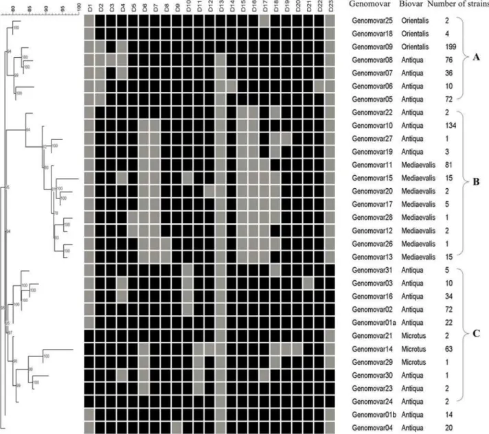
The 909 strains were grouped into 32 genomovars. The DFR profiles and Neighbor-Joining dendrogram of the 32 genomovars were shown in Figure 1. Most of the genomovars were clustered into 3 clusters, namely A, B and C, except for genomovar01b and genomovar04. All Orientalis strains (205 strains in 3 genomovars) were grouped together in cluster A, all Medievalis strains (122 strains in 8 genomovars) in cluster B and Microtus strains (66 strains in 3 genomovars) in cluster C. This clearly illustrated the close relationship of strains of the same biovar in China. However, Antiqua strains (516 strains in 18 genomovars) were distributed in different branches of all 3 clusters, revealing considerable genome diversities of Antiqua strains. This is not the first time to find this fact. SNPs(Single nucleotide polymorphisms) analysis has identi-fied 2 different molecular groups of Antiqua strains on 2 evolutionary lineages ofY. pestis(1.ANT and 2.ANT), which fitted into Orientalis and Medievalis branches, respectively [3]. CRIPSR analysis also identified 2 clusters of Antiqua strains (Asian and African)[22]
Biovar system is based on 2 phenotypes (nitrate reduction and glycerol utilization). Several studies have reported that, nitrate reduction negative stains might have different genetic mechanism for this phenotype[3,6]. As biovar strains are not genetically homogeneous, it seems that biovar typing system is no longer suitable for evolutionary or taxonomic purpose. Some genetics-based systems, such as SNP- and DFR-genetics-based ones, are alternatives as reliable typing methods forY. pestis[3,4,20].
Major genomovar and Minor genomovar
In China, there are 15 plague foci covering more than 1.4 million square kilometers now. A large number ofY. pestisstrains with clear background were isolated from different plague foci since the year of 1943. However, no strain was isolated since 1956 from animals during daily surveillance inMarmota sibiricaPlague Focus of the Hulun Buir Plateau in Inner Mongolia (Focus N), where used to have plague epidemics in early 19thcentury. There
was also no report of animal plague epidemics since then. We call Focus N a silent focus. The 909 strains in this study were carefully selected, considering their phenotypes, years and locations of isolation, vectors and hosts, etc. We assumed that they could represent the most abundant diversities of ChineseY. pestisstrains. Table 1 showed the distribution of genomovars in different plague foci. In our previous study, 260 strains were genotyped into 14 genomovars. Although we added nearly 650 strains and one new DFR marker in this study, we only got 18 new genomovars. More interestingly, most of our new strains fell into the previously identified genomovars (826/909, 90.9%), and most of the new genomovars contained only a few strains. Fourteen genomovars comprised more than 10 strains, which cover 93.8% (853/909) of all the tested strains. The DFR typing system is still open to new markers and new strains. It seems that, by adding more markers, we can get higher resolution without disturbing the framework of DFR typing.
From Table 1, we can also see the region-specific distribution of genomovars in different foci. For instance, genomovar13 strains were only found in Focus J, and genomovar15 ones exclusively in Focus O, while genomovar09 ones mainly in Focus F. On the other hand, strains in a specific focus always fell into a few genomovars. For example, all the 15 strains from Focus O belonged to genomovar15. While 191 of 198 strains from Focus F were identified as genomovar09, and the other 7 strains fell into other 5 genomovars, with no more than 3 strains each. The numbers of strains, belonging to different genomovars that predominated in certain plague foci, were italicized in Table 1.
especially the phenotypic effects of DFR loss-and-gain during microevolution ofY. pestis.
Notably, Major and Minor genomovars make sense only by combining with the concept of natural plague foci. The Major genomovar in one plague focus might be the Minor one in the other. For example, genomovar09 was the Major genomovar in Focus F, but the Minor one in Focus E. The distribution of the Major genomovar(s) in each plague focus of China was presented in Figure 2. Normally, each focus has its own characteristic Major genomovar(s). However, there were still some strains from several foci indistinguishable by the DFR profiles. For instance, strains from Foci G and H shared genomovar10, Foci K2 and I genomovar11 and Foci L and M genomovar14. This suggested the close relationship between the strains in the corresponding foci. These strains might be recently spread from one focus to another and there was no enough time for DFR varieties to accumulate. We might need other methods with higher resolution to differentiate strains
from these foci. Actually, based on CRISPR(clustered regularly interspaced short palindromic repeat) and MLVA(multiple-locus VNTR analysis) analysis we are able to differentiate strains from Foci L and M, as well as K2 and I.
Anyway, with this updated DFR typing system, we can roughly differentiate strains from most plague foci. In another word, we can tell the possible origin of certainY. pestisstrain by investigating their DFR profiles using a set of PCRs. We can estimate from the above data that this DFR-based genotyping system should be scientific sound because it correlates very well with the focus distribution of the pathogen and the conventional ecotyping system that is widely used by Chinese plague scientists for typingY. pestis[23]. We also performed genotyping analysis for one-third of the strains used in this study by MLVA, CRISPR, SNPs and IS-based method, validating the DFR-based method for genotyping plague bacteria (unpublished data). This inexpensive method can be developed as a source tracing protocol when unexpected plague outbreaks or bioterrorism attacks happen.
Figure 1. Neighbor-Joining Dendrogram of the 32 genomovars based on DFR profiles.The black and grey squares indicated the presence and absence of the corresponding DFRs, respectively. There are three clusters (A, B and C) for most of the genomovars.
doi:10.1371/journal.pone.0002166.g001
DFR Analysis of Y. pestis
Table 1.Distribution of genomovars in 909 strains ofY. pestisisolated from China
Focus or Subfocus
Number of
strains Genomovar*
01a 01b 02 03 04 05 06 07 08 09 10 11 12 13 14 15 16 17 18 19 20 21 22 23 24 25 26 27 28 29 30 31
A 13 0 0 0 0 11 0 0 0 0 0 0 0 0 0 0 0 1 0 0 0 0 0 0 0 0 1 0 0 0 0 0 0
B1 14 0 1 03 0 09 0 0 0 0 0 0 0 0 0 0 0 0 0 0 0 0 0 0 0 0 0 0 0 0 0 0 1
B2 46 2 0 43 0 0 0 0 0 0 0 0 0 0 0 0 0 1 0 0 0 0 0 0 0 0 0 0 0 0 0 0 0
B3 71 3 0 24 10 0 0 0 0 0 0 0 0 0 0 0 0 32 0 0 0 0 0 0 0 0 0 0 0 0 0 0 2
B4 17 15 0 2 0 0 0 0 0 0 0 0 0 0 0 0 0 0 0 0 0 0 0 0 0 0 0 0 0 0 0 0 0
C 152 0 9 0 0 0 63 10 10 48 0 8 0 0 0 0 0 0 0 0 1 0 0 0 1 2 0 0 0 0 0 0 0
D 33 0 2 0 0 0 3 0 0 27 0 0 0 0 0 0 0 0 0 0 0 0 0 0 1 0 0 0 0 0 0 0 0
E 31 0 0 0 0 0 0 0 23 0 8 0 0 0 0 0 0 0 0 0 0 0 0 0 0 0 0 0 0 0 0 0 0
F 198 0 0 0 0 0 0 0 1 1 191 0 0 0 0 0 0 0 0 3 0 0 0 0 0 0 1 1 0 0 0 0 0
G 44 0 1 0 0 0 0 0 0 0 0 39 0 0 0 0 0 0 1 0 1 0 0 2 0 0 0 0 0 0 0 0 0
H 91 0 0 0 0 0 0 0 1 0 0 87 0 0 0 0 0 0 1 1 0 0 0 0 0 0 0 0 1 0 0 0 0
I 74 0 0 0 0 0 0 0 0 0 0 0 69 2 0 0 0 0 0 0 1 2 0 0 0 0 0 0 0 0 0 0 0
J 17 0 0 0 0 0 0 0 1 0 0 0 0 0 15 0 0 0 1 0 0 0 0 0 0 0 0 0 0 0 0 0 0
K1 14 1 0 0 0 0 0 0 0 0 0 0 12 0 0 0 0 0 0 0 0 0 0 0 0 0 0 0 0 1 0 0 0
K2 11 1 1 0 0 0 6 0 0 0 0 0 0 0 0 0 0 0 1 0 0 0 0 0 0 0 0 0 0 0 0 0 2
L 22 0 0 0 0 0 0 0 0 0 0 0 0 0 0 20 0 0 1 0 0 0 0 0 0 0 0 0 0 0 1 0 0
M 46 0 0 0 0 0 0 0 0 0 0 0 0 0 0 43 0 0 0 0 0 0 2 0 0 0 0 0 0 0 0 1 0
O 15 0 0 0 0 0 0 0 0 0 0 0 0 0 0 0 15 0 0 0 0 0 0 0 0 0 0 0 0 0 0 0 0
Sum 909 22 14 72 10 20 72 10 36 76 199 134 81 2 15 63 15 34 5 4 3 2 2 2 2 2 2 1 1 1 1 1 5
*The corresponding genomovars of italic numbers were focus-specific Major genomovars
doi:10.1371/journal.pone.0002166.t001
DFR
Analysis
of
Y.
pestis
PLoS
ONE
|
www.plos
one.org
4
May
2008
|
Volume
3
|
Issue
5
|
In silico DFR profiling of the sequencedY. pestisandY. pseudotuberculosisgenomes
There are now 16 completed or draft genomes ofY. pestisand two completed genomes of Y. pseudotuberculosis, providing us valuable data to probe into the relationship between Y. pestis strains from China and other regions in the world. We investigated the presence or absence of the 23 DFRs in the 18 genomes by Blast searching, and the results were shown in Table 2.
Ten of the 16Y. pestisgenomes, 91001, Nepal516, B42003004, E1979001, Antiqua, KIM, K1973002, F1991016, CA88-4125 and CO92, fell into the 32 genomovars identified in this study. In these
strains, Nepal516, Antiqua[24], KIM[25], CO92[26] and CA88-4125[27] were isolated outside China. The other 6 strains failed to be classified into any of the 32 genomovars, and hence they should be grouped as new genomovars which need to be verified by using more strains. IP275[28], FV-1[29] and MG05-1020 were quite similar to genomovar09 with only one or two DFR differences. UG05-0454 was different from genomovar04 only by the absence of DFR02. Strains Angola and Pestoides F[30] were very different from Chinese strains by DFR profiling. Although the sequenced genomes were obviously not enough to cover the varieties of DFR profiles of Y. pestis strains worldwide, we tried to get some interesting results from these limited data.
Figure 2. The distribution Major genomovars in natural plague foci of China.There are 15 plague foci in China. Focus A:Marmota caudate Plague Focus of the Pamirs Plateau; Focus B:Marmota baibacina-Spermophilus undulatesPlague Focus of the Tianshan Mountains; Focus C:Marmota himalayanaPlague Focus of the Qinghai-Gansu-Tibet Grassland; Focus D:Marmota himalayanaPlague Focus of the Qilian Mountain; Focus E: Apodemus chevrieri-Eothenomys miletusPlague Focus of the highland of Northwestern Yunnan Province; Focus F:Rattus flavipectusPlague Focus of the Yunnan-Guangdong-Fujian provinces; Focus G:Marmota himalayanaPlague Focus of the Gangdisi Mountains; Focus H:Spermophilus dauricusPlague Focus of the Song-Liao Plain; Focus I:Meriones unguiculatusPlague Focus of the Inner Mogolian Plateau; Focus J:Spermophilus dauricus alaschanicus Plague Focus of the Loess Plateau in Gansu and Ningxia provinces; Focus K:Marmota himalayanaPlague Focus of the Kunlun Mountains; Focus L: Microtus brandtiPlague Focus of the Xilin Gol Grassland; Focus M:Microtus fuscusPlague Focus of the Qinghai-Tibet Plateau; Focus N:Marmota sibirica Plague Focus of the Hulun Buir Plateau of Inner Mongonia. Focus O:Rhombomys opimusPlague Focus of the Junggar Basin of Xinjiang. B1, B2, B3 and B4 are subfoci of Focus B, K1 and K2 are subfoci of Focus K. Focus N is a silent plague focus without strains available for this study.
doi:10.1371/journal.pone.0002166.g002
DFR Analysis of Y. pestis
Table 2.The DFR profiles of the sequenced and sequencing Y. pestis and Y.pseudotuberculosis
Strain name Isolate location Accession number Biovar
Genomovar or similar
Genomovar DFR profiles
01 02 03 04 05 06 07 08 09 10 11 12 13 14 15 16 17 18 19 20 21 22 23
Nepal516 Nepal CP000305 Antiqua 19 - + + + + - - + + + + + - + - - + - + + + + -B42003004 China AAYU00000000 Antiqua 01a - + + + + + + + + + + + - + + + + + + + + + +
E1979001 China AAYV00000000 Antiqua 07 - - + - + + + + + + + + - + + + + + + + + +
-UG05-0454 Uganda AAYR00000000 Antiqua 04* - - + + + + + + - + + + - + + + + + + + + + -Antiqua Congo CP000308 Antiqua 05 - - + + + + + + + + + + - + + + + + + + + + -Angola Angola AF167310 Antiqua 03* - - - - + + + + - - + - - - + + + - + + + + +
KIM Kurdistan AE009952 Medievalis 15 - + + - + - - + + - + + - + - - - - + + + + -K1973002 China AAYT00000000 Medievalis 17 - + + + + - - + + + + + - + - - - - + + + + -91001 China AE017042 Microtus 14 + + + + + - + + + + - - - + + + + - - - + +
-F1991016 China ABAT00000000 Orientalis 09 - - + - + + + + + + + + + + + + + + + + + + -MG05-1020 Madagascan AAYS00000000 Orientalis 09* - - + - + + + + + - + + + + + + + + + + + + -FV-1 Arizona.USA AAUB00000000 Orientalis 09* - - + - + + + + + + + + + + + - + + + + + +
-CA88-4125 California.USA ABCD00000000 Orientalis 09 - - + - + + + + + + + + + + + + + + + + + + -CO92 Colorado.USA AL590842 Orientalis 09 - - + - + + + + + + + + + + + + + + + + + + -IP275 Madagascan AAOS00000000 Orientalis 09* - - - - + - - - + - + + - + + + + + + + - +
-Pestoides F Former Soviet Union AF167309 Pestoides 30* + + + - + - - + - + + + - + + + + - + + + + +
IP 32953 France BX936398 Y. pseudotuberculosis 14* - - - + + - + + + + + - - + + + + - + + + + -IP 31758 Former Soviet Union CP000720 Y. pseudotuberculosis 14* - - - + + - + + + + + - - + + + - - - + + +
-*: The most similar genomovars to the corresponding strains
+: The presence of the DFR
-: The absence of the DFR
doi:10.1371/journal.pone.0002166.t002
DFR
Analysis
of
Y.
pestis
PLoS
ONE
|
www.plos
one.org
6
May
2008
|
Volume
3
|
Issue
5
|
Distribution of DFR23 inY. pestis
In our previous study, we identified a 383bp region (DFR4) specific for theY. pestisstrains from plague Focus B in China by suppression subtractive hybridization (SSH)[21]. We also con-firmed the presence of this fragment inY. pseudotuberculosisstrains 53518 (serotype I), 53519 (serotype II) and 29833 (serotype I)[21]. We renamed it as DFR23 in this study in order to maintain the consistency of our DFR nomenclature system.
DFR23 was thought to be specific toY. pestisfrom Focus B in our previous study by using limited number of strains[21]. In this study, DFR23 were found in all the isolates of Subfoci B2 (46 Antiqua strains), B3 (71 Antiqua strains) and B4 (17Antiqua strains). Whereas 11 of 14 Antiqua strains of Subfoci B1 did not possess this DFR. Meanwhile, 10 strains from Foci A, C, D, K and M harbored this region. Obviously, DFR23 was not only present in the strains from Focus B. The 4 strains from Foci C and D harboring DFR23 were thought to be the most ancient strains of China according to our MLVA results (unpublished data). As DFR23 was also present in some strains ofY. pseudotuberculosis, we presumed that the ancientY. pestisstrains should possess this region, and it was lost during the microevolution ofY. pestis. Interestingly, DFR23 also presented in the sequencedY. pestisstrain Angola and Pestoides F. Pestoides F was thought to be a very ancient strain, because it had a Y. pseudotuberculosis genomic region that was absent in all known Y. pestis strains. It seemed that DFR23 loses in the early phase of microevolutionary history ofY. pestis, and it might serve as a marker for identification of ancientY. pestisstrains.
The emergence and microevolution of the Orientalis strains
In this study, 205 Chinese Orientalis strains were grouped into 3 genotypes: genomovar 09, 18 and 25(Figure 1). 199 strains (98%) fell into genomovar09, whereas only 4 and 2 strains the genomovar18 and 25, respectively. These two Minor genomovars (genomovar18 and 25) were therefore not considered in inferring the relationships among Orientalis strains.
The third plague pandemic, caused presumably by the strains of biovar Orientalis, was believed to have originated from Yunnan Province, China, in 1855[31]. It then spread around the world with the aid of modern transportation[32]. Our studies strongly supported this notion. The Orientalis strains in China were mainly isolated from Focus F and grouped into genomovar09. Most Antiqua strains in Focus E, a neighbor to Focus F, fell into genomovar07, and only a few Orientalis strains genomovar09. It suggested that the Orientalis strains of genomovar09 be evolved from genomovar07, the Antiqua strains, in Focus E after acquiring DFR13 and other unknown genetic variations, and then expanded to Focus F even all over the world[20]. The sequenced Orientalis strain CO92[26] and CA88-4125[27], as well as F1991016 isolated from Focus F in China have identical DFR profiles with genomovar09. We hypothesized that Orientalis strains MG05-1020, IP275 and FV-1 came into being by losing certain DFRs from genomovar09.
By Comparing DFR profiles of the 6 sequenced Orientalis strains and the tested Orientalis strains in China, we deduced the microevolutionary pattern of Orientalis strains based reductionism (Figure 3). We also proposed a virtual genomovarX as the missing link of genomovar09 to MG05-1020 and IP275. Interestingly, MG05-1020 and IP275 were both isolated in Madagascar. It is hopeful that we can find this genomovar in Madagascar or elsewhere, which will be of great help for better understanding the spreading of the third plague pandemics.
The Orientalis is believed to be ‘‘young’’ biovar ofY. pestis, and the time for its spreading all over the world is no longer than
120 years. We can see that Orientalis strains in China were very homologous in DFR pattern (only 3 genomovars in 205 strains with 199 strains as genomovar09) and strains outside China showed considerable heterogeneity (4 genomovars in 5 strains). One possibility was that, when they expanded all around the world, the genome underwent mutations including parallel loss of DFRs for adapting themselves to various niches. The adaptive microevolution might lead to the discrete segregation between the progenitor and offspring strains. This genome reduction gradually caused the offspring strains to inhabit a more specific host niche, without overlapping with their progenitors.
Y. pestisMicrotus strains seems to be closely related toY. pseudotuberculosis
In our previous study, we assumed an ancestorY. pestisstrain as DFR12 positive[20]. However, we found in this study that, DFR12 was shared by almost allY. pestisstrains but genomovar14 (Microtus) and genomovar20 (Medievalis), as well as strain Angola. DFR12 was also absent from genomes ofY. pseudotuberculosis. Genomovar20 (only two strains) was a Minor genomovar which might contribute little to the microevolution of theY. pestis,and DFR12 in these two genomes might lose under certain unexpected conditions. Therefore, we neglected genomovar20 in discussing the DFR12 issue. If the ancestorY. pestisstrain was DFR12 positive, genomovar14 and strain Angola must abandon DFR12 from their genomes again, which can not be well explained by maximum parsimony principle in the evolution. So it might be more convincing to set a virtual ancestorY. pestisstrain as DFR12 negative.
Most Microtus strains in China were isolated from Foci L and M. In this study, 95.5% of Microtus strains as well as 91001 fell into genomovar14, which was Major genomovar of Foci L and M. From Table 2, we can see that, genomovar14 was quite similar to Y. pseudotuberculosisIP32953 and IP31758 (by only differing in 3 DFRs
Figure 3. The microevolution scenario of Orientalis strains based on the gain-and-loss of DFRs.Orientalis strains evolved from genomovar07 Antiqua strains of Focus E, by acquiring DFR13, and then evolved as different genomovars by losing certain DFRs. A virtual genomovarX was proposed to illustrate the step-by-step reductionism evolution.
doi:10.1371/journal.pone.0002166.g003
DFR Analysis of Y. pestis
except for the DFRs located in plasmid pMT1). We deduced that, from the point view of DFR profiling, strains belong to genomovar14 (including 91001) might be those most closely related to Y. pseudotuberculosis, which has been proved by SNPs analysis[3].
Diversity ofY. pestisstrains in Xinjiang province
Xinjiang is the biggest province of China. Its complex terrains and landforms as well as a wide variety of ecological systems have created a natural paradise for biodiversity. The Y. pestis in this region presented highly diversity in their genomes according to the DFR profiles. Of the 32 genomovars, 13 were identified in this province and 7 of them were Major genomovars (Table 3). The significant diversity implied a long evolutionary history ofY. pestisin this region, or theY. pestisin China might be originated from this region.
There are 4 foci found in Xinjiang to date, including Foci A, B (Subfoci B1 to B4), O and K (Subfoci K1 and K2). The first 3 are geographically linked to the plague foci of the Central Asia (See Figure S1 ) [7]. Due to the lack of the strains outside China, it is still very difficult to provide a detailed and integrated relationships between the strains in Xinjiang and those of the Central Asia. The Figure S1 showed that Plague Foci in the Desert of Central Asia (labeled with ‘‘1’’ ) stretched eastward directly to China, and jointed with the newly identified Focus O,Rhombomys opimus(great gerbil) Plague Focus of the Junggar Basin of Xinjiang [33]. The Foci B (Subfoci B1–B4) and A in China adjoin Plague Foci of Western Section of Tianshan Mountains (labeled with ‘‘3’’) and Plague Foci of Pamirs-Alai (labeled with ‘‘2’’), respectively. The close geographical relationships implied that, Xinjiang province might act as an exchange access ofY. pestis between China and Central Asia. In 2005, 15 strains were isolated from theRhombomys opimus in Focus O[33]. All of them were biovar Medievalis and grouped into genomovar15, the same as the sequenced Medievalis strain KIM isolated from Kurdistan[25]. It is very unusual for strains apart so far away to share the same DFR profile. It is still difficult to tell the direction of foci expansion, and we need strains from former USSR to figure out the exact evolution scenario ofY. pestisin this region.
By comparing the DFR profiles of Xinjiang strains, the microevolution of the genomovars seemed to be consistent with the expansion of plague foci. Subfocus B4 situates in the west section of the Northern Tianshan Mountain (NTM) and joins with the plague foci of Kazakstan, where the Major genomovar is genomovar01a (similar to the hypothetical ancestor of Y. pestis). The strains isolated from Subfocus B3, the mid-section of the
NTM, fell into genomovar 02 and 16, and they two were equally defined as the Major genomovars. The east section of the NTM was designated as Subfocus B2 where the Major genomovar is genomovar02. There is no geographical obstacle between these 3 subfoci, which might account for the spreading ofY. pestisfrom west to east. By reductive evolution hypothesis, we proposed that when the strains of genomovar01a transmited from B4 to B3, DFR10 was lost in their genomes to adapt the new niches and colonized as genomovar02, then expanded to B2 and stably existed as the Major genomovar there. Genomovar16 came into being after losing DFR04 and played an important role together with genomovar02 within Subfocus B3. Then different genomo-vars evolved continuously by losing or acquiring certain DFRs to adapt to new niches of the host.
The great genomic diversities of Xinjiang strains make them an ideal collection to study the microevolution ofY. pestis, some other markers such as SNP, VNTR and CRISPR might help us better understand the myth of its evolution.
Distribution of DFR 13 in 4Y. pestisbiovars
DFR13 potentially encodes a prophage (YpfW), which contains 13 putative open reading frames (ORFs) (YPO2271–2281)[34]. It was previously suggested that DFR13 was restricted to the Orientalis strains[19,20,35]. However, recent studies indicated that it was acquired by the Y. pestis ancestor, and its genome presented in the threeY. pestisbiovars[34]. Our results in this study strongly support this notion. Of the 377 strains amplified with 3 primer pairs targeting different regions of DFR13, all 52 Orientalis strains were positive for all 3 loci. Meanwhile, 6 out of 222 Antiqua strains, 1 out of 60 Medievalis and 1 out of 43 Microtus strains were also positive for at least 2 loci, although the amplicons were somehow faint (Table 4). The sequences of the positive products were highly homologous to the corresponding region of CO92 by sequencing analysis (data not shown). It was supposed that the phage may not be stably integrated in the genomes of non-Orientalis strains and the proportion of phage-positive cells within the bacterial population may be unequal[34]. The serial dilution was used for confirming the content of this phage DNA inY. pestis. If its content is lower than the chromosomal DNA it should become negative by PCR after a certain dilutions. After diluting the DNA template 8 times, we failed to identify any amplicon from these 8 non-Orientalis strains. This suggested that the signal variations detected among variousY. pestisstrains may be due to a difference in the proportion of phage-positive cells within the
Table 3.The distribution of genomovars ofY. pestisin the foci of Xinjiang
Focus or Subfocus
Number of
strains Genomovar*
01a 01b 02 03 04 05 11 15 16 17 25 28 31
A 13 0 0 0 0 11 0 0 0 1 0 1 0 0
B1 14 0 1 3 0 9 0 0 0 0 0 0 0 1
B2 46 2 0 43 0 0 0 0 0 1 0 0 0 0
B3 71 3 0 24 10 0 0 0 0 32 0 0 0 2
B4 17 15 0 2 0 0 0 0 0 0 0 0 0 0
K1 14 1 0 0 0 0 0 12 0 0 0 0 1 0
K2 11 1 1 0 0 0 6 0 0 0 1 0 0 2
O 15 0 0 0 0 0 0 0 15 0 0 0 0 0
*: The corresponding genomovars of the italic numbers are dominant in a certain focus, and determined as Major genomovars in certain focus.
doi:10.1371/journal.pone.0002166.t003
bacterial population. It also implied that, in contrast to Orientalis strains, the phage is unstable in the other three biovars and easy to lose under laboratory conditions. One Microtus strain M1997002 was identified to harbor the phage in this study, which strongly supported the suggestion that the YpfW had been acquired horizontally, as an unstable episome by theY. pestisancestor after its divergence from Y. pseudotuberculosis. The phage then became stable in the Orientalis strains, upon permanent integration of its genome into the bacterial chromosome[34].
Concluding remarks
In this study, 909 strains ofY. pestisfrom China were grouped into 32 genomovars. Orientalis, Medieavalis and Microtus strains showed biovar specific DFR profiles, and were clustered into three distinct groups. But genomovars of Antiqua strains distributed among these three groups. Genomovars distribution was somehow focus-specific in China, and we proposed Major and Minor genomovars for explaining their distribution and roles played in microevolution of Y. pestis. By in silico DFR profiling of the sequenced genomes, we were able to compare Chinese strains and those outside China as well. Orientalis strains in China turned out to be more ancient than those aboard according to the DFR profiles, supporting the notion that the Orientalis strains were originated from China. Xinjiang province could be an access ofY. pestisspreading between China and Central Asia. It is the first time that we systematically classified a large amount of strains in China based on the profiling gene acquisition/loss in their genomes. Data presented here will be of great help to develop a genomic polymorphism database of Y. pestisfor tracing the origin of this agent when the plague outbreak or bioterrorism attack occurs. Hopefully, DFR analysis can be modified for genotyping other bacteria that have similarly plastic genomes.
Materials and Methods Strains and DNA
912 strains isolated from 15 plague foci from the year of 1943 to 2005 were included in this study, which presumably represented the most abundant diversity ofY. pestis strains in China. All the strains were collected by Qinghai Institute for Endemic Diseases Prevention and Control, the Center for Disease Control and Prevention of Xinjiang Uygur Autonomous Region and the
Yunnan Institute for Endemic Disease Control and Prevention. The bacteria were cultivated in nutrient agar at 28uC for 48 hours, and then the genome DNAs were extracted by using conventional SDS lysis and phenol-chloroform extraction method.
Genotyping based on DFR profiling
As three (DFR 01-03) of the 23 DFRs are located on plasmid pMT1, all the strains were screened by pMT1-specific primers before genotyping. Primers AP-YPMT1.44F (59 AACACTATCT-CATTCCGCAGTAAAG39) and AP-YPMT1.44R (59 AGTG-GATGATGAAGTAGACCGAG39) were used to screen the presence or absence of this plasmid. The primers used for amplify the 23 DFRs were provided in the Table S1. The composition of PCR mix and the reaction conditions were describe else-where[20,21]. The DNA mixture of strains 91001 and EV76 were used as positive control. Negative control was also set in each plate to monitor the amplification.
The data were processed with Bionumerics 5.00 (Applied Math NV. Belgium). Dendrogram was constructed by the Neighbor-Joining method with Dice means.
In silicoDFR typing of the published genomes
Sequence blasting was performed on NCBI to compare the PCR target genes of the 23 DFRs with the genomes of the sequenced and sequencingY. pestisandY. pseudotuberculosis. The gene was thought to be present if the identities between the query and subject sequences were above 98%, with 95% coverage of the gene.
Distribution of DFR13 in 4Y. pestisbiovars
To evaluate the distribution of DFR13 in the 4 biovars strains, 377 strains (60 Medievalis, 43 Microtus, 222 Antiqua and 52 Orientalis) were tested with the primers for DFR13 amplification. Primers targeting YPO2273, YPO2274 and YPO2277 were used[34].
For the positive amplicons obtained from biovars other than Orientalis, they were sequenced and compared with the corresponding regions of CO92. Furthermore, a two-fold serial dilution (starting from 2 ng/ml) of DNA from the positive strains and strain EV76 were prepared and used as templates for amplification as mentioned above in order to evaluate the relative contents of the specific target.
Table 4.Non-Orientalis strains amplified with DFR13 identification primers
Strain Biovar Isolate location PCR(2ng/ml)* PCR(0.25ng/
ml)*
YPO2274 YPO2277 YPO2273 YPO2274 YPO2277 YPO2273
D0000002 Antiqua Qinghai qilian + + - - -
-K21985006 Antiqua Xinjiang ruoqiang + + + - -
-A1956001 Antiqua Xinjiang wuqia + + + - -
-B11979001 Antiqua Xinjiang atushi + + - - -
-B31989002 Antiqua Xinjiang wusu + + - - -
-M1997002 Microtus Sichuan shiqu + + - - -
-I1978002 Medievalis Inner Mongolia guoqianqi + + - - -
-H1955008 Antiqua Inner Mongolia keyouqianqi + + + - -
-EV76 Orientalis Its parent strain was isolated from a patient in Madagascar
+ + + + + +
*: Indicated the template concentration
The ‘‘+’’ and ‘‘-’’ indicated the positive and negative results, respectively. EV76 was the positive control.
doi:10.1371/journal.pone.0002166.t004
DFR Analysis of Y. pestis
Supporting Information
Table S1 Primers used for DFR analysis
Found at: doi:10.1371/journal.pone.0002166.s001 (0.09 MB DOC)
Figure S1 The geographic relationship between the foci in Xinjiang and Central Asia. 1. Plague Foci in Desert of Central Asia. 2. Plague Foci of Pamirs-Alai. 3. Plague Foci of Western Section of Tianshan Mountains. Foci A, B1–B4 and O see Figure 2 Found at: doi:10.1371/journal.pone.0002166.s002 (3.17 MB TIF)
Acknowledgments
Due to the limited space of this paper, we cannot include all the scientists from the Qinghai Institute for Endemic Diseases Prevention and Control,
the Center for Disease Control and Prevention of Xinjiang Uygur Autonomous Region, and the Yunnan Institute for Endemic Disease Control and Prevention for their help to identify theY. pestisisolates and extract DNAs from them. Ms Jin Wang was especially appreciated for her kind help throughout the experiments.
Author Contributions
Conceived and designed the experiments: RY YL ED YC DZ JZ YS. Performed the experiments: YL ED YC. Analyzed the data: YL ED YC YS. Contributed reagents/materials/analysis tools: ML YZ MW ZG XD BC ZW HW XD ZS ZQ. Wrote the paper: RY YL ED YC YS.
References
1. Perry RD, Fetherston JD (1997)Yersinia pestis–etiologic agent of plague. Clin Microbiol Rev 10: 35–66.
2. Broussard LA (2001) Biological agents: weapons of warfare and bioterrorism. Mol Diagn 6: 323–333.
3. Achtman M, Morelli G, Zhu P, Wirth T, Diehl I, et al. (2004) Microevolution and history of the plague bacillus,Yersinia pestis. Proc Natl Acad Sci U S A 101: 17837–17842.
4. Achtman M, Zurth K, Morelli G, Torrea G, Guiyoule A, et al. (1999)Yersinia pestis, the cause of plague, is a recently emerged clone ofYersinia pseudotuberculosis. Proc Natl Acad Sci U S A 96: 14043–14048.
5. Song Y, Tong Z, Wang J, Wang L, Guo Z, et al. (2004) Complete genome sequence ofYersinia pestisstrain 91001, an isolate avirulent to humans. DNA Res 11: 179–197.
6. Zhou D, Tong Z, Song Y, Han Y, Pei D, et al. (2004) Genetics of Metabolic Variations betweenYersinia pestisBiovars and the Proposal of a New Biovar, microtus. J Bacteriol 186: 5147–5152.
7. Anisimov AP, Lindler LE, Pier GB (2004) Intraspecific diversity ofYersinia pestis. Clin Microbiol Rev 17: 434–464.
8. Guiyoule A, Grimont F, Iteman I, Grimont PA, Lefevre M, et al. (1994) Plague pandemics investigated by ribotyping ofYersinia pestisstrains. J Clin Microbiol 32: 634–641.
9. Huang XZ, Chu MC, Engelthaler DM, Lindler LE (2002) Genotyping of a homogeneous group ofYersinia pestisstrains isolated in the United States. J Clin Microbiol 40: 1164–1173.
10. Adair DM, Worsham PL, Hill KK, Klevytska AM, Jackson PJ, et al. (2000) Diversity in a variable-number tandem repeat from Yersinia pestis. J Clin Microbiol 38: 1516–1519.
11. Klevytska AM, Price LB, Schupp JM, Worsham PL, Wong J, et al. (2001) Identification and characterization of variable-number tandem repeats in the
Yersinia pestisgenome. J Clin Microbiol 39: 3179–3185.
12. Pourcel C, Andre-Mazeaud F, Neubauer H, Ramisse F, Vergnaud G (2004) Tandem repeats analysis for the high resolution phylogenetic analysis ofYersinia pestis. BMC Microbiol 4: 22.
13. Guiyoule A, Grimont F, Iteman I, Grimont PA, Lefevre M, Carniel E (1994) Plague pandemics investigated by ribotyping ofYersinia pestisstrains. J Clin Microbiol 32: 634–641.
14. Yu DZ, Hai R, Dong XQ, Li M, Xia LX, Shi XM, Wei JC, Cui BZ, Wang P, Sun LZ, Zhang ZK, Hu Y, Zhang EM (2003) Genetic analysis ofYersinia pestis
strains isolated in China. Zhonghua Liu Xing Bing Xue Za Zhi 24: 1005–1009. 15. Huang F, Yu D, Hai R, Cai H (2000) Study on the application of random amplified polymorphic DNA inYersinia pestisgenotyping. Zhonghua Liu Xing Bing Xue Za Zhi 21: 424–426.
16. Guiyoule A, Rasoamanana B, Buchrieser C, Michel P, Chanteau S, et al. (1997) Recent emergence of new variants ofYersinia pestis in Madagascar. J Clin Microbiol 35: 2826–2833.
17. Hai R, Yu DZ, Wei JC, Xia LX, Shi XM, Zhang ZK, Zhang EM (2004) Molecular biological characteristics and genetic significance ofYersinia pestisin China. Zhonghua Liu Xing Bing Xue Za Zhi 25: 509–513.
18. Radnedge L, Agron PG, Worsham PL, Andersen GL (2002) Genome plasticity inYersinia pestis. Microbiology 148: 1687–1698.
19. Hinchliffe SJ, Isherwood KE, Stabler RA, Prentice MB, Rakin A, et al. (2003) Application of DNA microarrays to study the evolutionary genomics ofYersinia pestis andYersinia pseudotuberculosis. Genome Res 13: 2018–2029.
20. Zhou D, Han Y, Song Y, Tong Z, Wang J, et al. (2004) DNA microarray analysis of genome dynamics inYersinia pestis: insights into bacterial genome microevolution and niche adaptation. J Bacteriol 186: 5138–5146.
21. Dai E, Tong Z, Wang X, Li M, Cui B, et al. (2005) Identification of different regions among strains ofYersinia pestisby suppression subtractive hybridization. Res Microbiol 156: 785–789.
22. Pourcel C, Salvignol G, Vergnaud G (2005) CRISPR elements inYersinia pestis
acquire new repeats by preferential uptake of bacteriophage DNA, and provide additional tools for evolutionary studies. Microbiology 151: 653–663. 23. Zhang G, Zhang G, Wang S, SUn Q, Shan G (2002) Survey of natural plague
foci and epidemic situation in China. Chin J Ctrl Endem Dis 71: 101–103. 24. Chain PS, Hu P, Malfatti SA, Radnedge L, Larimer F, et al. (2006) Complete
genome sequence ofYersinia pestisstrains Antiqua and Nepal516: evidence of gene reduction in an emerging pathogen. J Bacteriol 188: 4453–4463. 25. Deng W, Burland V, Plunkett G 3rd, Boutin A, Mayhew GF, et al. (2002)
Genome sequence ofYersinia pestisKIM. J Bacteriol 184: 4601–4611. 26. Parkhill J, Wren BW, Thomson NR, Titball RW, Holden MT, et al. (2001)
Genome sequence ofYersinia pestis, the causative agent of plague. Nature 413: 523–527.
27. Auerbach RK, Tuanyok A, Probert WS, Kenefic L, Vogler AJ, et al. (2007)
Yersinia pestis evolution on a small timescale: comparison of whole genome sequences from North America. PLoS ONE 2: e770.
28. Welch TJ, Fricke WF, McDermott PF, White DG, Rosso ML, et al. (2007) Multiple antimicrobial resistance in plague: an emerging public health risk. PLoS ONE 2: e309.
29. Touchman JW, Wagner DM, Hao J, Mastrian SD, Shah MK, et al. (2007) A North AmericanYersinia pestisdraft genome sequence: SNPs and phylogenetic analysis. PLoS ONE 2: e220.
30. Garcia E, Worsham P, Bearden S, Malfatti S, Lang D, et al. (2007) Pestoides F, an atypicalYersinia pestisstrain from the former Soviet Union. Adv Exp Med Biol 603: 17–22.
31. Devignat R (1951) [Varieties of Pasteurella pestis; new hypothesis.]. Bull World Health Organ 4: 247–263.
32. Wren BW (2003) The yersiniae–a model genus to study the rapid evolution of bacterial pathogens. Nat Rev Microbiol 1: 55–64.
33. Jiang W (2005) First isolation ofYersinia pestisfromRhombomys opimus in the Junggar Basin of Xinjiang. Chinese Journal of Zoonoses 21: 1051–1051. 34. Derbise A, Chenal-Francisque V, Pouillot F, Fayolle C, Prevost MC, et al. (2007)
A horizontally acquired filamentous phage contributes to the pathogenicity of the plague bacillus. Mol Microbiol..
35. Chain PS, Carniel E, Larimer FW, Lamerdin J, Stoutland PO, et al. (2004) Insights into the evolution ofYersinia pestisthrough whole-genome comparison withYersinia pseudotuberculosis. Proc Natl Acad Sci U S A 101: 13826–13831.