Arteriovenous Fistula and Pseudoaneurysm: Two Important But
Rare Complications of Percutaneous Renal Biopsy and Safety of the
16 G Automated Biopsy Needle
Arteriovenöz Fistül ve Psodoanevrizma: Böbrek Biyopsinin Nadir
Fakat Önemli
İ
ki Komplikasyonu ve 16 G Otomatik Biyopsi
İğ
nesinin
Güvenilirli
ğ
i
Kenan TURGUTALP1
Tolga KÖŞECİ1
Demir APAYDIN2
Ahmet KIYKIM1
1 Mersin University, Faculty of Medicine, Department of Internal Medicine, Division of Nephrology, Mersin, Turkey
2 Mersin University, Faculty of Medicine, Department of Radiology,
Mersin, Turkey
Correspondence Address:
Kenan TURGUTALP
Mersin Üniversitesi, İç Hastalıkları
Anabilim Dalı, Nefroloji Bilim Dalı, Mersin, Turkey
Phone : + 90 532 492 68 83 E-mail : k.turgutalp@hotmail.com Received : 06.05.2013
Accepted : 12.05.2013
ABSTRACT
OBJECTIVE: The aim of this study was to evaluate the safety of 16 G automated biopsy needles, and
the incidence of AVF and pseudoaneurysm in patients undergoing renal biopsy.
MATERIAL and METHODS: Patients who underwent renal biopsy from January 2010 to December
2012 were prospectively evaluated. Color Doppler USG, hemoglobin and platelet counts, spot urine analysis were performed perormed before and after the biopsy procedure, AVF and pseudoaneurysm that developed secondary to percutaneous renal biopsy were evaluated by Doppler USG at 12 h, and 24 h, and 6 and 12 months after biopsy.
RESULTS: Postbiopsy small perirenal, subcapsular, or parenchymal bleeding without clinical relevance
were detected in 59 patients (89.2 %) 12 and 24 hours after procedure. RBC counts per high power fi eld increased signifi cantly after the procedure in all subjects (p<0.05). Signifi cant retroperitoneal bleeding, macroscopic hematuria, arteriovenous malformation, and pseudoaneurysm were not detected in our cohort. Glomeruli counts were acceptable in all subjects.
CONCLUSION: To optimize safety, patient comfort, and diagnostic yield, a 16 G automated biopsy
needle with real-time U/S guidance may be prefered as the standard approach.
KEY WORDS: Renal biopsy, Arteriovenous fi stula, Pseudoaneurysm
ÖZ
AMAÇ: Bu çalışmanın amacı, nefrotik sendrom nedeni ile böbrek biyopsisi yapılan hastalarda,
psödoanevrizma ve A-V fi stül sıklığını, 16 G otomatik biopsi iğnesinin güvenilirliğini değerlendirmektir.
GEREÇ ve YÖNTEMLER: Çalışmaya Ocak 2010 ile Aralık 2012 yılları arasında nefrotik sendrom
tanısı nedeni ile böbrek biyopsisi yapılan hastalar alındı. Biyopsi işleminden sonra psödoanevrizma ve A-V fi stül gelişip gelişmediğini değerlendirmek için işlem sonrası 12. saat, 24. saat, 6. ay ve 12. ayda hastalar renkli Doppler USG, hemogram, trombosit değerleri ve spot idrar tahlil sonuçları ile değerlendirildi.
BULGULAR: Biyopsi işlemi sonrasında 12. Saat ve 24. saatlerde 59 hastada (% 89.2) klinik önem
taşımayan perirenal, subkapsüler ve parenkimal kanama saptandı. 12. ve 24. saatlerde gözlenen mikroskobik hematüri oranı 6. ay ve 12. aylarda görülen mikroskobik hematüri oranına göre istatistiksel olarak anlamlı oranda yüksek saptandı (p<0.05). Ancak klinik olarak anlamlı kanama, hemogramda anlamlı düşme, makroskobik hematüri saptanmadı. Hastaların işlem sonrası takibi sırasında psödoanevrizma ve de A-V fi stül saptanmadı.
SONUÇ: Böbrek biyopsi işlemi sonrasında A-v fi stül görülme oranı nadir olmamakla birlikte genellikle
sessiz seyretmektedir. Klinisyenler yeterli doku elde etmek için ideal iğne kalınlığı ve görüntüleme yöntemi konusunda dikkatli olmalıdırlar. Güvenli işlem, hasta rahatlığı ve yeterli tanı materyali için USG eşliğinde 16-G otomatize böbrek biyopsi iğnesi kullanmalıdırlar.
INTRODUCTION
Renal biopsy is the most sensitive tool to evaluate causes of kidney diseases (1). Possible complications after renal biopsy are usually related to hemorrhage and include hematoma, pseudoaneurysm, hematuria, and formation of arteriovenous
fi stula (AVF) (2). Minor complications of renal biopsy such as microscopic hematuria and post-biopsy hematoma are common features and usually resolve within weeks after biopsy (3). However, an incidence of AVF that varies between 0.5 (4) and 16 % after renal biopsy (5) and complications developing up to 25 years later have been reported. The time period between the creation of the fi stula and the clinical presentation can be very variable (6), ranging from 7 to 365 days post-biopsy (6). Some of the complications such as AVF and pseudoaneurysm may be due to the needle size (7). Using ultrasound-guided automated biopsy guns has lowered the complication rate in a signifi cant way (8) so that the procedure is now carried out more frequently and renal biopsy can be considered a safe procedure (3). Color Doppler sonography (CDS) permits a safe, inexpensive, and noninvasive diagnosis and follow-up of postbiopsy AVF and pseudoaneurysm (7).
In the literature, only a few case reports have been presented with AVF in patients with nephrotic syndrome that underwent renal biopsy (9, 10, 11). The aim of this prospective study was to evaluate the safety of the 16 G automated biopsy needle, and to obtain the incidence of AVF and pseudoaneurysm in patients undergoing percutaneous renal biopsy.
PATIENTS and METHODS
The study was approved by our institutional review board. The investigation included only data of biopsy procedures performed for a clinical indication of the patients.
Patients who underwent renal biopsy from January 2010 to December 2012 were prospectively evaluated. After renal biopsy, the patients were placed under absolute bed-rest and monitored for at least 6 hours to reduce the risk of any bleeding. No patient died during the 12 months. Color Doppler USG, hemoglobin and platelet count, spot urine analysis were performed during the biopsy and after procedure. AVF and pseudoaneurysm that developed secondary to percutaneous renal biopsy were evaluated by Doppler USG at 12 h, and 24 h, and 6 and 12 months after biopsy.
The indications for renal biopsy were several renal diseases: clinically asymptomatic proteinuria with or without hematuria, to account for causes of glomerulonephritis or nephrotic syndrome.
Exclusion criteria included patients with bleeding diatheses such as those with an elevated International Normalized Ratio (INR), activated partial thromboplastin time (aPTT), signifi cant thrombocytopenia, or elevated bleeding time or symptomatic cardiovascular disease including coronary artery disease, cerebral vascular disease, peripheral artery disease, and aortic
aneurysm, patients with rapidly progressive glomerulonephritis, acute nephritic syndrome, renal failure as well as hypertensive patients. Patient undergoing renal surgery or had a neoplasm and patients taking anticoagulants or antiaggregant were also excluded.
One radiologist, who had 10 years of experience in US-guided biopsy, performed the procedures. Biopsies were performed under ultrasound guidance using a 16G automated needle (C.R. Bard, Covington, UK). A needle guidance device in metal was attached to the transducer and the needle path was indicated on the screen by two parallel lines to facilitate correct needle insertion. The path was put along the cortex as close to the surface of the kidney as possible and major vessels were avoided by CDS evaluation. The skin entrance site was marked and the area washed with 5% chlorhexidine. Under local anesthesia with lidocaine, and with sterile dressing, the needle was inserted with the bevel pointing away from the kidney to avoid needle deviation into the central part of the kidney. The biopsy was preferably performed from the peripheral cortex either at the upper or lower pole of the kidney. Three biopsy specimens were removed. Procedural success was assessed according to Banff 97 criteria (12). An adequate specimen was defi ned as one that has 10 or more glomeruli and two or more arteries and a minimal specimen was defi ned as one that has at least seven glomeruli and at least one artery.
All of the Doppler US assessments were performed with a color Doppler US device (Aplio, Toshiba Tokyo/Japan) with the help of 3-6 MHz broadband convex probe (PLT 3.75 Toshiba Tokyo, Japan) prior to, during, and immediately after the procedure by radiologist. A comparison with the other kidney was attempted in all patients. Additional US examinations were performed only if clinically indicated, e.g., in cases of pain, fever, and a drop in serum hemoglobin, indicating hemorrhagic complications.
All data and fi ndings were evaluated for prevalence of complications (bleeding and hematoma, AVF).
Demographical Data
Systolic and diastolic blood pressures (SBP, DBP) were obtained in all subjects. Arterial blood pressure measurements in “mmHg” were performed with the oscillometric system using the device in a seated position by taking the mean of two consecutive measurements performed within 5 minutes (OMRON M6 Comfort, Kyoto, Japan).
Laboratory Tests
groups involved, 1-way analysis of variance (ANOVA) was applied. Multiple comparisons were done with Tukey Honestly Signifi cant Difference (HSD) test. For the assessment of the effects of interactions of 2 different variables on the mean values, the factorial ANOVA test was used. Linear association between 2 variables was evaluated with the Pearson correlation test. Type I error level was determined to be 0.05 in all of the comparisons.
RESULTS
A total of 65 patients (34 males, 31 females) with a mean age of 40.25 ± 14.2 years were included in the study. All biopsies were successful and yielded suffi cient material for diagnostic histologic workup. The mean number of glomeruli per patient was found to be 8.9 ± 1.0. The fi nal diagnoses were various types of FSGS in 19 patients, minimal change disease in 12 patients, MPGN in 10 patients, membranous glomerulonephritis in 9 patients, and others in 15 patients.
All patients’ blood pressure was normal (mean systolic pressure was 117.6 ± 10.7 mmHg, mean diastolic pressure was 77.9 ± 5.1 mmHg) and none of patients used antihypertensive medication.
Postbiopsy small hematomas, mainly small perirenal, subcapsular, or parenchymal bleeding without clinical relevance were detected in 59 patients (89.2 %) in the fi rst 12 and 24 hours after the biopsy procedure. Red blood cell counts per high power fi eld increased signifi cantly after the procedure in all subjects (p<0.05). Signifi cant bleeding with clinical fi ndings, macrohematuria and signifi cantly decreasing hemoglobin and platelet levels were not observed. Some biochemical laboratory
fi ndings during and following renal biopsy were shown in Table I. Neither AVF nor pseudoaneurysm was detected at the 12 and 24th hours and in the 6 and 12th months of after the procedure. minutes after the blood samples were drawn. Serum creatinine,
fasting plasma glucose (FPG), albumin, total cholesterol (TC), high-density lipoprotein-cholesterol (HDL-C), and triglyceride (TG), were evaluated (Olympus AU 640, Japan). Low-density lipoprotein cholesterol (LDL-C) level was assessed with the Friedewald formula which can be formulated as LDL =TC - (HDL-C) – (TG/5) (13). Estimated glomerular fi ltration rate (eGFR) was evaluated with the “Modifi cation of Diet in Renal Disease” MDRD formula which can be shown as MDRD: GFR=170 x [Scr]– 0.999 x [age]– 0.176 x (0.762 if the subject is women) x (1.180 if the patient is black) x [BUN]– 0.170] (14). APTT (reference interval, 23–36 seconds) was obtained by a using the HemoTech device (Medtronic, Minneapolis, Minnesota), INR (reference interval, 0.9–1.2) was measured by a BCS-XP device (Siemens-Germany), and the bleeding time (reference interval, 2-9 minutes) was also measured.
Spot urine analysis performed by using the dipstick test. Microscopic hematuria was considered in the case of two of three urine samples with 3 or more red blood cells per high powered fi eld (RBCs/hpf). Visible hematuria was defi ned as a macroscopic or gross hematuria.
Proteinuria was evaluated by 24 h urinary protein excretion. Samples were obtained at two different times and assessed with the Olympus AU 640 device by immunoturbidimetric methods (SOR). The average of proteinuria values at two different times was calculated.
STATISTICAL ANALYSIS
MedCalc were used for statistical analysis. To test the normal distribution for permanent data, the Shapiro-Wilk test was applied. According to the results gathered from this test, parametric methods were used if necessary. The mean comparisons of binary groups were done with Student’s t test. For the mean comparisons of the groups where more than 2
Table I: During and following renal biopsy,comparison of laboratory data’s and detect of both AVF and pseudoaneurysm in CDU
Prebiopsy 12 hours AB 24hours AB 6months AB 12 months AB p value
Hb, g/dL 14.25 ± 1.3 14.14 ± 1.2 14.21 ± 1.2 14.33 ± 1.3 14.28 ± 1.3 NS
PLT, x103/mL 275.85 ± 80.7 270.25 ± 79.6 302.54 ± 83.3 298.74 ± 73.2 301.23 ± 68.9 NS
RBCs in spot urine,
hpf 0.82 ± 0.5 122.13 ± 8.3 113.24±5.4 0.7±0.3 0.6±0.3 <0.05
AVF ND ND ND ND ND NS
PSD ND ND ND ND ND NS
Creatinine, mg/dL 0.79 ± 0.2 0.78 ± 0.2 0.80 ± 0.3 0.76 ± 0.2 0.77 ± 0.2 NS
SBP, mm Hg 117.62 ± 10.7 116.53 ± 10.4 118.32 ± 11.1 120.63±11.3 119.49 ± 11.2 NS
DBP, mm Hg 77.91 ± 5.1 76.56 ± 4.9 78.85 ± 5.3 79.12 ± 5.3 79.10 ± 5.6 NS
automated). Although the larger manual needles provided more glomeruli per fi eld (16), they were related with an increased rate of complications (17). The only comparative study of the same gauge (14 G) removed more glomeruli with the automated needle without an alteration in complications (18). Recently, a prospective study using 14-G and 16-G automated needles showed no difference in the incidence of complications or in the number of glomeruli (19). In another prospective study (20) 16-G and 18-G automated needles were investigated. There was no difference in major complications, but patients had more pain in the 16-G group. The biopsy specimen was signifi cantly better in the 16-G group, with more glomeruli and less artifact. Additionally, a prospective randomized controlled trial using automated needles of 3 different sizes (14, 16, and 18 G) in kidney allograft biopsies revealed no difference in complication rates between the 3 sizes, but a higher diagnostic adequacy and more pain in the group with the largest needle (21). The mean number of glomeruli per subject in our study was found as 8.09 ± 1.1 (range=0–86) and was signifi cantly higher by use of automated 16 G needle. In our subjects, glomeruli count was comparable as reported in literature.
Although the ultrasonography-guided procedure is simple to perform, it carries the risk of vascular complications such as AVF and pseudoaneurysm (22). While the reported incidence of AVF has been between 5 and 10 % so far (23), the incidence of symptomatic post-biopsy AVF is between 0.3 and 4 % (24). Seventy percent of AVF are asymptomatic, and usually resolve spontaneously within weeks or months (25).
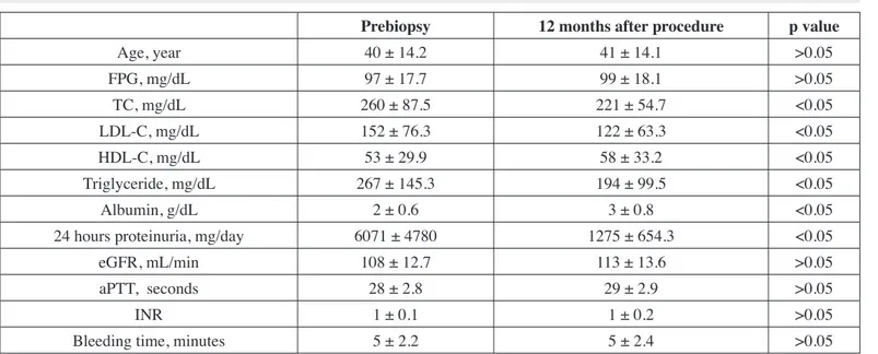
The statistical signifi cance of demographic and laboratory data’s prebiopsy and 12th month of after the procedure were shown in Table II. Gender, age, systolic blood pressure, diastolic blood pressure, creatinine levels, fasting plasma glucose levels, bleeding time levels, INR levels, aPTT levels, and eGFR prebiopsy were not found to be signifi cantly different as compared to 12th month of after the renal biopsy (p>0.05). The total cholesterol, low-density lipoprotein cholesterol, high-density lipoprotein cholesterol, 24 hours proteinuria, and albumin values prebiopsy were signifi cantly different from the values 12 months after biopsy procedure (p<0.05).
DISCUSSION
This study is a rare prospective observational study to investigate the reliability of 16 G sizes of automated biopsy needle, and risk of AVF and pseudoaneurysm in patients undergoing percutaneous renal biopsy. In upper and lower pole of kidney where removed biopsy specimen is safety procedure in patient with normotensive and with normal kidney function. If biopsy specimen was removed in these regions with 16 G needle, AVF and pseudoaneurysm were not occurring in short and long time duration. The most common iatrogenic biopsy-related vascular injuries in native kidneys as well as in renal allografts are AVF and pseudoaneurysm. Size of needle and location are important factors that determine presentation. It has been suggested that the complication rate is lower with the use of thin biopsy needles (15). However, the previous studies that compared needle sizes (14 and 18 G) had confl icting results due to the use of different types of needles (manual and
Table II: Demographic and laboratory data during and after renal biopsy.
Prebiopsy 12 months after procedure p value
Age, year 40 ± 14.2 41 ± 14.1 >0.05
FPG, mg/dL 97 ± 17.7 99 ± 18.1 >0.05
TC, mg/dL 260 ± 87.5 221 ± 54.7 <0.05
LDL-C, mg/dL 152 ± 76.3 122 ± 63.3 <0.05
HDL-C, mg/dL 53 ± 29.9 58 ± 33.2 <0.05
Triglyceride, mg/dL 267 ± 145.3 194 ± 99.5 <0.05
Albumin, g/dL 2 ± 0.6 3 ± 0.8 <0.05
24 hours proteinuria, mg/day 6071 ± 4780 1275 ± 654.3 <0.05
eGFR, mL/min 108 ± 12.7 113 ± 13.6 >0.05
aPTT, seconds 28 ± 2.8 29 ± 2.9 >0.05
INR 1 ± 0.1 1 ± 0.2 >0.05
Bleeding time, minutes 5 ± 2.2 5 ± 2.4 >0.05
Abbreviations: SBP, systolic blood pressure; DBP diastolic blood pressure; FPG, fasting plasma glucose; TC, total cholesterol;
arteriography is essential (4). There was none of these fi ndings or signifi cant bleeding post-biopsy in our patients so we did not consider an angiography procedure.
It is suggested that the particular route of the biopsy track, precise position of the real-time ultrasonography during biopsy (43), renal medullary disease, nephrocalcinosis, hypertension, renal insuffi ciency, and number of attempts and depth of the biopsy are more relevant to the incidence of complications than the size of the needle used (44). Any core-cutting biopsy needle introduced through a large renal artery or vein is likely to cause a serious complication irrespective of the needle size. There is a well-documented range of AVF and pseudoaneurysm after percutaneous core biopsies in both native and transplanted kidneys ranging from 0.9 to 15 % (45). Selim et al. (46) reported that parenchymal pseudoaneurysm of a renal allograft may appear after core needle biopsy. They suspected that the size of the core biopsy needle which was 14 G was a likely contributor as well as the biopsy region which was the corticomedullary junction. In contrast, our biopsy specimens were usually removed from the lower and upper pole with an ultrasonographic automated 16 G biopsy needle. While ultrasound guidance is helpful in avoiding major vascular injuries of this sort, it is more diffi cult to avoid penetrating the renal medulla even with the help of imaging. Bleeding would be expected to be more likely when the biopsy needle penetrates the renal medulla, as this is the site of the larger renal blood vessels.
CONCLUSION
AVF occurring after renal biopsies are not rare, but are mostly silent. Clinicians should be aware of the ideal needle size and method of imaging to obtain tissue. To optimize safety, patient comfort, and diagnostic yield, they can use to a 16-G automated needle with real-time U/S guidance as a standard approach. Future studies evaluating AVF as a complication of renal biopsy should focus methods and needle size to modify this risk for patients who require this procedure.
REFERENCES
1. Vogler C, Wang Y, Brink DS, Wood E, Belsha C, Walker PD: Renal pathology in the pediatric transplant patient. Adv Anat Pathol 2007; 14: 202-216
2. Furness PN, Philpott CM, Chorbadjian MT, Nicholson ML, Bosmans JL, Corthouts BL, Bogers JJ, Schwarz A, Gwinner W, Haller H, Mengel M, Seron D, Moreso F, Cañas C: Protocol biopsy of the stable renal transplant: A multicenter study of methods and complication rates. Transplantation 2003;76: 969-973
3. Feneberg R, Schaefer F, Zieger B, Waldherr R, Mehls O, Schärer K: Percutaneous renal biopsy in children: A 27-year experience. Nephron 1998; 79: 438-446
4. Matsell DG, Jones DP, Boulden TF, Burton EM, Baum SL, Tonkin IL: Arteriovenos fi stula after biopsy of renal transplant kidney: Diagnosis and treatment. Pediatr Nephrol 1992; 6: 562-564 In fi ve series totaling 1,042 native kidney biopsies, 31 AVF
(3 %) were detected after the renal biopsy (26-30). It should be noted that their frequency was quite low in the more recent series; the higher incidence in the old series may have been due to cruder methods of localizing the biopsy site. As for renal allografts, 22 AVF complicated 309 biopsies (7 %) in two series (31, 32). AVF and pseudoaneurysm complications were not seen in the patients described here but have been recorded elsewhere. The incidence of these AVF and pseudoaneurysm has been reduced by the introduction of ultrasound guidance, and blind renal biopsy should always be avoided. However, Riccabona et al. reported that despite ultrasonography-guided renal biopsy, postbiopsy renal AVF and pseudoaneurysm are more common than expected (7). Unfortunately, they performed the biopsy in the prone position and with 14 G core biopsy Bio-Cut needle.
AVF is important complication of renal biopsy because it may causes to fl ank pain, gross hematuria, hypertension, arterial bruits, congestive heart failure, or even renal failure (11). If macrohematuria occurs after percutaneous renal biopsy it should be considered to diagnose as AVF. Although rates of macrohematuria as low as 3% or less have also been reported (33-35) signifi cant macrohematuria leading to anemia rate was not seen our study suggesting that all needle sizes can be used with equal safety.
Biopsy-induced AVF and pseudoaneurysm are known to be correctly identifi ed by CDU imaging, which can distinguish intrarenal blood fl ow abnormalities (36) and by selective arteriography, which can evaluate size and localization (37). Although angiography is known as a gold standard for the diagnosis of postbiopsy AVF and pseudoaneurysm, CDU has been used effectively to detect AVF and pseudoaneurysm after biopsy (26, 31, 32, 37-39). In a series of 208 biopsies of renal allografts, Renowden et al. detected 16 AVF and found none false positive or false-negative outcomes with CDU as compared with angiography (32).
19. Manno C, Strippoli GF, Arnesano L, Bonifati C, Campobasso N, Gesualdo L, Schena FP: Predictors of bleeding complications in percutaneous ultrasound-guided renal biopsy. Kidney Int 2004; 66:1570-1577
20. Arora K, Punia RS, D’Cruz S: Comparison of diagnostic quality of kidney biopsy obtained using 16g and 18g needles in patients with diffuse renal disease. Saudi J Kidney Dis Transpl 2012;23:88-92
21. Nicholson ML, Wheatley TJ, Doughman TM, White SA, Morgan JD, Veitch PS, Furness PN: A prospective randomized trial of three different sizes of core-cutting needle for renal transplant biopsy. Kidney Int 2000; 58: 390-395
22. Orons PD, Zajako AB: Angiography and interventional aspects of renal transplantation. Radiol Clin North Am 1995; 33: 461-471
23. Boschiero LB, Saggin P, Galante O, Prati GF, Dean P, Longo M, Ancona G: Renal needle biopsy of the transplant kidney: Vascular and urologic complications. Urol Int 1992; 48(2): 130-133
24. Bilge I, Rozanes I, Acunas B, Minareci O, Nayir A, Oktem F, Tonguç E, Kozok Y, Emre S, Ander H, Sirin A, Poyanli A: Endovascular treat-ment of arteriovenous fi stulas complicating percutaneous renal biopsy in three paediatric cases. Nephrol Dial Transplant 1999; 14: 2726-2730
25. Beaujeux R, Boudjema K, Ellero B, Rimmelin A, Roy C, Dietemann JL, Wolf P, Cinqualbre J, Bourjat P: Endovascular treatment of renal allograft post-biopsy arteriovenous fi stula with platinum microcoils. Transplantation 1994; 57: 311-314
26. Rollino C, Garofalo G, Roccetello D, Sorrentino T, Sandrone M, Basolo B, Quattrocchio G, Massara C, Ferro M, Picciotto G, Rendine C, Piccoli G: Color coded Doppler sonography in monitoring native kidney biopsies. Nephrol Dial Transplant 1994; 9: 1260-1263
27. Tung KT, Downes MO, O’Donnell PJ: Renal biopsy in diffuse renal disease: Experience with a 14-gauge automatic biopsy gun. Clin Radiol 1992; 46:111-113
28. Altebarmakian VK, Guthinger WP, Yakub YN, Guitierrez OH, Linke CA: Percutaenous kidney biopsies: Complications and their management. Urology 1981; 18:118-122
29. Ekelund L, Lindholm T: Arteriovenous fi stulae following percutaneous renal biopsy. Acta Radiol Diagn (Stockh) 1971; 11(1): 38-48
30. Bennett AR, Wiener SN: Intrarenal arteriovenous fi stula and aneurysm: A complication of percutaneous renal biopsy. Am J Roentgenol Radium Ther Nucl Med 1965; 95:372-382
31. Hubsch PJS, Mostbeck G, Barton P, Gritzmann N, Fruehwald FXJ, Schurawitzki H, Kovarik J: Evaluation of arteriovenous fi stulae and pseudo aneurysm in renal allograft following percutaneous needle biopsy. J Ultrasound 1990; 9: 95-100
32. Renowden SA, Blethyn J, Cochlin DL: Duplex and color fl ow sonography in diagnosis of post biopsy arteriovenous fi stulae in the transplant kidney. Clin Radiol 1992; 45:233-237
33. Pillay VK, Kurtzman NA: Percutaneous biopsy of the transplanted kidney. JAMA 1973; 226:1561-1562
5. Brandenburg VM, Frank RD, Riehl J: Color-coded duplex sonography study of arteriovenous fi stulae and pseudoaneurysms complicating percutaneous renal allograft biopsy. Clin Nephrol 2002; 58: 398-404
6. Tarif N, Dunne PM, Parachuru PR, Bakır AA: Life-threatening hematuria from an arteriovenous fi stula complicating an open renal biopsy. Nephron 1998; 80: 66-70
7. Riccabona M, Schwinger W, Ring E: Arteriovenous fi stula after renal biopsy in children. J Ultrasound Med 1998; 17: 505-508
8. Riehl J, Maigatter S, Kierdorf H, Schmitt H, Maurin N, Sieberth HG: Percutaneous renal biopsy: Comparison of manual and automated puncture techniques with native and transplanted kidneys. Nephrol Dial Transplant 1994; 9: 1568-1574
9. Rüth EM, Dittrich K, Jüngert J, Uder M, Rascher W, Dötsch J: Successful interventional treatment of arteriovenous fi stula after kidney biopsy in paediatric patients-a report of three cases. Nephrol Dial Transplant 2008; 23: 3215-3218
10. Omoloja AA, Racadio JM, McEnery PT: McEnery: Post-biopsy renal arteriovenous fi stula. Pediatr Transplantation 2002; 6: 82-85
11. Alcázar R, de la Torre M, Peces R: Symptomatic intrarenal arteriovenous fi stula detected 25 years after percutaneous renal biopsy. Nephrol Dial Transplant 1996; 11: 1346-1348
12. Racusen LC, Solez K, Colvin RB, Bonsib SM, Castro MC, Cavallo T, Croker BP, Demetris AJ, Drachenberg CB, Fogo AB, Furness P, Gaber LW, Gibson IW, Glotz D, Goldberg JC, Grande J, Halloran PF, Hansen HE, Hartley B, Hayry PJ, Hill CM, Hoffman EO, Hunsicker LG, Lindblad ES, Marcussen N, Mihatsch MJ, Nadasdy T, Nickerson P, Olsen TS, Papadimitriou JC, Randhawa PS, Rayner DC, Roberts I, Rose S, Rush D, Madrigal LS, Salomon DR, Sund S, Taskinen E, Trpkov K, Yamaguchi Y: The Banff 97 working classifi cation of renal allograft pathology. Kidney Int 1999;55:713
13. Friedewald WT, Levy RI, Fredrickson DS: Estimation of the concentration of low-density lipoprotein cholesterol in plasma, without use of the preparative ultracentrifuge. Clin Chem 1972; 18: 499-502
14. Levey AS, Coresh J,Greene T, Marsh J, Stevens LA, Kusek JW, Van Lente F; Chronic Kidney Disease Epidemiology Collaboration: Expressing the Modifi cation of Diet in Renal Disease Study equation for estimating glomerular fi ltration rate with standardized serum creatinine values. Clin Chem 2007; 53: 766-772
15. Korbet SM: Percutaneous renal biopsy. Semin Nephrol 2002; 22: 254-267
16. Kim D, Kim H, Shin G, Ku S, Ma K, Shin S, Gi H, Lee E, Yim H: A randomized, prospective, comparative study of manual and automated renal biopsies. Am J Kidney Dis 1998; 32: 426-431
17. Nyman RS, Cappelen-Smith J, al Suhaibani H, Alfurayh O, Shakweer W, Akhtar M: Yield and complications in percutaneous renal biopsy. A comparison between ultrasound-guided gun-biopsy and manual techniques in native and transplant kidneys. Acta Radiol 1997; 38:431-436
41. Escolar G, Dı´az-Ricart M, Cases A: Uremic platelet dysfunction: Past and present. Curr Hematol Rep 2005; 4: 359-367
42. De Beukelaer MM, Schreiber MH, Dodge WF, Travis LB: Intrarenal arteriovenous fi stulas following needle biopsy of the kidney. J Pediatr 1971; 78: 266
43. Jiang WX, Wang HF, Ma J, Han HJ.: Arteriovenous fi stula and pseudoaneurysm as complications of renal biopsy treated with percutaneous intervention. Chin Med J 2010; 123 (19):2736-2738
44. Wickre CG, Golper TA: Complications of percutaneous needle biopsy of the kidney. Am J Nephrol 1982; 2: 173-178
45. Orons PD, Zajko AB: Angiography and interventional aspects of renal transplantation. Radiol Clin North Am 1995; 33:461
46. Selim M, Goldstein MJ: Goldstein Case report: Parenchymal pseudoaneurysm of a renal allograft after core needle biopsy: A rare cause of allograft injury. Transplantation Proceedings 2011; 43: 2781-2783
34. Buselmeier TJ, Schauer RM, Mauer SM, Goetz FC, Simmons RL, Najarian JS, Kjellstrand CM, Najarian JS, Kjellstrand CM: A simplifi ed method of percutaneous allograft biopsy. Nephron 1976; 16:318-321
35. Nagar D, Wathen RL: An improved method for renal allograft biopsy. Kidney Int 1979; 16: 519-521
36. Bach AM, Merton DA, Burke JFJr, Halpern EJ: Sonographic diagnosis of arteriovenous fi stula and pseudoaneurysm after biopsy of a transplanted kidney. J Ultrasound Med 1993; 12: 545-547
37. Hubsch P, Schurawitzki H, Traindl O, Karnel F: Renal allograft arteriovenous fi stula due to needle biopsy with late onset of symptoms – diagnosis and treatment. Nephron 1991; 59: 482-485
38. Harrison KL, Nghiem HV, Coldwell DM, Davis CL: Renal dysfunction due to an arteriovenous fi stula in a transplant patient. J Am Soc Nephrol 1994; 5: 1300-1306
39. Pall AA, Reid AW, Allison ME: Renal artery aneurysm six years after percutaneous renal biopsy: Successful treatment by embolization. Nephrol Dial Transplant 1992; 7: 883