Ixodes
Ticks from North America and Europe
Chris D. Crowder1, Heather E. Matthews1, Steven Schutzer5, Megan A. Rounds1, Benjamin J. Luft3, Oliver Nolte4, Scott R. Campbell2, Curtis A. Phillipson1, Feng Li1, Ranga Sampath1, David J. Ecker1, Mark W. Eshoo1*
1Ibis Biosciences, Carlsbad, California, United States of America,2Suffolk County Department of Health Services, Yaphank, New York, United States of America, 3Department of Medicine, State University of New York at Stony Brook, Stony Brook, New York, United States of America,4Laboratory of Dr. Brunner, Constance, Germany,5Department of Medicine, University of Medicine and Dentistry of New Jersey, Newark, New Jersey, United States of America
Abstract
Background:Lyme disease, caused by various species ofBorrelia, is transmitted byIxodesticks in North America and Europe. Studies have shown the genotype ofBorrelia burgdorferisensu stricto (s.s.) or the species ofB. burgdorferisensu lato (s.l.) affects the ability of the bacteria to cause local or disseminated infection in humans.
Methodology/Principal Findings:We used a multilocus PCR electrospray mass spectrometry assay to determine the species and genotype Borrelia from ticks collected in New York, Connecticut, Indiana, Southern Germany, and California and characterized isolates from parts of the United States and Europe. These analyses identified 53 distinct genotypes ofB. burgdorferisensu stricto with higher resolution than ospC typing. Genotypes of other members of theB. burgdorferisensu lato complex were also identified and genotyped includingB. afzelii, B. garinii, B. lusitaniae, B. spielmanii, and B. valaisiana. While each site in North America had genotypes unique to that location, we found genotypes shared between individual regions and two genotypes found across the United States. SignificantB. burgdorferis.s. genotypic diversity was observed between North America and Europe: only 6.6% of US genotypes (3 of 45) were found in Europe and 27% of the European genotypes (3 of 11) were observed in the US. Interestingly, 39% of adultIxodes scapularisticks from North America were infected with more than one genotype ofB. burgdorferis.s. and 22.2% ofIxodes ricinusticks from Germany were infected with more than one genotype ofB. burgdorferis.l.
Conclusions/Significance:The presence of multipleBorreliagenotypes in ticks increases the probability that a person will be infected with more than one genotype ofB. burgdorferi, potentially increasing the risks of disseminated Lyme disease. Our study indicates that the genotypic diversity of Borrelia in ticks in both North America and Europe is higher then previously reported and can have potential clinical consequences.
Citation:Crowder CD, Matthews HE, Schutzer S, Rounds MA, Luft BJ, et al. (2010) Genotypic Variation and Mixtures of LymeBorreliainIxodesTicks from North America and Europe. PLoS ONE 5(5): e10650. doi:10.1371/journal.pone.0010650
Editor:Laurent Re´nia, BMSI-A*STAR, Singapore
ReceivedDecember 10, 2009;AcceptedApril 21, 2010;PublishedMay 14, 2010
This is an open-access article distributed under the terms of the Creative Commons Public Domain declaration which stipulates that, once placed in the public domain, this work may be freely reproduced, distributed, transmitted, modified, built upon, or otherwise used by anyone for any lawful purpose.
Funding:The authors acknowledge the Lyme Disease Association, the Tami Fund, National Institute of Allergy and Infectious Diseases (grant#1R43AI077156-01 and AI47553) and the CDC (contract 200-2008-M-27504) for financial support for this work. The content is solely the responsibility of the authors and does not necessarily represent the official views of the National Institute of Allergy and Infectious Diseases or the National Institutes of Health. The funders had no role in study design, data collection and analysis, decision to publish, or preparation of the manuscript.
Competing Interests:Many of the authors are employed by Ibis Biosciences a subsidiary of Abbott Molecular.
* E-mail: meshoo@ibisbio.com
Introduction
Borrelia burgdorferisensu stricto is a bacterial spirochete that is one of the causative agents of Lyme disease[1,2,3]. Human risk for the disease may be dependent on a complex interaction between environment and vector [1] with transmission of the resultant bacterial genotypes an essential determinant of disease.
B. burgdorferi is transmitted by Ixodes ticks: I. scapularis and I. pacificusin North America andI. ricinusin Europe [2,3,4]. The tick often acquires the spirochetes during its larval stage by feeding on an infected host, usually small rodents [5]. After molting, the nymphs feed on small mammals or birds; adults primarily on deer. It is during these feedings as a nymph or adult that the spirochetes from an infected tick can be transmitted to a new host. Humans, although not part of the natural enzootic cycle, can become
infected withB. burgdorferiafter being bitten by an infected tick, primarily by nymphs due to their small size. Lyme disease in humans may be localized to the site of the tick bite and is often characterized by the almost pathognomonic bull’s-eye-like lesion called erythema migrans (EM). In other cases, the infection may disseminate and produce systemic pathology affecting other skin sites, the nervous system, the joints, or the heart [6,7,8,9].
in patients with systemic disease, suggesting that the ospC type may be one marker for disease dissemination in humans [10].
Multilocus sequence typing (MLST) and multilocus variable-number tandem repeat (VNTR) analysis has been used to distinguishB. burgdorferi sensu stricto and sensu lato isolates and examine their evolutionary lineage [13,14,15,16]. MLST has been shown to have a higher resolving power than other methods [15]. However, traditional MLST requires sequencing of each locus to define the genotype and cannot resolve mixtures of genotypes. Multilocus PCR followed by electrospray ionization mass spectrometry (PCR/ESI-MS) has been used to genotype a number of medically relevant organisms [17,18,19,20,21,22] and to detect tick-borne pathogens in clinical specimens [23]. To efficiently identify the different species and genotypes ofB. burgdorferis.s. and s.l., we used multilocus PCR/ESI-MS genotyping assay to characterizeBorreliapopulations in individual ticks.
Using the PCR/ESI-MS genotyping assay on a diverse collection of culture isolates and tick extracts we identified 53 different genotypes ofB. burgdorferis.s.. Furthermore the assay was able to characterize members of theB. burgdorferis.l. group to the species and genotype levels. Results of this analysis demonstrated significant B. burgdorferi s.s. genotypic diversity among different regions in the United States and even more diversity between North American and European strains. The PCR/ESI-MS genotyping assay further identified a high prevalence of ticks containing multipleB. burgdorferigenotypes in the US and Europe. Exposure to multipleBorreliagenotypes from a single tick bite may further increase the risk of disseminated Lyme disease.
Materials and Methods
Multilocus PCR/ESI-MS genotyping assay
The PCR/ESI-MS genotyping assay employs eight PCR primer pairs that target seven Borrelia genes (Table 1). The primers are designed to amplify and distinguish all known Borrelia species by targeting conserved regions of DNA that amplify variable regions. All primers used in this study had a thymine nucleotide at the 59-end to minimize the addition of non-templated adenosines during
amplifi-cation with Taq polymerase [24]. An internal positive control (calibrant) made from synthetic DNA (BlueHeron Biotechnology, Bothell, WA) was included in each PCR reaction at 50 copies per PCR reaction. The calibrant sequence contains a five-base pair (bp) deletion within the amplicon so that the calibrant amplicons can be resolved from the bacteria-derived amplicons; the calibrant serves as an internal positive PCR control and allows quantification of amplicons in the PCR reaction. Nucleic acid extract from individual ticks was added to each of eight wells of 96-well plates containing everything required for PCR except the template DNA. PCR was performed in a 40-mL reaction containing 1mL nucleic acid, 1 unit of
Immolase Taq polymerase (Bioline USA, Taunton, MA), 20 mM Tris (pH 8.3), 75 mM KCl, 1.5 mM MgCl2, 0.4 M betaine, 200mM
dATP, 200mM dCTP, 200mM dTTP (each dNTP from Bioline
USA), and 200mM 13C-enriched dGTP (Cambridge Isotope
Laboratories, Andover, MA), 20 mM sorbitol (Sigma-Aldrich, St. Louis, MO), 2mg/mL sonicated poly-A RNA (Sigma-Aldrich),
500mg/mL of ultrapure BSA (Invitrogen, Carlsbad, CA), and
250 nM of each primer. The following PCR cycling conditions were used on an MJ Dyad 96-well thermocycler (Bio-Rad Inc., Hercules, CA): 95uC for 10 min, followed by 8 cycles of 95uC for 30 s, 48uC for 30 s, and 72uC for 30 s, with the 48uC annealing temperature increasing 0.9uC for each cycle. The PCR was then continued for 37 additional cycles of 95uC for 15 s, 56uC for 20 s, and 72uC for 20 s. The PCR cycle ended with a final extension of 2 min at 72uC followed by a 4uC hold. The cycled PCR plates were then processed on the Ibis Bioscience (Carlsbad, CA) T5000 ESI-MS system.
ESI-MS and base composition analysis
The base composition of the PCR amplicons was determined by ESI-MS using the Ibis Biosciences T5000 system as previously described [21,25,26].
Borreliaisolates and nucleic acid extraction
Borrelia isolates were characterized from nucleic acid extracted from cultured isolates, field-collected ticks, and a clinical specimen. Adult and nymphal Ixodes ticks were collected in New York,
Table 1.Primers used in genotyping assay.
Primer Pair ID Primer code Gene target Primer sequence (59-39)
BCT3511 BCT8229F gyrB TGCATTTGAAAGCTTGGCATTGCC
BCT8230R TCATTTTAGCACTTCCTCCAGCAGAATC
BCT3514 BCT8235F rpoC TTTGGTACCACAAAGGAATGGGA
BCT8236R TGCGAGCTCTATATGCCCCAT
BCT3515 BCT8237F rplB TCCACAAGGTGGTGGTGAAGG
BCT8238R TCGGCTGTCCCCAAGGAG
BCT3516 BCT8239F leuS TCATGTTGGTCATCCGGAAGG
BCT8240R TTGCATAACTTTCAGCAGGAAGTCC
BCT3517 BCT8241F flaB TGCTGAAGAGCTTGGAATGCA
BCT8242R TACAGCAATTGCTTCATCTTGATTTGC
BCT3518 BCT8243F ospC TGACGGTATTTTTATTTATATCTTGTAATAATTCAGG
BCT8244R TTTGCTTATTTCTGTAAGATTAGGCCCTTT
BCT3519 BCT8245F hbb TCGAATAATGTTATTGAGTTTAGATCTTTTGGTAC
BCT8246R TGGACGAAAATACGCAACATGATGATC
BCT3520 BCT8247F hbb TGTCTTTTCCAAGAAGACCAAAGGTTACTAA
BCT8248R TACCCTTAAGCTCTTCAAAAAAAGCATC
Connecticut, California, Indiana, and southern Germany (Table 2). The species of the tick was determined by a trained entomologist. Ticks from the United States were collected through use of a drag cloth. Total nucleic acids were extracted from ticks from North American and from skin biopsy as previous described [27]. Briefly, the specimens were homogenized by bead-beating with zirconia/ yttria beads (Glen Mills, Clifton, NJ) and the nucleic acids were extracted using a modified Qiagen MinElute Virus spin kit protocol. Nucleic acids were eluted in AVE buffer (Qiagen) and stored at
280uC.
Cultured isolates were provided by Barbara J. Johnson at the CDC in Ft. Collins, CO. DNA was extracted from these isolates by diluting the sample in water and heating to 95uC for 10 min. DNA from isolates previously characterized by their ospC type was provided by the laboratory of Benjamin Luft at Stony Brook University, NY [28].
Ixodes ricinusticks were collected either in the area of Constance by flagging (Lake Constance, Southern Germany) or obtained from patients living in the southwestern parts of Germany. All ticks were
visually identified asI. ricinusby an entomologist. DNA from large Ixodes ricinusticks was obtained by slicing the ticks open using a sterile single-use scalpel blade and transferring the material to a 1.5 mL reaction tube, containing 200mL QIAGEN’s ATL-buffer (QIAamp DNA Mini Kit; QIAGEN, Hilden, Germany). Nymphs and larvae were transferred into 1.5 mL reaction tubes, containing 200mL
ATL-buffer and squeezed with a single use, sterile PP pistil (Carl Roth, Karlsruhe, Germany). The crude lysates were digested for a minimum of 3 h (nymphs and larvae) and a maximum overnight (adults) after addition of 20mL Proteinase K. After brief centrifuga-tion, supernatants were transferred to sterile 2.0 mL screw capped tubes (Sarstedt, Nu¨mbrecht, Germany). DNA was extracted in a QIAcube instrument, applying the protocol ‘‘QIAamp DNA Mini– Blood or body fluid–Manual Lysis’’, available from the QIAGEN homepage. The elution volume was set to 200mL.
Data analysis and interpretation
The basecount signature was used to determine the genotype of aBorrelia-containing specimen. If two isolates differed by a single
Table 2.Sample origin locations and prevalence ofBorreliainfected ticks.
State County Collection Site Sample type Samples (n)
Borrelia infected (i)
B. burgdorferis.s. Genotypes found
California Marin China Camp SP, Olompali SP Adult 141 2 2
Napa Bothe SP, Skyline Regional Park Adult 186 2 1
Sonoma Glen Ellen Nymphal 44 3 3, 4
Glenn Elk Creek Adult 69 0
Santa Cruz Nisene Marks SP, Felton Adult 64 0
Placer Colfax, Foresthill, Lake Arthur, Drivers Flat Adult 250 6 32, 34
Lake Clear Lake SP Adult 128 2
Humbolt Arcata Adult 14 0
San Joaquin Lodi Adult 10 0
Mendocino Russian Gulch SP Adult 5 0
Del Norte Patrick’s Creek Six Rivers National Forest Adult 15 0
Alameda Oakland Adult 22 0
Total Ticks 948 15
Connecticut Fairfield Bridgeport Adult 99 67 1, 6, 7, 8, 10, 11, 12,
13, 22, 23, 33, 34, 36, 37, 38, 39, 40, 41, 42
Indiana Pulaski Tippecanoe River State Park Adult 81 19 2, 10, 11, 22, 25, 26,
32, 34, 39
New York Dutchess Nymphal 74 11 31, 32
Suffolk Shelter Island, Connetquot, Mastic Adult 179 109 1, 6, 7, 8, 10, 11, 12, 13, 15, 22, 23, 33, 34, 35, 40, 41, 44, 45, 46, 47, 48
Nymphal 24 11 1, 11, 22, 23, 33, 41,
43, 53
Westchester Franklin D. Roosevelt State Park - Ben Luft Adult 44 24 1, 6, 7, 10, 11, 15, 23, 25, 30, 32, 34, 44, 52
Total 321 155
Germany Konstanz unknown 178 34 9
NJ skin biopsy 1 1 33
CDC culture isolate 9 N/A N/A
Stony Brook culture isolate 79 N/A N/A
base count they were considered to be unique genotypes. Primer pairs BCT3514 and BCT3515 were principally used to determine the species ofBorreliaand the remaining primer pairs were used to genotype the organisms. When more than one allele was detected for any of the gene targets, the sample was considered to contain a mixture of Borrelia burgdorferi genotypes. If a mixture differed at more than one of the eight loci, we required a 1.5-fold difference in the amplitudes to assign the alleles to the major and minor contributors. When the mixtures were present at equal levels the genotypes could not be resolved.
Results
Multilocus PCR/ESI-MS genotyping ofBorrelia
The multilocus PCR/ESI-MS genotyping assay was evaluated by testing 351Borrelia burgdorferis.l. positive specimens: 61 cultured isolates from North America and Europe, 256 B. burgdorferi s.s. infected field-collected ticks from North America and 34 ticks infected with Borrelia from Europe (Table 2). These analyses
resulted in the identification of 53 distinct B. burgdorferi s.s. genotypes as shown in Table 3. Primer pairs BCT3514 and BCT3515 produced a consistent signature forB. burgdorferis.s., but distinguish other members of the B. burgdorferi s.l. complex: B. afzelii, B. garinii,B. valaisiana, B. andersonii, B. bissettii, and B. lusitaniae (Table S1). The allelic diversity of the loci ranged from a high of 21 alleles for primer pair BCT3518 (targeting theOspCgene) to a low of four alleles for primer pair BCT3514, which was used to identify the Borrelia species. The multilocus PCR/ESI-MS genotyping assay identified nine genotypes ofB. afzelii and four genotypes ofB. garinii. All eight primer pairs detected a unique base count signature forB. valaisiana, B. andersonii, B. bissettii, and B. lusitaniae, but because there was only one isolate of each we could not demonstrate the assay’s ability to genotype these species (Table 4). One sample ofB. spielmanii was also detected on six of the eight primer pairs. Additionally, the assay was able to genotype B. burgdorferidirectly from a skin biopsy from a patient presenting with erythema migrans. Results of this analysis showed the patient was infected with our genotype 33 (specimen NJ1, Table 3).
Table 3.Borrelia burgdorferiisolates and genotype.
Specimen ID1 Genotype Specimen ID1 Genotype
109A, 160B, CA3, CT13, CT20, CT32, CT38, CT39*, CT45, NY5, NY13*, NY33, NY74, NY76, NY83, NY87, CT23*, CT6
1 MI418 27
CA1, CA2, IN9* 2 168a 28
CA92-0953, CA6 3 Bol26 29
CA92-1096, CA4, CA5, CA11 4 NY85* 30
NC92-0972 5 NY4, NY51 31
B31, 297, 136b, 132a, 163b, 132b,IP2, PKa2, IP3, L65, H3, IP1, HB1, Lenz, HII, Ho, OEA11, 132A, Z11, CT28*, CT42*, CT48, NY24, NY27, NY40, NY55, NY75*
6 CA8, IN6, IN12*, NY84, NY85*, IN9* 32
CT8, CT19, CT31, CT34, CT46, NY23*, NY48, NY73* 7 CT42*, NY2, NY58, NJ1 33
NC-2, JD1, CT11, NY20, NY22, NY25, NY45, NY46, NY53, NY59*, NY41* 8 CA7, IN10, CT6, CT24, NY10*, NY15*, NY32, NY57, NY71*, NY78
34
si-4,217-5, Bol6, Z6, GER_204 9 CA9, NY69 35
WI-MCI, IN5, CT41*, CT49*, NY50, NY70, NY79 10 CT30, NY1 36
IN2, IN8, CT4, CT10, CT12, CT16*, CT17*, CT21, CT23*, CT25, CT26, CT27, CT33, CT35, CT36*, CT37, CT40, NY8, NY16, NY18, NY29, NY34, NY36*, NY37, NY39, NY42*, NY44, NY47, NY52, NY64, NY66, NY72, CT39*, NY59*, CT16*, CT36*, NY36*, NY52, NY71*
11 CT18 37
94a, CT1, CT5, CT9, CT47, NY12, NY17, NY26, NY30, NY35, NY41*, NY65, NY67, NY68 12 CT29 38
CT14, CT44, NY54 13 CT41*, IN12*, IN1, IN11 39
Bol12, Bol24, Lx36, ZS7, DK7, Y32, Y2, Y3, HY19, Y20, Y9 14 NY13*, CT28*, CT17* 40 N40,JS91, KL55, SD91, NP14, 88a,KI93, GI71, NY28*, NY38, NY49*, NY56, NY60, NY82 15 CT2*, NY7*, NY15* 41 Bol29, Bol30, MT30, KD13, SF3, KI29, BL31679, VR26 16 CT50 42
Z9 17 NY14 43
Bol27, Bol15, FR-93/1, Bol25, Bol2 18 NY75*, NY10* 44
VS219 19 NY11* 45
Y1, Y10, Ri9, Y14 20 NY11* 46
Ri5 21 NY19 47
86b, 97b, IN3, CT2*, CT15*, NY7*, NY61*, NY63 22 NY28* 48
80a, 72a, CT3*, CT7, CT22, CT43, NY3, NY6, NY9, NY21, NY43, NY62, NY77, NY80 23 NY31 49
MI407 24 NY23* 50
156a, IN7, NY81*, NY86 25 NY42* 51
MI415, IN4* 26 NY73* 52
CA10 53
1Italicized samples are European in origin; unitalicized samples are United States in origin.
Comparison of multilocus PCR/ESI-MS genotyping with ospCtyping
A previously characterized collection of 52 isolates representing 18 ospC types were analyzed using the multilocus PCR/ESI-MS genotyping assay. For the 18 different ospC types examined, we observed 21 different multilocus PCR/ESI-MS genotypes. The results of this analysis are summarized in Table 5.
Characterization ofBorrelia burgdorferi s.s. inIxodesticks Nucleic acids were extracted from the ticks and screened forB. burgdorferi using primer pair BCT3515. A total of 501 Ixodes scapularis ticks (403 adults and 98 nymphs) were collected and analyzed from one site in Indiana (adults), five sites in New York (adults and nymphs), and one site in Connecticut (adults) during the 2008 and 2009 tick seasons. During the same years, 948Ixodes pacificus ticks (904 adults and 44 nymphs) were collected and analyzed from 18 sites in California (Table 2). New York State had aB. burgdorferiinfection rate of 59.6% in adult ticks and 45.8% in nymphs. It should be noted that the nymphal ticks obtained from Dutchess county NY were dead and dessicated at the time of testing, potentially affectingB. burgdorferidetections, and therefore were excluded from the infection rate determination. Connecticut had a higher infection rate with 67 of the 99 adult ticks positive for B. burgdorferi(67.7%). In Indiana 19 of 81 of adults ticks sampled (23.5%) were infected withB. burgdorferi. California had the lowest infection rate with 12 of 904 adult I. pacificusticks and 3 of 44 nymphs (1.4% and 6.8% respectively) positive for B. burgdorferi, (Table 2).
We were able to determine the genotypes of 139 of the 219B. burgdorferi s.s. positive adult I. scapularis ticks. The remaining 80 samples contained either un-typableB. burgdorferimixtures (n = 30), or there was insufficient DNA from the specimen to enable a full
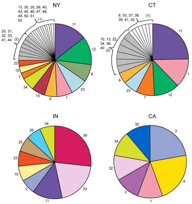
genotype analysis (n = 50). The predominant genotypes by region were: New York (8, 11, 12 and 23), Connecticut (11, 1, 12, and 7) and Indiana (39, 32 and 11) and represented 40%, 57% and 64% of the infected ticks, respectively (Figure 1). Nine of 15B. burgdorferi s.s. positive adultI. pacificusticks from Northern California were genotyped and found to predominately contain genotypes 2 and 4 (Figure 1).
B. burgdorferis.s. genotypic diversity by geographic region
We compared the genetic diversity ofB. burgdorferiin our field-collected specimens and culture isolates at the local level (New York vs. Connecticut), the regional level (Northeast vs. Indiana vs. California) and the continental level (North America vs. Europe), as shown in Figure 2A, B, and C respectively. Single specimens containing a resolvable mixture of genotypes were treated as separate counts for determining genotype frequency for the area.
In each region studied, a few genotypes were responsible for the majority of theB. burgdorferiinfections (Figure 1). Proximal Connecti-cut and New York shared the highest percentage of common genotypes: 79% of CT genotypes were also observed in NY and 48% of NY genotypes were also observed in CT (Figure 2A). In examining North America, we grouped CT and NY (designated NE for Northeastern US) together due to their close proximity and the extent of shared genotypes. We found two genotypes (32 and 34) common to California, Indiana, and the Northeast (Figure 2B). Indiana shared 77% of its genotypes with the Northeast while 57% of California’s genotypes were also found in the Northeast.
Table 4.OtherBorrelia speciesgenotypes.
Species Specimen ID1 Genotype
Borrelia afzelii ACA1 (UK-814), GER_169 1 pKo, GER_106, GER_139* 2 GER_033, GER_038 3
GER_005* 4
GER_005*,GER_139*,
GER_176, GER_178*,GER_200
5
GER_119, GER_178* 6 GER_022, GER_116 7
GER_125 8
GER_004 9
Borrelia garinii VSBM 1
NT29 HS9 2
GER_158 3
GER_020 4
B. valaisiana GER_143 1
B. andersonii 19857 1
B. bissettii DN127 1
B. lusitaniae PotiB1 1
B. spielmanii GER_173 1
Italicized samples are European in origin; non-italicized samples are United States in origin.
*Denotes a mixture of more then one genotype. doi:10.1371/journal.pone.0010650.t004
Table 5.Comparision ofospCtyping vs. PCR/ESI-MS genotyping.
Strain ospC Type Genotype
B31, IP2, PKa2, IP3, Lenz, L65, HII, Ho, IP1, HB1, B31, 132a, 132b
A 6
MI415 B1 26
109a, 160b B1 1
Bol12, Lx36, ZS7 B2 14
VS219 B2 19
JD1 C 8
SD91, NP14, N40, 88a E 15
MI407 F 24
72a, 80a G 23
MI411, 156a H 25
86b, 97b I 22
118a J 28
297, OEA11, 136b, 163b K 7
Y1, Y10 L 20
217-5, Bol6, Z6 L 9
MI418 N 27
Bol15, Bol25, Bol27, Fr-93/1 Q 18
Z9 S 17
94a U 12
Bol29, Bol30 V 16
Ri5 W 21
In the comparison of North American to European isolates (Figure 2C), only three genotypes were found on both continents. Of the B. burgdorferi s.s. genotypes observed in North America and Europe, only 6.6% of US genotypes (3 out of 45) were found in Europe and 27% of the European genotypes (3 of 11) were observed in the US.
Characterization of genotypicBorrelia mixtures inIxodes ticks
Of the 169 B. burgdorferi-positive adult I. scapularis ticks we characterized, 62% (n = 104) had a single genotype, 34% (n = 57) contained two genotypes and 5% (n = 8) had three or more genotypes (Figure 3). In those specimens with mixtures of more than one genotype, we were able to determine the individual B. burgdorferigenotypes in 52% of the specimens (34 of 65) based upon difference in the allele amplitude as described above in the
methods section and shown in Figure 3A. Additionally, one of elevenB. burgdorferiinfected nymphs from Suffolk County, NY was found to be infected with a mixture of twoB. burgdorferigenotypes (genotypes 22 and 41). The proportion of B. burgdorferi s.s. genotypes per infected adult Ixodes scapularis ticks from Indiana, Connecticut, and New York is shown in Figure 3B.
Borrelia burgdorferis.l. genotyping ofI. ricinusticks from Southern Germany
178I. ricinusticks were tested for the presence ofBorreliaon the genotyping assay and 36 were found to haveBorrelia(20.2%). The majority of the positive samples wereB. afzelii(n = 23) followed by B. garinii(n = 6) andB. burgdorferis.s. (n = 3),B. valaisiana(n = 2),B. lusitaniae(n = 1),B. spielmanii(n = 1). In total 20 of the 36 samples positive for Borrelia were genotyped. The remaining samples contained un-genotypable mixtures or had limited levels of DNA. Figure 1.B. burgdorferis.s. genotypes detected in NY, CT, IN, and CA.The number indicates the genotype and the pie size is relative to the proportion that the genotype was observed compared to all the genotypes in that region. The number ofB. burgdorferisamples genotyped in each region was 104 for NY, 61 for CT, 14 for IN, and 9 for CA.
Mixtures ofBorreliagenotypes were also observed in theI. ricnius ticks with 8 (22.2%) of the 36 Borreliapositive specimens having two or more genotypes present. Five of the eight had a mixture of two genotypes while the remaining samples had 3 or more genotypes present. Three of the mixtures were resolved to their individual genotypes. Five of the mixtures were of B. afzelii genotypes, one a mixture ofB. garinii, one was a mixture of B. lusitaniaegenotypes, and one a mixture ofB. valaisiana.
Discussion
Lyme disease, caused byB. burgdorferi, has symptoms which are usually nonspecific, consisting of fever, fatigue, chills, headache, and muscle aches. In some cases, signs of the disease remain localized to an EM. In other cases there are signs of dissemination into systemic disease such as multiple erythema migrans lesions, central and peripheral neurologic abnormalities, cardiac impair-Figure 2. Venn diagrams displaying number of genotypes shared between (A) local, (B) regional, and (C) continental areas.The local and regional diagrams represent data from field-collectedIxodesticks containingB. burgdorferis.s. that were genotyped. The continental Venn diagram incorporates data from all samples (ticks and culture isolates) whose origin was known. The size of the circles is proportional to the number of genotypes in the region.
doi:10.1371/journal.pone.0010650.g002
Figure 3. Detection of multipleB. burgdorferigenotypes in adultI. scapularisticks.A) Example of assignment of alleles to a genotype in a mixture. Analysis of sample NY71 from a tick collected in Connetquot, Suffolk County, New York is shown. B) Proportions of the minimum number of
ment, and swelling of large joints. Different strains ofB. burgdorferi have been associated with varying pathogenicities in the human host, with some strains more often observed in disseminated infections [10,29]. Previous studies have shown that certain strains ofB. burgdorferiare more likely to cause an erythema migrans and/ or disseminate through the body[10,30]. The ability to quickly identify theBorreliaspecies and associate particular genotypes with certain symptoms can have important patient-management implications. For example, the clinical observation of a single erythema migrans lesion where at least one of the recovered spirochetes is associated with an invasive phenotype might alert the treating physician to consider the use of an antibiotic, such as an intravenously administered one, that will reliably penetrate the central nervous system [31,32] or, at the very least, the physician would monitor the patient for development of neurologic symptoms even with administration of oral antibiotics.
Previous studies have also shown culturing spirochetes from ticks or clinical specimens can alter the genotypes observed as compared to those obtained from direct PCR [33,34]. Using our rapid multilocus PCR/ESI-MS genotyping assay, we have demonstrated that we can genotype B. burgdorferi directly from adult or nymphal ticks without the need for culture. Additionally, we have shown that our PCR/ESI-MS method can be used to detect and genotypeB. burgdorferidirectly from a clinical specimen. The PCR/ESI-MS genotyping assay has the added benefit of being able to identify the species and genotype a variety ofBorrelia that can cause Lyme disease.
Studies have shown that the multilocus sequence typing (MLST) approach provides a finer separation of strains than does typing based on theospCgene alone [15]. Using our multi-locus PCR/ESI-MS genotyping assay, severalospC groups were subdivided into multiple genotypes. In North America we observed 44 different genotypes of B. burgdorferi, 35 of which were found at sites within 100 miles of each other, which could indicate a much more diverse population of B. burgdorferi than previously described.
In this study, we found thatBorreliainfection rates in adultI. scapularis ticks from the northeastern US were similar to those observed previously[4,35,36]; B. burgdorferi infection rates were relatively lower in Indiana and California. An earlier study utilizing an MLST approach reported that no genotypes were shared between the Northeastern United States and the Midwest (with Ohio being the dividing state) [37]. In contrast to this observation, we found two genotypes in all three regions of the United States. Although our sample sizes from the Midwest and California were lower, the presence of these genotypes in each region may indicate that there was a widespread expansion of a few clonal isolates across North America followed by subsequent local diversification.
Past studies employing either single-strand conformation polymorphism (SSCP) or reverse line blotting (RLB) have shown that ticks can be infected with more than one strain ofB. burgdorferi [4,38]. Due to the limits of DNA sequencing, MLST approaches cannot resolve mixtures without culture. Due to the capability of mass spectrometry to resolve mixtures, our PCR/ESI-MS genotyping assay could detect and frequently resolve specimens containing a mixture of more than one genotype. Of the 169B.
burgdorferipositiveI. scapularisticks characterized from the US, 39% contained two or more genotypes. Of the 36Borrelia positiveI. ricinusticks from Europe, we found 22.2% containing more than one pathogenic species/member of Borrelia burgdorferi sensu lato complex. These findings indicate genotype mixtures in ticks are common in both Europe and North America. Our finding of a nymph (which has fed only once) with multiple genotypes and 5% of the adult ticks (which have fed twice), with three or more genotypes indicates that ticks can become infected with more than one genotype ofB. burgdorferifrom a single feeding. Further studies are needed to determine if certain combinations of genotypes are more likely to co-infect a tick or cause disease in humans.
In this study we developed a very high resolution Borrelia genoptying assay that can genotype directly from a variety of sample types and discriminate Borrelia mixtures. We found a diversity of genotypes across geographic regions. This capability to rapidly identify and genotype Borrelia may facilitate better management of patients with Lyme disease and provide a better understanding of the association ofBorreliagenotypes and disease. Our findings also underscore the importance of collaborations between scientists with environmental and microbial ecology expertise [1,12] along with those in health related research. While our findings, which were not designed to be comprehensive, provides the impetus and shows the feasibility to explore other areas where Lyme is being to reported such as the southern US and northern Mexico. Such studies can contribute to a broader epidemiologic assessment of the breadth of potential Lyme disease in North America. Future studies focusing onBorreliagenotypes in nymphs will further define their genotypic co-infection rates and determine if certain genotypes tend to preferentially co-infect. The findings here indicate that the diversity of genotypes in ticks known to cause Lyme disease is greater then previously described and that Borrelia may frequently exist as genotypic mixtures in the environment.
Supporting Information
Table S1 Table showing the detected A, G, C, T nucleotides detected for the different genotypes.
Found at: doi:10.1371/journal.pone.0010650.s001 (0.04 MB XLS)
Acknowledgments
We would like to thank Angella Falco, Ann Donohue, David James, Jack Cavier, Jamesina Scott, Jianmin Zhong, Paul Binding, Rob Cummings, Ronald Keith, Stacy Berden, Sylviane Schwarz, Wakoli Wekesa, Kirby Stafford, Richard Ostfeld, Keith Clay, and Benedikt Mothes for providing
ticks and Heike Haag for excellent technical assistance with theI. ricnius
ticks. Additionally we would like to thank Barbara J. Johnson at the CDC in Ft. Collins for providing us with variousBorreliaculture isolates.
Author Contributions
Conceived and designed the experiments: CDC MWE. Performed the experiments: CDC MAR ON CAP. Analyzed the data: CDC HEM MAR MWE. Contributed reagents/materials/analysis tools: SES BJL ON SC FL RS. Wrote the paper: CDC SES ON MWE.
References
1. Brownstein JS, Holford TR, Fish D (2003) A climate-based model predicts the spatial distribution of the Lyme disease vector Ixodes scapularis in the United States. Environ Health Perspect 111: 1152–1157.
2. Falco RC, McKenna DF, Daniels TJ, Nadelman RB, Nowakowski J, et al. (1999) Temporal relation between Ixodes scapularis abundance and risk for Lyme disease associated with erythema migrans. Am J Epidemiol 149: 771–776.
3. Hulinska D, Votypka J, Kriz B, Holinkova N, Novakova J, et al. (2007) Phenotypic and genotypic analysis of Borrelia spp. isolated from Ixodes ricinus ticks by using electrophoretic chips and real-time polymerase chain reaction. Folia Microbiol (Praha) 52: 315–324.
vector (Ixodes scapularis) in the Northeastern United States. Genetics 160: 833–849.
5. Hanincova K, Ogden NH, Diuk-Wasser M, Pappas CJ, Iyer R, et al. (2008) Fitness variation of Borrelia burgdorferi sensu stricto strains in mice. Appl Environ Microbiol 74: 153–157.
6. Schutzer SE (1994) Diagnosing Lyme disease. Hosp Pract (Off Ed) 29: 8, 11. 7. Coyle PK, Dattwyler R (1990) Spirochetal infection of the central nervous
system. Infect Dis Clin North Am 4: 731–746.
8. Coyle PK, Schutzer SE (2002) Neurologic aspects of Lyme disease. Med Clin North Am 86: 261–284.
9. Schutzer SE, Coyle PK, Krupp LB, Deng Z, Belman AL, et al. (1997) Simultaneous expression of Borrelia OspA and OspC and IgM response in cerebrospinal fluid in early neurologic Lyme disease. J Clin Invest 100: 763–767. 10. Seinost G, Dykhuizen DE, Dattwyler RJ, Golde WT, Dunn JJ, et al. (1999) Four clones of Borrelia burgdorferi sensu stricto cause invasive infection in humans. Infect Immun 67: 3518–3524.
11. Wang IN, Dykhuizen DE, Qiu W, Dunn JJ, Bosler EM, et al. (1999) Genetic diversity of ospC in a local population of Borrelia burgdorferi sensu stricto. Genetics 151: 15–30.
12. Brisson D, Dykhuizen DE (2004) ospC diversity in Borrelia burgdorferi: different hosts are different niches. Genetics 168: 713–722.
13. Farlow J, Postic D, Smith KL, Jay Z, Baranton G, et al. (2002) Strain typing of Borrelia burgdorferi, Borrelia afzelii, and Borrelia garinii by using multiple-locus variable-number tandem repeat analysis. J Clin Microbiol 40: 4612–4618. 14. Qiu WG, Schutzer SE, Bruno JF, Attie O, Xu Y, et al. (2004) Genetic exchange
and plasmid transfers in Borrelia burgdorferi sensu stricto revealed by three-way genome comparisons and multilocus sequence typing. Proc Natl Acad Sci U S A 101: 14150–14155.
15. Margos G, Gatewood AG, Aanensen DM, Hanincova K, Terekhova D, et al. (2008) MLST of housekeeping genes captures geographic population structure and suggests a European origin of Borrelia burgdorferi. Proc Natl Acad Sci U S A 105: 8730–8735.
16. Vitorino LR, Margos G, Feil EJ, Collares-Pereira M, Ze-Ze L, et al. (2008) Fine-scale phylogeographic structure of Borrelia lusitaniae revealed by multilocus sequence typing. PLoS ONE 3: e4002.
17. Sampath R, Russell KL, Massire C, Eshoo MW, Harpin V, et al. (2007) Global surveillance of emerging Influenza virus genotypes by mass spectrometry. PLoS One 2: e489.
18. Ecker DJ, Massire C, Blyn LB, Hofstadler SA, Hannis JC, et al. (2009) Molecular genotyping of microbes by multilocus PCR and mass spectrometry: a new tool for hospital infection control and public health surveillance. Methods Mol Biol 551: 71–87.
19. Hall TA, Sampath R, Blyn LB, Ranken R, Ivy C, et al. (2009) Rapid molecular genotyping and clonal complex assignment of Staphylococcus aureus isolates by PCR coupled to electrospray ionization-mass spectrometry. J Clin Microbiol 47: 1733–1741.
20. Hannis JC, Manalili SM, Hall TA, Ranken R, White N, et al. (2008) High-resolution genotyping of Campylobacter species by use of PCR and high-throughput mass spectrometry. J Clin Microbiol 46: 1220–1225.
21. Ecker JA, Massire C, Hall TA, Ranken R, Pennella TT, et al. (2006) Identification of Acinetobacter species and genotyping of Acinetobacter baumannii by multilocus PCR and mass spectrometry. J Clin Microbiol 44: 2921–2932.
22. Blyn LB, Hall TA, Libby B, Ranken R, Sampath R, et al. (2008) Rapid detection and molecular serotyping of adenovirus by use of PCR followed by electrospray ionization mass spectrometry. J Clin Microbiol 46: 644–651.
23. Eshoo MW, Crowder CD, Li H, Matthews HE, Meng S, et al. (2009) Detection and Identification of Ehrlichia species in Blood Using PCR and Electrospray Ionization Mass Spectrometry. J Clin Microbiol.
24. Brownstein MJ, Carpten JD, Smith JR (1996) Modulation of non-templated nucleotide addition by Taq DNA polymerase: primer modifications that facilitate genotyping. Biotechniques 20: 1004–1006, 1008–1010.
25. Ecker DJ, Sampath R, Blyn LB, Eshoo MW, Ivy C, et al. (2005) Rapid identification and strain-typing of respiratory pathogens for epidemic surveil-lance. Proc Natl Acad Sci U S A 102: 8012–8017.
26. Ecker DJ, Sampath R, Massire C, Blyn LB, Hall TA, et al. (2008) Ibis T5000: a universal biosensor approach for microbiology. Nat Rev Microbiol 6: 553–558. 27. Crowder CD, Rounds MA, Phillipson CA, Picuri JM, Matthews H, et al. (2009) Extraction of Total Nucleic Acids from Ticks for the Detection of Bacterial and Viral Pathogens. J Med Entomol In Press.
28. Qiu WG, Bruno JF, McCaig WD, Xu Y, Livey I, et al. (2008) Wide distribution of a high-virulence Borrelia burgdorferi clone in Europe and North America. Emerg Infect Dis 14: 1097–1104.
29. Fikrig E, Coyle PK, Schutzer SE, Chen M, Deng Z, et al. (2004) Preferential presence of decorin-binding protein B (BBA25) and BBA50 antibodies in cerebrospinal fluid of patients with neurologic Lyme disease. J Clin Microbiol 42: 1243–1246.
30. Wormser GP, Brisson D, Liveris D, Hanincova K, Sandigursky S, et al. (2008) Borrelia burgdorferi genotype predicts the capacity for hematogenous dissemination during early Lyme disease. J Infect Dis 198: 1358–1364. 31. Coyle PK, Deng Z, Schutzer SE, Belman AL, Benach J, et al. (1993) Detection
of Borrelia burgdorferi antigens in cerebrospinal fluid. Neurology 43: 1093–1098.
32. Wormser GP, Dattwyler RJ, Shapiro ED, Halperin JJ, Steere AC, et al. (2006) The clinical assessment, treatment, and prevention of lyme disease, human granulocytic anaplasmosis, and babesiosis: clinical practice guidelines by the Infectious Diseases Society of America. Clin Infect Dis 43: 1089–1134. 33. Norris DE, Johnson BJ, Piesman J, Maupin GO, Clark JL, et al. (1997)
Culturing selects for specific genotypes of Borrelia burgdorferi in an enzootic cycle in Colorado. J Clin Microbiol 35: 2359–2364.
34. Liveris D, Varde S, Iyer R, Koenig S, Bittker S, et al. (1999) Genetic diversity of Borrelia burgdorferi in lyme disease patients as determined by culture versus direct PCR with clinical specimens. J Clin Microbiol 37: 565–569.
35. Tokarz R, Jain K, Bennett A, Briese T, Ian Lipkin W (2009) Assessment of Polymicrobial Infections in Ticks in New York State. Vector Borne Zoonotic Dis.
36. Tsao JI, Wootton JT, Bunikis J, Luna MG, Fish D, et al. (2004) An ecological approach to preventing human infection: vaccinating wild mouse reservoirs intervenes in the Lyme disease cycle. Proc Natl Acad Sci U S A 101: 18159–18164.