Evaluation of
In Vitro
Anticancer Activity
of
Ocimum Basilicum, Alhagi Maurorum,
Calendula Officinalis
and Their Parasite
Cuscuta Campestris
Mandana Behbahani*
Department of Biotechnology, Faculty of Advanced Sciences and Technologies, University of Isfahan, Isfahan 81746-73441, Islamic Republic of Iran
Abstract
The present investigation was carried out to study the relationship between presence of cytotoxic compounds in Ocimum basilicum, Alhagi maurorum, Calendula officinalisand their parasiteCuscuta campestris. The cytotoxic activity of the pure compounds was performed by MTT assay against breast cancer cell lines (MCF-7 and MDA-MB-231) and normal breast cell line (MCF 10A). The induction of apoptosis was measured by the expression levels of p53, bcl-2, bax and caspase-3 genes using quantitative Real Time PCR. Three active fractions were detected by nuclear magnetic resonance as lutein, lupeol and eugenol, respectively, inC. officinalis,A. maurorumandO. basilicum. These compounds and their epoxidized forms were also detected in their parasite C. campestris. The cytotoxic activity of lutein epoxide, lupeol epoxide and eugenol epoxide was significantly more than lutein, lupeol and eugenol. The mRNA expression level of p53, caspase-3 and bax genes were increased in both cancer cells treated with all pure compounds. However, bcl-2 gene expression decreased in treated breast cancer cells. In conclusion, all the data indicated that the epoxide forms of lupeol, lutein and eugenol are potential drug candidates for inducing apoptosis in human breast cancer cells.
Introduction
Breast cancer is one of the most common diseases among women worldwide and the majority of cases have been reported in Asian countries over the past two
OPEN ACCESS
Citation:Behbahani M (2014) Evaluation ofIn VitroAnticancer Activity ofOcimum Basilicum, Alhagi Maurorum, Calendula Officinalisand Their ParasiteCuscuta Campestris. PLoS ONE 9(12): e116049. doi:10.1371/journal.pone.0116049
Editor:Yuan-Soon Ho, Taipei Medical University, Taiwan
Received:September 4, 2014
Accepted:November 30, 2014
Published:December 30, 2014
Copyright:ß2014 Mandana Behbahani. This is an open-access article distributed under the terms of theCreative Commons Attribution License, which permits unrestricted use, distribution, and reproduction in any medium, provided the original author and source are credited.
Data Availability:The author confirms that all data underlying the findings are fully available without restriction. All relevant data are within the paper.
Funding:This study was financially supported by the University of Isfahan, Isfahan, Iran. The funder had no role in study design, data collection and analysis, decision to publish, or preparation of the manuscript.
decades [1]. Currently, many studies have been carried out worldwide to isolate the active novel compounds from plants for cancer treatment. Several secondary metabolites including alkaloids, polyphenols, flavonoids and triterpenes were purified from medicinal plants [2]. These products cause apoptosis which is modulated by direct or indirect modulating expression of some genes such as p53, bcl2 and caspase-3 [3,4]. However, it is important to develop the natural chemopreventive agents which evaluate cytotoxicity and apoptosis induction in cancer cell lines. Therefore, we became eager to assess Iranian species with respect to their anti-cancer activity and determine their active constituents. The genus
Cuscuta (Convolvulaceae) also known as dodder is an obligate stem parasite with
a worldwide distribution. Cuscuta campestrisis the most common species in the
genus Cuscuta and grown on wide range of host species [5]. In recent decades,
through extracting natural compounds from cuscuta specious a number of
compounds such as cuscutin, amarbelin, b-sitosterol, stigmasterol, kaempferol,
dulcitol, myricetin, quercetin, coumarin and oleanolic acid have been isolated [6,7]. C. campestrishas been proved to have analgesic, antipyretic,
anti-inflammatory and anti-cancer properties on different hosts [8]. Some studies have reported that some secondary metabolites transferred from host to the parasite and responsible for medical properties of parasite [9,10]. Three Iranian common
hosts for C. Campestrisare Alhagi maurorum, Calendula officinalis and Ocimum
basilicum. Medicinal activity of these plants has been also subjected by several investigations [11–13]. So far, cytotoxic activity of C.campestris and the
relationship with its hosts has not been investigated yet. Hence, the present study
is focused to evaluate anticancer potential of pure compounds isolated from A.
maurorum, C. officinalis, O. basilicum and their parasiteC. campestris against breast cancer cell lines (MCF7 and MDA-MB231) and human normal breast cancer cell line (MCF 10A).
Materials and Methods
Ethics statement
No ethics statement was necessary for this study. The study was only carried outin vitro on an immortalized cell line. None of these species is an endangered or protected species and did not need a permit.
Plant material
Extraction and isolation of compounds
Methanol extracts of the powdered aerial parts of O. basilicum, A. maurorum, C. officinalis and their parasite C. campestris (50 g) were prepared. The extraction
was done three times at room temperature (25–28
˚
C) by the maceration method(3624 h). The crude methanol extract of each sample was concentrated by a
rotary evaporator (Steroglass, Italy) and freeze dried (Zirbus, Germany). Silica-gel column fractionation chromatography was separately performed with 5 g of each methanol extract and eluted with hexan: acetone: methanol (1:0:0 to 0:0:10, v/v). The following fractions have been obtained from these three plants and their parasite. The anticancer activity of all fractions were verified by MTT assay.
Fractions 1–17 (0.31, 0.20, 0.21, 0.25, 0.29, 0.22, 0.31, 0.21, 0.25, 0.36, 0.20, 0.21, 0.35, 0.19, 0.20, 0.18 and 0.20 g) fromO. basilicumand fractions 1–23 from its parasite C. campestris(0.37, 0.18, 0.14, 0.35, 0.28, 0.36, 0.17, 0.21, 0.18, 0.23, 0.22, 0.28, 0.30, 0.21, 0.21, 0.14, 0.22, 0.30, 0.20, 0.17, 0.18, 0.17 and 0.27 g) were
obtained. Fraction 13 from O. basilicum and fractions 17 and 23 from C.
campestris were the most active fractions.
Fractions 1–25 (0.25, 0.15, 0.18, 0.22, 0.20, 0.13, 0.21, 0.14, 0.25, 0.24, 0.21, 0.19, 0.18, 0.2, 0.21, 0.21, 0.16, 0.2, 0.18, 0.18, 0.21, 0.15, 0.20, 0.15 and 0.20 g) from A. maurorum and fractions 1–27 from its parasiteC. campestris (0.19, 0.15, 0.21, 0.2, 0.18, 0.20, 0.21, 0.20, 0.15, 0.19, 0.3, 0.32, 0.25, 0.20, 0.2, 0.18, 0.18, 0.19, 0.22, 0.20, 0.25, 0.16, 0.16, 0.25, 0.21, 0.20, and 0.20 g) were achieved. Fraction 15 fromA. maurorumand fractions 16 and 21 from its parasiteC. campestriswere the most active fractions.
Fractions 1–26 (0.20, 0.21, 0.16, 0.20, 0.22, 0.2, 0.18, 0.17, 0.15, 0.19, 0.21, 0.21, 0.21, 0.21, 0.22, 0.21, 0.21, 0.15, 0.20, 0.19, 0.23, 0.24, 0.22, 0.2, 0.21, and 0.20 g) from C. officinalis and fractions 1–24 from its parasite C. campestris(0.15, 0.19, 0.21, 0.23, 0.24, 0.23, 0.21, 0.23, 0.24, 0.22, 0.2, 0.21, 0.21, 0.22, 0.24, 0.21, and 0.20 g) were obtained. Fraction 19 of C. officinalis and fractions 14 and 17 of
C.campestris were the most active fractions. The active fractions were detected with Nuclear Magnetic Resonance (NMR).
Instrumental analysis
NMR screening was used to identify trace compounds in each fraction. 1H NMR
and 1C NMR data were recorded on Bruker 400 MHz spectrometer by use of
CDCl3and DMSO as residual solvent with chemical shifts expressed in parts per
million (ppm).
Culture medium and cell lines
The cell line MCF 10A, MCF-7 and MDA-MB-231 were acquired from National Cell Bank of Pasture Institute, Tehran, Iran. Cell lines were cultured in Dulbecco’s Modified Eagle Medium (DMEM) complemented with 10% heat-inactivated Fetal
Bovine Serum (FBS), 100 U/ml penicillin and 100 mg ml2streptomycin and
atmosphere containing 5% CO2. All reagents and cell culture media were
purchased from the Gibco Company (Germany).
Cytotoxicity assay
The cytotoxic activity of active fractions isolated from O. basilicum, A.maurorum, C. officinalis andtheir parasiteC. campestrison cultured cells were measured using MTT assay [14]. The cells were grown in 96-well plates at a density of 56104cells
per well. After incubation for 24 h, the cells were treated with different
concentrations of samples and incubated for 48 h. Later, the MTT solution (25 ml
of 5 mg/ml, Roche) was added to each well, and the plate was re-incubated for an
additional 4 h. Finally, the medium was removed and 100 ml of DMSO was added
to solubilize the formed formazan crystals. The amount of formazan crystal was determined by measuring the absorbance at 492 nm using a microplate
spectrophotometer (Awareness Technology Inc., stat fax 2100). The 50% cytotoxic concentration (CC50) of all pure compounds was calculated. All assays were done in triplicate.
Determination of the expression levels of apoptosis-regulatory
genes
Expression levels of four widely established apoptotic-related mRNAs, p53, bcl-2, bax and caspase-3 were analyzed using Real Time PCR assay as described
previously [15,16]. MCF-7 and MDA-MB-231 cells were treated with all pure
compounds at 1/4 of CC50 values for 6 and 12 h periods. Total cellular RNA was isolated from the untreated and treated cells using the Tri-Pure Isolation Reagent (Roche, USA), according to the manufacturer’s instructions.
Quantitative real-time polymerase chain reaction assay for p53,
bcl-2, bax and caspase-3
Real Time PCR was performed to quantify the amount of mRNA in untreated and
treated cells. A PCR reaction mixture of 50 ml containing 5ml of dH2O, 25 mL of
Taq Man Universal PCR MasterMix, 5 ml of primer forward, 5 ml primer reverse,
5 ml FAM- TAMRA probes, 0.5ml of reverse transcriptase, 2ml random hexmer
and 2 ml of purified RNA was used. Five pairs of primers were separately used:
four pairs to amplify the p53, bcl2, bax and caspase-3 genes, the other pair for the endogenous control gene, gapdh. The primers and probes have been shown in
Table 1. Real-time PCR was carried out on Corbett Cycler. Cycling conditions were as follows: initial reverse transcription at 55
˚
C for 45 min, 1 cycleWestern blot analysis
The expression levels of p53, caspase-3, bcl-2 and bax proteins in MCF-7 and MDA-MB-231 cells were assessed by western blot method as described by Fido et al. [17]. Both cells (56106 cells/ml) were treated with lutein epoxide, lupeol
epoxide, eugenol epoxide, lutein, lupeol and eugenol at 1/4 of CC50 values for 48 h at 37
˚
C. Cells were lysed with 10 ml of lyses buffer (120 mmol/L Tris-HCl, 2 mmol/L N-ethylmaleimide, 2 mmol/L phenylmethyl sulfonylfluoride, 4% sodium dodecylsulfate, 4% dithiothreitol, 20% glycerol, 0.01% bromophenolblue, 2 mol/L urea and 10 mmol/L Na-EDTA at pH56.8. Cell lysates were
centrifuged at 16000 rpm/min for 20 min at 4
˚
C. 50 mg of each sample wasseparately resolved by SDS-PAGE and move onto a nitrocellulose membrane overnight at 30 mA. Membranes were blocked with 2% BSA diluted in PBS for
1 h at 37
˚
C. Membranes were incubated with saturating concentrations ofprimary antibody (anti-p53; anti-caspase-3, anti-bcl2, anti-bax) for 2 h under gentle agitation at room temperature. The blots were washed three times and incubated with horse reddish peroxdase-conjugated anti-mouse IgG antibody for
1 h at 37
˚
C. Then the signal was detected with an enhanced chemiluminescencedetection kit (Roche, Mannheim, Germany). Diaminobenzidine reagent was used to develop the immunoblots.
Statistical analysis
The data of three independent experiments are presented as mean¡SD. The
CC50 values were calculated by Microsoft Excel 2003. The student’s unpaired t-test was used to evaluate significance between the t-test sample and solvent control. One-way analysis of variance (ANOVA) followed by a Dunnett post hoc test was
Table 1.The primers and probe sequences used in real-time PCR assay.
Gene Sequence
p53 Forward: 59-AGAGTCTATAGGCCCACCCC-39
Reverse: 59-GCTCGACGCTAGGATCTGAC-39 Probe: 59-(FAM) TTGGGCAGTGCTCGCT-MGB-3
bcl-2 Forward: 59-TTCGATCAGGAAGGCTAGAGTT-39
Reverse: 59-TCGGTCTCCTAAAAGCAG GC-39
Probe: 59-(FAM)CCCAGAGCATCAGGCCGCCAC(TAMRA)-39
gapdh Forward: 59-CATGGGGAAGGTGAAGGTCGA-39
Reverse: 59-TTGGCTCCCCCCTGCAAATGAG-39 Probe: 59-(JOE)CCGACTCTTGCCCTTCGAC(TAMRA)-39
caspase-3 Forward: 59-TGCGCTGCTCTGCCTTCT-39
Reverse: 59-CCATGGGTAGCAGCTCCTTC-39
Probe: 59-FAM-AGCTTCTTCATTTGTGTGCTCCGCTTTCA(TAMRA)-39
bax Forward: 59-CATGTTTTCTGACGGCAACTTC -39
Reverse: 59- AGGGCCTTGAGCACCAGTTT-39
Probe: 59-(FAM) CCGGGTTGTCGCCCTTTTCTACTTTG(TAMRA)-39
performed to evaluate significance differences among different groups. P value
,0.05 was shown to be statistically significant.
Results
NMR Analysis
The most active fraction obtained from O. basilicum was fraction 13, which
analyzed by NMR experiment as eugenol. Fractions 17 and 23 were also isolated
from its parasite C.campestris and determined as eugenol and eugenol epoxide.
Fraction 15 ofA.maurorumand fractions 16 and 21 ofC.campestriswere analyzed as lupeol, lupeol and lupeol epoxide respectively. Fraction 19 ofC. officinalisand fractions 14 and 17 of C.campestris were analyzed as lutein, lutein and lutein epoxide.
Eugenol epoxide
1
HNMR (CDCl3, 400 MHz): d6.80 (d, 1H, J57.9 Hz), 6.75 (d, 1H, J51.8 Hz),
6.64 (dd, 1H, J51.8, 7.9 Hz), 5.60 (s, 1H), 3.85 (s, 3H), 3.02 (m, 1H), 2.80 (dd, 1H,J155.5,J2514.7 Hz), 2.73 (dd, 1H,J152.4,J254.9 Hz), 2.68 (dd, 1H,J155.6,
J2514.8 Hz), 2.50 (dd, 1H, J154.9, J252.4 Hz).
13CNMR (CDCl
3, 100 MHz) d147.46, 145.28, 133.14, 122.15, 115.22, 112.78,
56.24, 54.76, 48.32, 41.34.
Eugenol
1
HNMR (CDCl3, 400 MHz):d7.12 (s, 1H), 6.78 (d, 2H,J57.2 z), 6.32–6.42 (m,
2H), 5.45–5.65 (m, 2H), 3.85 (s, 3H), 3.26 (m, 2H)
13
CNMR (CDCl3, 100 MHz) d147.23, 145.01, 138.23, 133.14, 122.15, 117.01,
114.52, 112.36, 56.24, 41.03.
Lupeol
1H NMR (CDCl
3, 400 MHz):d 4.84 (d, 1H,J52.4 Hz), 4.62 (d, 1H,J52.4 Hz),
3.18 (1H, m), 2.42 (m, 1H), 1.80 (m, 1H), 1.71 (t, 1H, J55.6Hz), 1.60 (s, 3H), 1.57 (m, 2H), 1.40 (m, 2H), 1.36 (m, 2H), 1.20 (m, 1H), 1.03 (m, 1H), 0.99 (s, 3H), 0.97 (s, 3H), 0.95 (s, 3H), 0.83 (s, 3H), 0.79 (s, 3H), 0.75 (s, 3H).
13C NMR (CDCl
3, 100 MHz):d149.12, 111.25, 80.24, 57.15, 51.63, 49.44, 48.16
41.23, 41.06, 41.02, 40.62, 39.85, 38.94, 38.23, 37.36, 36.21, 34.56, 32.04, 29.21, 28.01, 27.15, 24.36, 22.13, 19.85, 19.01, 18.45, 17.14, 16.78, 15.27, 14.42.
FT-IR [KBr,n(cm21)]: 3359, 3075, 2948, 2868, 1651, 1461
Lupeol epoxide
1
HNMR (CDCl3, 400 MHz): d3.23 (1H, m), 2.59 (d, 1H, J54.8 Hz), 2.42 (m,
1.57 (m, 2H), 1.40 (m, 2H), 1.36 (m, 2H), 1.20 (m, 1H), 1.03 (m, 1H), 0.99 (s, 3H), 0.97 (s, 3H), 0.95 (s, 3H), 0.83 (s, 3H), 0.79 (s, 3H), 0.75 (s, 3H).
13CNMR (CDCl
3, 100 MHz):d79.42, 64.32, 58.14, 53.21, 50.36, 48.65, 48.01,
42.47, 42.01, 41.74, 40.59, 39.84, 38.94, 38.25, 37.34, 36.21, 34.56, 32.12, 30.15, 28.16, 28.02, 24.03, 22.13, 19.86, 19.03, 18.56, 17.17, 16.78, 15.27, 14.42.
Lutein
1
HNMR (DMSO, 400 MHz):d6.21–6.76 (m, 13H), 5.52 (m, 1H), 3.82 (m. 1H),
3.41 (m, 1H), 3.68 (s, 1H), 2.95 (s, 1H), 2.71 (d, 1H, J59.2 Hz), 1.58–2.36 (m, 6H), 1.69 (s, 12H), 1.71 (s, 6H), 1.01 (s, 6H), 0.85 (s, 6H).
13CNMR (DMSO, 100 MHz):d138.11, 137.82, 137.65, 136.81, 136.50, 135.06,
131.24, 131.01, 130.65, 130.01, 129.85, 128.06, 126.66, 125.02, 68.35, 65.01, 57.32, 48.02, 43.91, 42.98, 37.65, 32.34, 28.01, 26.62, 24.31, 21.93, 14.35, 13.78
Lutein epoxide
1HNMR (DMSO, 400 MHz):d 6.18–6.73 (m, 13H), 5.61 (m, 1H), 3.61 (s, 2H),
3.55 (m, 1H), 3.21 (m, 1H), 2.43 (d, 1H,J59.2 Hz), 1.89 (s, 12H), 1.55–1.85 (m, 6H), 1.29 (s, 6H), 0.92 (s, 12H).
13
CNMR (DMSO, 100 MHz):d137.64, 136.95, 136.14, 135.24, 133.61, 132.58,
132.34, 130.45, 130.23, 129.41, 128.02, 71.02, 68.32, 75.05, 67.23, 65.12, 55.15, 52.18, 47.24, 43.34, 41.27, 35.03, 29.87, 28.31, 27.62, 24.32, 20.14, 13.65, 13.46
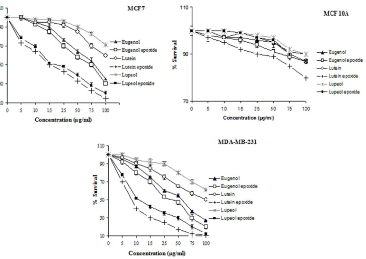
Cytotoxicy assay
Different concentrations of lutein, lupeol, eugenol, lutein epoxide, lupeol epoxide, eugenol epoxide were tested for cytoxicity against MCF7, MDA-MB-231 and MCF 10A cells. These compounds were tested under comparable conditions at different
concentrations (5, 10, 15, 25, 50, 75 and 100 mg/ml). All pure compounds showed
dose-dependent cytotoxic activity on MCF7 and MDA-MB-231 cell lines. The 1/4 of CC50 values of lutein, lupeol, eugenol, lutein epoxide, lupeol epoxide and eugenol epoxide were, respectively,.100,.100, 84, 15, 17.5, 75 mg/ml for MCF7
and 100, .100, 55, 7.5, 11.5, 48 mg/ml for MDA-MB-231. As a result, cytotoxic
activity of lupeol, lutein, lupeol epoxide and lutein epoxide against MDA-MB cancer cell line was rather than MCF7 cancer cell line. The cytotoxic activity of lupeol, lutein and eugenol were significantly lower than epoxide forms of these compounds. The results also showed that cytotoxic activity of these extracts on
MCF7 and MDA-MB cells were significantly more than MCF 10A cells (Fig 1).
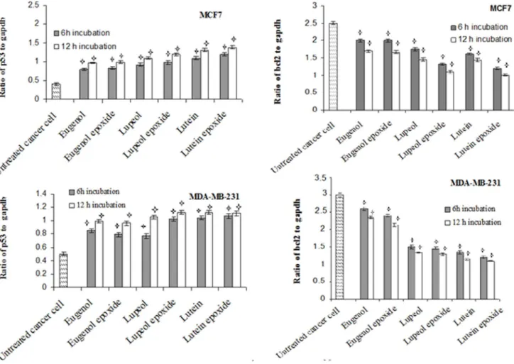
Expression level of apoptosis-related genes
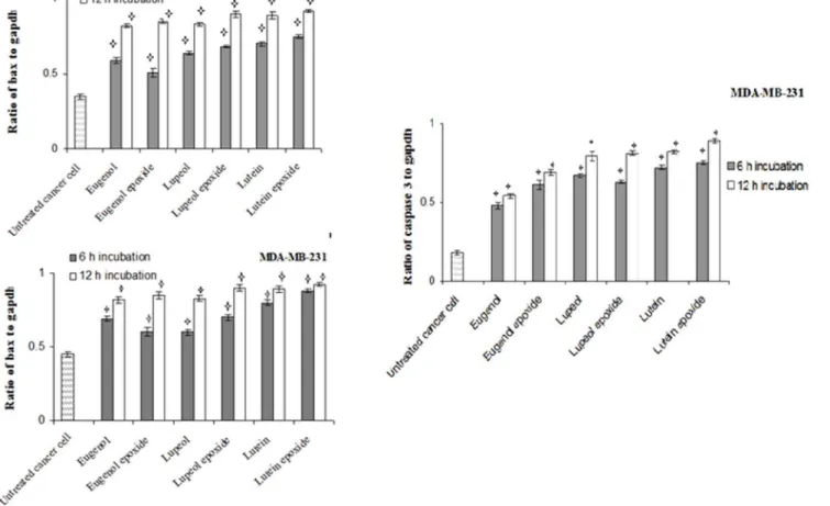
The expression levels of apoptosis-related genes in MCF7 and MDA-MB 231 cells which induced by with 6 separate pure compounds at 1/4 of CC50 values were determined. The mRNA levels of p53, caspase3, bax and bcl-2 were evaluated by
respectively, increased and decreased in both cells treated with lupeol, lupeol epoxide, lutein, lutein epoxide, eugenol and eugenol epoxide for 6 and 12 h incubation compared to untreated cells. The expression level of p53 and bcl2 in
both breast cancer cells treated with these compounds was time dependent.Fig. 3
indicated that the relative expression of bax was increased in cells treated with all pure compounds for 6 and 12 h incubation compared to untreated cells. The relative expression of caspase-3 in MDA-MB-231 cancer cells treated with these extracts was also increased as time-dependent to reach the maximum level at 12 h after stimulation. The absence of caspase-3 in MCF-7 cell leads to lack of any gene expression in treated and untreated cells (data not shown).
Fig. 1. Cytotoxic activity of lutein epoxide, lupeol epoxide, eugenol epoxide, lutein, lupeol and eugenol against MCF7, MDA-MB231 and MCF 10A cell lines.
Western blot analysis
The expression level of bcl2, bax and p53 proteins in MCF7 and MDA-MB23 cells treated with lutein, lupeol, eugenol, lutein epoxide, lupeol epoxide, eugenol
epoxide along with b-actin as an internal control are shown by western blotting
analysis. p53 and bax genes expressed, respectively, 53-kda and 21-kda proteins on western blotting. MDA and MCF7 cells also encode a 32-kDa and 24-kda proteins
whose association with caspase-3 and bcl2 proteins. As shown in Fig. 4, Western
blot analysis showed the increase in band intensity of p53 and bax proteins in
MCF7 and MDA cells when compared to the internal control b-actin. caspase-3
was also increased in MDA cells compared to b-actin. The absence of caspase-3
gene in MCF-7 cell leads to lack of any caspase-3 protein in treated and untreated cells. bcl2 protein was decreased in cancer cells treated by all compounds at 1/4 of CC50 values.
Fig. 2. Time dependency effects of p53 and bcl-2 mRNA levels in human breast cancer cell line, MCF7, incubated withlutein, lupeol, eugenol, lutein epoxide, lupeol epoxide, eugenolepoxide at 1/4 of CC50 values for 6 h and 12 h incubation.gapdh was used as an endogenous control gene. The stars indicate that the data are significantly different (p,0.05) from the untreated control.
Fig. 3. Time dependency effects of caspase 3 and bax mRNA levels in human breast cancer cell line, MCF7 and MDA-MB 231 incubated with
lutein, lupeol, eugenol, lutein epoxide, lupeol epoxide, eugenolepoxide at 1/4 of CC50 values for 6 h and 12 h incubation.gapdh was used as an endogenous control gene. The stars indicate that the data are significantly different (p,0.05) from the untreated control.
doi:10.1371/journal.pone.0116049.g003
Fig. 4. Western blot analysis of MDA-MB-231 and MCF7 treated with lutein (A), lupeol (B), eugenol (C), lutein epoxide (D), lupeol epoxide (E) and eugenol epoxide (F).Western blot analysis was performed with monoclonal antibodies to human bcl-2, p53 and bax.b-actin was used as loading control.
Discussion
Medicinal plants have been used widely in traditional medicine for cancer treatment. In this study, lutein epoxide, lupeol epoxide and eugenol epoxide
isolated from C. campestris were considered as potential anticancer compounds.
Lutein and lutein epoxide are plant cartenoids which is widely distributed in leaves and fruits of many plants such as tangerine, olive fruits, peas and pumpkins [18,19]. Antioxidant and cytotoxic activity of lutein and oxidized lutein in Hela cells have been report previously [20]. Lupeol and lupeol epoxide are naturally occurring triterpenoids which found in many plants, including olive, mango, crataeva and strawberry [21]. However, lupeol and its derivatives have also been reported to possess a wide spectrum of medicinal properties such as anticancer, antibacterial and antifungi properties [22–24]. Eugenol and its derivatives have been identified in various plants, such as basil, cinnamon, lemon balm and clove [25]. In the present study, the active compounds oflutein,lupeolandeugenolwere observed, respectively, in C. officinalis,A. maurorum and O. basilicum. The
epoxide forms of these compounds were also detected in their parasite C.
campestris. The mRNA expression level of p53, caspase-3 and bax genes were increased and bcl-2 gene expression decreased in breast cancer cells treated with these six compounds. The bcl-2 proteins are able to inhibit programmed cell death. So the expression of bcl-2 corresponds with the status of p53 in cells. Bax is another pro-apoptotic protein, which increases apoptosis in cells [26]. The result of this study demonstrated that p53 is involved in apoptosis which induced by lutein, lupeol, eugenol, lutein epoxide, lupeol epoxide and eugenol epoxide. There would be decreased expression of bcl-2 and increased expression of bax proteins in treated cells compared with non–treated cells. Importantly, the ratio of bax/bcl-2 protein expression after treatment was dose-dependently increased which indicated the susceptibility of MCF-7 and MDA-MB-231 cells toward apoptosis. This study concluded that some anticancer compounds transferred from host to
the parasite C. campestris and change to epoxide forms.
Conclusions
The results of detection of bioactive compounds signified that eugenol epoxide, lutein epoxide and lupeol epoxide constituted the most active fractions in the
crude methanol extract of C. campestris and displayed the cytotoxic effects on
breast cancer cell.
Author Contributions
References
1. Matsuda T, Matsuda A (2013) Burden of cancer incidence below the age of 40 in Asia 2002 extrapolated from the cancer incidence in five continents. Jpn J Clin Oncol 43: 449–50.
2. Rates SMK(2001) Plants as source of drugs. Toxicon, 39, 603–613.
3. Kobayashi S, Iwase H, Ito Y, Yamashita H, Iwata H, et al.(1997) Clinical significance of bcl2 gene expression in human breast cancer tissues. Breast Cancer Res Treat 42: 173–181.
4. Miyashita T, Reed JC(1995) Tumor suppressor p53 is a direct transcriptional activator of the human bax gene. Cell 80: 293–299.
5. Dawson JH, Musselman LJ, Wolswinkel P, Do¨rr I(1994) Biology and control of Cuscuta. Rev Weed Sci 6: 265–317.
6. Gilani AUH, Aftab K(1992) Pharmacological action ofCuscuta reflexa.Int JPharma 30: 296–302.
7. Borole SP, Oswal RJ, Antre RV, Kshir sagar SS, Bagul YR(2011) Evaluation of anti-epileptic activity of Cuscuta reflea Roxb. Res J Pharm Biol Chem Sci 2: 657–663.
8. Azza MA, Essam AS, Ahmed G(1996) Pharmacological Study ofCuscuta campestrisYuncker.Phyto Res10: 117–120.
9. Ghule RS, Venkatanarayan R, Thakare SP, Jain H, Ghule PR(2011) Analgesic activity ofCuscuta campestrisYuncker a parasitic plant grown on Nerium indicum Mill. J Adv Pharm Tech Res 1: 45–51.
10. Jadhav RB, Anarthe SJ, Surana SJ, Gokhale SB(2005) Host-hemiparasite transfer of the C-glucosyl xanthone mangiferin betweenMangifera indicaandDendrophthoe falcata. J Plant Interact 1: 171–177.
11. Jime´nez-Medina E, Garcia-Lora A, Paco L, Algarra I, Collado A, et al.(2006) A new extract of the plant calendula officinalis produces a dual in vitro effect: cytotoxic anti-tumor activity and lymphocyte activation. BMC. Cancer 6: 1–14.
12. Laghari AH, Ali Memon A, Memon S, Nelofar A, Khan KM, et al.(2012) Determination of free phenolic acids and antioxidant capacity of methanolic extracts obtained from leaves and flowers of camel thorn (Alhagi maurorum). Nat Prod Res 26: 173–176.
13. Qamar KS, Dar AS, Bina S, Kabir N, Aslam H, et al.(2010) Anticancer activity ofOcimum basilicum
and the effect of ursolic acid on the cytoskeleton of MCF-7 human breast cancer cells. Lett in Drug Des Discov 7: 726–736.
14. Behbahani M, Shanehsaz Zadeh M, Mohabatkar M(2013) Evaluation of antiherpetic activity of crude extract and fractions ofAvicenna marina, in vitro, Antivir Res 97: 376–380
15. Suzuki K, Kazui T, Yoshida M, Uno T, Kobayashi T, et al.(1999) Drug-induced apoptosis and p53, bcl-2 and bax expression in breast cancer tissues in vivo and in fibroblast cellsin vitro. Jpn J Clin Oncol 29: 323–331.
16. Bong I, Lim P, Balraj P, Sim Ui Hang E, Zakaria Z.(2006) Quantitative analysis of the expression of p53 gene in colorectal carcinoma by using real-time PCR. Trop Biomed 23: 53–59
17. Fid RJ, Tatham AS, Shewry PR(1995) Western blotting analysis. Methods Mol Biol, 49, 423–437.
18. Edelenbos M, Christensen LP, Grevsen K(2001) HPLC determination of chlorophyll and carotenoid pigments in processed green pea cultivars (Pisum sativumL). J Agric Food Chem 49: 4768–4774.
19. Humphrieset JM, Khachik F(2003) Distribution of lutein, zeaxanthin, and related geometrical isomers in fruit, vegetables, wheat, and pasta products. J Agric Food Chem 51: 1322–1327.
20. Lakshminarayana R, Aruna G, Sathisha UV, Dharmesh SM, Baskaran V(2013) Structural elucidation of possible lutein oxidation products mediated through peroxyl radical inducer 2, 29-Azobis (2-methylpropionamidine) dihydrochloride: antioxidant and cytotoxic influence of oxidized lutein in HeLa cells. Chem Biol Interact 203: 448–55.
21. Asif Saeed M, Sabir AW(2002) Irritant potential of triterpenoids from Ficus carica leaves. Fitoterapia 73: 417–420.
23. Shai LJ, McGaw LJ, Aderogba MA, Mdee LK, Eloff JN(2008) Four pentacyclic triterpenoids with antifungal and antibacterial activity fromCurtisia dentata(Burm. F) C.A. Sm. leaves. J Ethnopharmacol 119: 238–44.
24. Siddique HR, Saleem M (2011) Beneficial health effects of lupeol triterpene: A review of preclinical studies. Life Sci 88: 285–293.
25. Deans SG, Nobl RC, Hiltunen R, Wuryani W, Pe´nzes LG (1995) Antimicrobial and antioxidant properties of syzygium aromaticum (L.) Merr. & Perry: impact upon bacteria, fungi and fatty acid levels in ageing mice. J flav frag 10: 323–328.