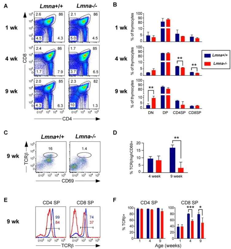
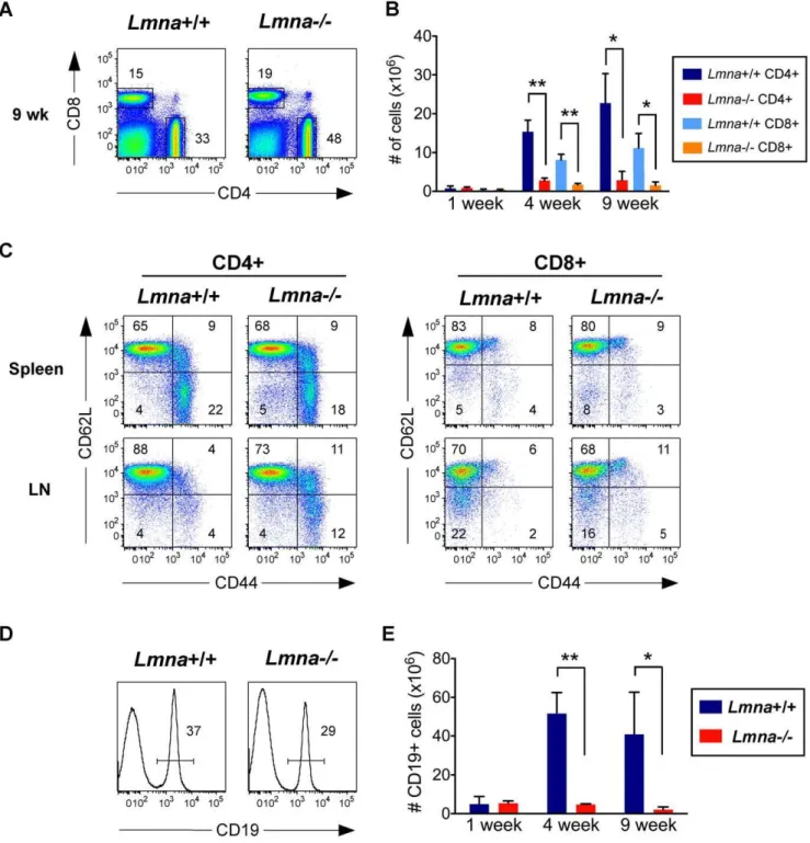
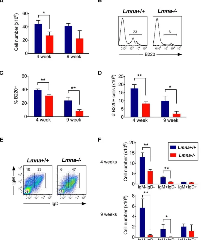
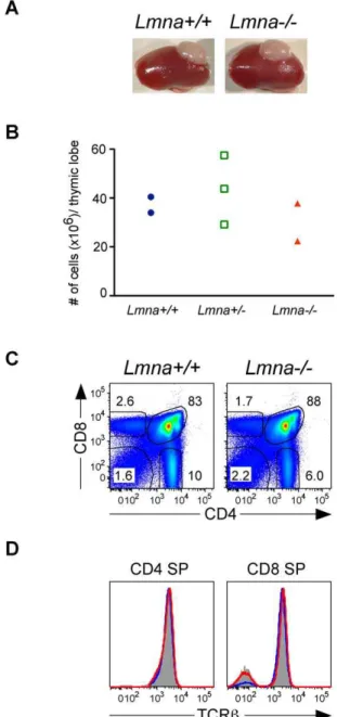
Cell-extrinsic defective lymphocyte development in Lmna(-/-) mice.
Texto
Imagem
Documentos relacionados
Increased proliferation of human synovial mesen- chymal stem cells with autologous human serum: compari- sons with bone marrow mesenchymal stem cells and with fetal bovine serum.
Our large fertility study showed no age related differences in the group of ex cryptorchid patients having defective germ cell development (no Ad spermatogonia) indicating that in
Our observation of defective development of the smooth musculature in the epididymis in Insl3 homozygous mutant mice, combined with its high intraabdominal undescended
Given that we observed defects in the survival of migrating cranial neural crest cells together with defects in placode development, we carefully examined the interactions between
T cells. Cells from A20 tumor-bearing mice were examined.. primed in secondary lymphoid tissues, are the major subset of Tregs in tumors. Additionally, the results provide insights
Our data indicate that while the development of single positive CD4 + and CD8 + T cells in the thymus remained intact in granzyme B-deficient mice, the peripheral pool of CD8 + T
counterparts in part due to mitotic defects. Defects in mitosis have been reported previously for other LMNA mutant cells [6]. Here, we further show that Lmna Dhe/ + mitotic
Studies had shown that Dfg5p and its homologous defective cell wall protein (Dcw1p) of Saccharomyces cerevisiae are required for cell growth and cell wall biogenesis and are
