Assessment of pharyngeal airway space
using Cone-Beam Computed Tomography
Sabrina dos Reis Zinsly*, Luiz César de Moraes**, Paula de Moura***, Weber Ursi****
Introduction: Evaluation of upper airway space is a routine procedure in orthodontic
di-agnosis and treatment planning. Although limited insofar as they provide two dimensional images of three-dimensional structures, lateral cephalometric radiographs have been used routinely to assess airway space permeability. Cone-Beam Computed Tomography (CBCT) has contributed to orthodontics with information concerning the upper airway space. By producing three-dimensional images CBCT allows professionals to accurately determine the most constricted area, where greater resistance to air passage occurs. Objectives: The purpose of this article is to enlighten orthodontists on the resources provided by CBCT in the diagnosis of possible physical barriers that can reduce upper airway permeability.
Abstract
Keywords: Cone-Beam Computed Tomography. Pharynx. Upper airway space.
* Specialist in Orthodontics, PROFIS/Bauru. MSc in Oral Biopathology, area of Dental Radiology, UNESP - São José dos Campos. ** Head Professor of Dental Radiology, UNESP.
*** Specialist in Dental Radiology. MSc in Oral Biopathology, area of Dental Radiology, UNESP.
**** MSc and PhD in Orthodontics, Bauru, USP. Chairman - UNESP - São José dos Campos. Head of the Specialization Program in Orthodontics, APCD - São José dos Campos, Brazil.
INTRODUCTION
Clinicians and researchers involved in the treatment of dentofacial deformities have sought to elucidate the determinants of facial morphol-ogy. The relationship between respiratory pattern disorders and changes in facial morphology has been extensively debated in the literature1,2 and remains controversial. Conflicting opinions can be divided into two camps: One that considers breathing pattern an important etiological factor in producing the long face syndrome (LFS) and one which believes that LFS expresses an inher-ited pattern and breathing pattern would act only as an aggravating factor. Currently the prevailing
view is that skeletal morphology is a result of ge-netically determined growth superimposed by the action of its functional matrix. And, according to this view, the action of soft tissue genotype would continue during growth.
Several factors may be associated with mouth breathing, among which are constriction of the nasal passage, narrow or obstructed nasopharynx, hypertrophic nasal membranes, enlarged turbi-nates, hypertrophic palatine or pharyngeal tonsils, nasal septal deviation, choanal atresia and tumors in the nose or nasopharynx.
or due to the narrow anatomical structure of the nasopharynx—the resulting functional imbal-ance can impact craniofacial growth and develop-ment, reflected in a tendency toward vertical fa-cial growth, which leads to the stereotype of the adenoid face or long face syndrome (LFS). This syndrome is characterized by lip incompetence, underdeveloped nostrils, maxillary atresia with the presence of deep palate and posterior crossbite, increased anterior inferior facial height, increased gonial angle and mandibular retrognathism.2,3,4 Because LFS is a multifactorial syndrome it is not always easy to diagnose and, to be successful, treat-ment requires a multidisciplinary approach.
The upper airway space can be described in terms of height, width and depth. It is known that the limiting factor determining respiratory capac-ity is a reduced cross-sectional air passage area5,6 anywhere in the pharyngeal path.
Over the past century extensive research1,7-10 was conducted to elucidate the relationship between craniofacial morphology and breath-ing pattern. Most studies were based on lateral cephalometric radiographs because such radio-graphs are part of the records used for proper planning of orthodontic treatment. Although it can provide a wealth of information, cephalo-metric radiography is limited in the sense that it produces two-dimensional images (height and depth) of a three-dimensional structure, there-fore hindering accurate assessment of the size and complexity of this structure.
Cone-Beam Computed Tomography has made it possible to acquire 3D image volumes of all structures in the maxillofacial complex. With the use of specific software and acquisition protocols based on individual needs, these digi-tal volumetric scans can be turned into multiple planar view images (axial, coronal and sagittal). Software tools also allow bone structure mea-surements to be obtained as well as 3D assess-ment of soft tissues, and the shapes, volumes and features of the face and upper airways.
Currently, assessment of upper airway space is a routine procedure in orthodontic diagnosis and treatment planning. Cone-Beam CT equipment has become more efficient, reducing acquisition time and developing specific software, which provides improved image processing and analy-sis of three-dimensional images of the structures comprised in the maxillofacial region. This infor-mation may provide clinical benefits and a foun-dation for rational decision-making regarding the appropriate treatment to be administered to growing individuals with decreased pharyngeal airway space in order to minimize the etiological influence of breathing pattern on the develop-ment of malocclusion.
AssessINg UppeR AIRwAy spACe
Understanding the morphology and func-tion of the skeletal structures and soft tissue that make up the upper airway space is essential for an understanding of the physiology and patho-genesis of obstruction. Assessment is complex however because of its location, which does not allow direct visualization. Different forms of im-age-based exams have been used to evaluate the upper airway space, skeletal structures and adja-cent soft tissues. Each method has inherent ad-vantages and disadad-vantages, and there is no con-sensus regarding the gold standard procedure for evaluation. Among the methods used are acous-tic rhinometry, fluoroscopy, nasopharyngoscopy, MRI, cephalometry and tomography.11
Over the last century a large number of tests were suggested for evaluation of upper air-way space from lateral radiographs using linear and angular measurements, and sagittal areas between cephalometric landmarks.12-15 These points are defined by superimposing projections of different structures.
the radiographs and the volume obtained from CBCT, although the latter showed greater vari-ability in patients with similar airway space in lat-eral cephalometric radiographs. This is expected since cephalometric analysis of conventional later-al radiographs only measures pharynx height and depth and therefore does not allow cross-sectional (i.e., width) examination.
Clinically, orthodontists can assess obstructed airway space in conventional cephalometric ra-diography. When this obstruction is considered severe, the patient is referred to an otolaryngolo-gist. It is imperative that more accurate diagnostic tools be employed that inform otolaryngologists and orthodontists on the proper procedures to be adopted, thereby averting obstacles in the air pas-sage that can affect dentition, speech, and cranio-facial development.
ACQUIRINg CBCT sCANs FOR AIRwAy AssessMeNT
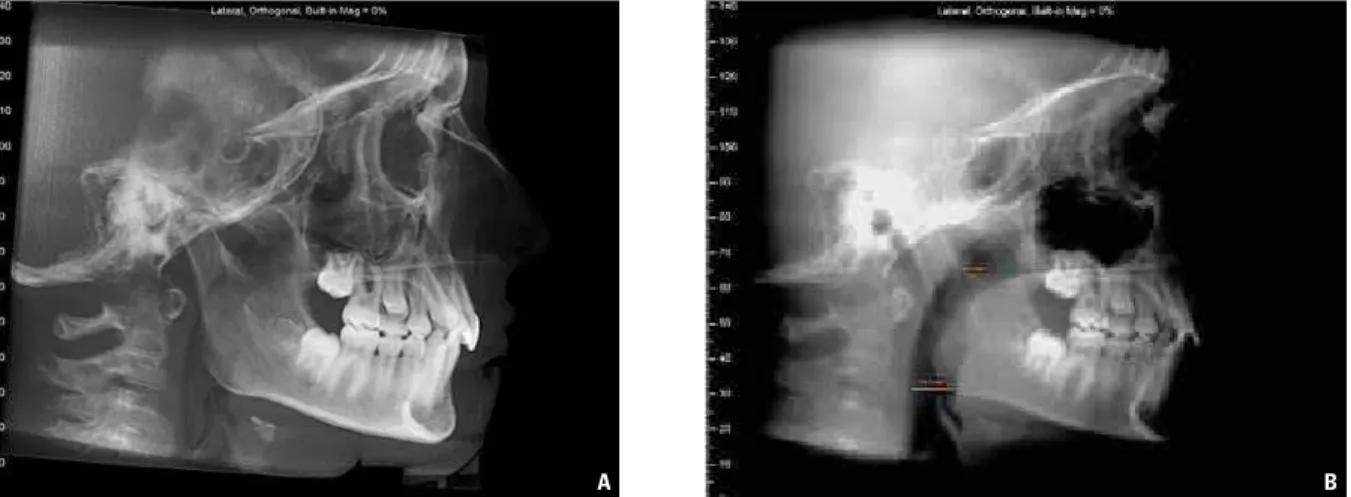
CT examinations for assessing the airways have a specific image acquisition protocol. Pa-tients must be sitting, in maximum intercuspa-tion, with the midsagittal plane perpendicular to the horizontal plane and Frankfort plane par-allel to the horizontal plane. An extended field of view (EFOV) of 17X 23 cm should be used; 0.25 mm voxel size; 40 seconds. Upon comple-tion of the CBCT examinacomple-tion, some manipu-lations can be performed using the software provided by the scanner manufacturer. The raw image (raw data) is reconstructed to enable vi-sualization of 3D reconstruction and multiple planar cross-sections. These two-dimensional images of the pharynx can be examined from any direction. The most commonly used are sag-ittal, coronal and axial (Fig 1).
Images can be better observed using specific tools. Images can be rotated and magnified to al-low better assessment of a given region. Images can also be rendered from any angle, and in any scale or position. Different filters can be applied,
allowing differentiation between tissues of differ-ent densities and the use of transparency, which enables hard tissue to be viewed through soft tis-sue. A linear measurement tool is also available, which can measure height, width and depth of any portion of the pharynx (Fig 2).
These images can also be converted to DICOM (Digital Imaging and Communications in Medi-cine) files that can be exported to other 3D assess-ment software, which in turn enables a wider range of resources useful in airway space evaluation.
VIewINg THe UppeR AIRwAy spACe UsINg CONe-BeAM COMpUTeD TOMOgRApHy
Software is available for assessment of the up-per airway space, such as InVivoDental, 3dMD-vultus and Dolphin Imaging.17
Dolphin Imaging program version 11.0 is an airway space analysis tool that not only enables the evaluation of the shape and contour of the up-per airway space in three dimensions, but also cal-culates volume, sagittal area and the smallest pre-defined cross-sectional area in the airway space. It provides segmentation of the upper airway space through images that can be rotated and magnified. The program features two threshold filters: For hard tissue and soft tissue, displaying the airway space together with skeletal tissue or separately.
Once a tool is selected for evaluating the air-way space it is necessary to define, in the sagit-tal cross-section, the area of interest in the airway space. The program automatically provides the area and total volume of any predefined region as well as location and dimensions of the most con-stricted airway space area (Fig 5).
CReATINg TwO-DIMeNsIONAl pROjeCTIONs FROM A
THRee-DIMeNsIONAl IMAge
Most of these cephalometric landmarks created for two-dimensional images cannot be viewed or are difficult to trace on the curved surface of
three-dimensional images. Currently, for ethical reasons, longitudinal growth records are forbidden, and there are as yet no normative standards for these three-dimensional dimensions. However, the parameters established for two-dimensional images can be com-pared with three-dimensional records.18,19 Softwares have been developed using algorithms that allow projections to be generated similarly to radiographs. These projections can show morphological changes in maxillofacial structures in the 3 orthogonal planes, which might contribute to air passage obstruction.
To create these radiographic projections from a volumetric CT using Dolphin 3D Imag-ing program version 11.0 (Dolphin ImagImag-ing and
FIGURE 1 - Opening screen of the XoranCat software provided by the man-ufacturer of the i-CAT scanner, showing the multiple planar views (MPV) (sagittal, coronal and axial) obtained from volumetric reconstruction. The cursor, represented by two intersecting lines, indicates the precise loca-tion in virtual space, making it possible to go through these two-dimen-sional images of the pharynx in any direction.
FIGURE 2 - XoranCat software screen, where anatomy can be evaluated and measurements of the pharyngeal structure performed in any slice.
FIGURE 3 - Dolphin 3D software object orientation screen. In frontal view, the midsagittal plane should coincide with the individual’s median plane, and the axial plane must be tangent to the infraorbital rim.
B A
Management Solutions, Chatsworth, CA), it is first necessary that the image be properly oriented. In the radiographic projection construction module, the program lets one choose an orthogonal projec-tion or perspective. The upper and lower limits of the image must be set, as well as its thickness. Once the projection has been created, different types of display filters can be applied. Ray-sum is the filter that provides the best visualization of upper airway space (Figs 6 and 7).
The program also features a measurement tool and cephalometric analysis tool, providing
linear and angular measurements in these two-dimensional images, which enable the evalua-tion of craniofacial factors that may contribute to the obstruction of the upper airway space (retrognathism, crossbite, asymmetries, hyper-trophic tonsils).
AssessINg MORpHOlOgy IN 3D ReCONsTRUCTIONs
3D reconstructions also allow assessment of airway space morphology. Resistance to air flow is related to airway space size and shape. Airway
FIGURE 5 - Using Dolphin Imaging Program version 11.0 airway space assessment tool one can obtain the sagittal area, volume and smallest cross-sectional area of a predefined pharyngeal airway space. To this end, one must choose the area of interest by moving the markers that define the green line, starting from the sagittal cross-section.. The yellow marker is then placed within the airway space, and the program performs the calculation of sagittal area and volume. In order to obtain the smallest cross-sectional area, one should drag the red reference lines delimiting the area to be evaluated.
FIGURE 6 - Dolphin Imaging program’s radiograph creation tool. One must choose the type of projection desired. In this case, a right lateral projec-tion was selected with the applicaprojec-tion of Dolphin filter 1, which allows better definition of skeletal structures.
B A
space can be large, but a winding path can offer considerable effective resistance to air flow and affect respiratory function. Studies using CBCT have established a correlation between airway space and facial pattern. The oropharyngeal air-way space of individuals with Class III anteropos-terior skeletal pattern appears to be wider and more flattened,20 displaying a more vertical orien-tation relative to the sagittal plane.17 Individuals with Class II anteroposterior skeletal pattern, on the other hand, showed a more anterior superior airspace.17 Abransom et al21 also evaluated chang-es in the shape of the pharynx and argued that with age the airway space becomes wider in the transverse direction and therefore more elliptical. Ogawa et al23 associated the shape of the airway space with Obstructive Sleep Apnea Syndrome (OSAS). OSAS patients had a more elliptical or concave air space, unlike non-OSAS individuals, who exhibited a more rounded or square shape.
UppeR AIRwAy spACe AssessMeNT AND OsAs
Obstructive Sleep Apnea Syndrome (OSAS) is a disease characterized by the collapse of the pharyngeal airway space resulting in repeated
episodes of air passage obstruction, decreased oxygen saturation and sleep disruption. The anatomy of the upper airway space seems to play a critical role in the pathogenesis respon-sible for upper airway space collapse in OSAS patients. Collapse may occur at different spots in the upper airway space of OSAS patients. The retroglossal and retropalatal regions are most fre-quently involved.22 It is known that the pharynx is bounded by a musculomembranous wall sup-ported by a skeletal framework, so that the loca-tion of the most constricted area depends on the relationship between craniofacial skeletal struc-tures and surrounding soft tissue. Therefore, the tonsils and adenoids, soft palate, uvula, tongue and lateral pharyngeal walls are soft tissue struc-tures crucial in defining the upper airway space. Moreover, the mandible and hyoid bone are the major skeletal determinants of the airway space. Any abnormality in these structures can affect the airway space and cause SAOS.22
SOAS has a multifactorial etiology involving among others a reduced upper airway space, nasal cavity obstruction, distributed body fat mass and muscle tone. The upper airway space is significantly constricted in OSAS compared with non-OSAS
patients, although the most constricted region var-ies from OSAS patient to OSAS patient.
Treatment of OSAS is primarily geared towards airway space maintenance, which is achieved with the use of a ventilation therapy device named CPAP—continuous positive airway pressure— which provides a constant air flow while keeping the airways open.
Secondarily, treatment seeks to make the air-way space less likely to collapse. Increased pharyn-geal airway space can be obtained in a reversible manner, with the use of removable appliances, or permanently, with surgery. When secondary treatments are needed, the most constricted oro-pharyngeal area must be identified in order to determine an appropriate treatment solution. To be able to assess upper airway space morphology, determine the degree and location of constric-tion and evaluate the effectiveness of treatment, examinations such as nasopharyngoscopy with Muller maneuver, fluoroscopy, cephalometry, rhi-nomanometry, MRI and CT have been employed. Cephalometric studies have shown that indi-viduals with OSAS have smaller, retruded man-dibles, narrowing of the posterior airway space, larger tongues, more inferiorly positioned hyoid bone and retropositioned maxilla when compared with non-OSAS individuals23. Although this in-formation is valuable, it does not enable clinicians to have access to the complex morphology of the upper airway space.
Because CBCT is three-dimensional, it allows clinicians to assess the airway space and surround-ing structures, and determine three-dimensional naso-, oro- and hypopharyngeal measurements, such as the most constricted area, volume and the smallest anteroposterior and lateral pharyngeal di-mensions in OSAS patients. One can also evaluate changes that might potentially be induced by the treatment modality itself, and identify which pa-tients would benefit from such treatment (Fig 9). Haskell et al24 asserted that it was possible to pre-dict the amount of increase in total volume and
in the cross-sectional area of the oropharynx ob-tained through appliance-induced mandibular ad-vancement, since the most constricted area could move to any higher or lower point in the pharynx. They argued therefore that CT evaluation would be necessary prior to installing the appliance to determine whether the patient would benefit from its use. They further stressed that, in treating OSAS, it is more important to achieve improve-ment in the most constricted area than to increase the volume of the pharynx as a whole.
ClINICAl IMplICATIONs AND lIMITATIONs OF CBCT IN AssessINg THe UppeR AIRwAy spACe
Besides the anatomy of the skeletal and soft tissue, airway space depends on some dynamic variables such as lung volume, intraluminal and extraluminal pressure, muscle tone and head po-sition.21 Since the soft palate and the tongue are structures composed of soft tissue with no rigid support, they are greatly affected by gravitational forces. Therefore, in CT scans and other exami-nations performed in the supine position, these structures move further toward the posterior pharyngeal wall, which results in changes in the dimensional measurements of the upper airway space, as demonstrated by Lowe et al,25 Huang et al,26 Abramson et al21 and Ono et al.27 Thus, scan results obtained with the patient sitting cannot be extrapolated or even directly compared to those obtained with the individual in the supine posi-tion. The latter position is recommended for in-dividuals with OSAS. Lohse et al28 suggest that in assessing OSAS patients a modification be made to the CBCT acquisition technique, namely, re-moving the chin positioner so that the patient can hold their head in a natural position.
time will be faster in order to prevent patient movements (breathing, swallowing and involun-tary movements) from interfering with the results.
CONClUsIONs
Although no normative data are available regarding information gained through CBCT, a
host of scientific studies have been conducted for this purpose, which leads us to believe that soon CBCT will be able to guide orthodontic diagnosis and planning by enlightening clini-cians about the effects caused by mechanother-apy applied to the airway space and the conse-quences of these effects.
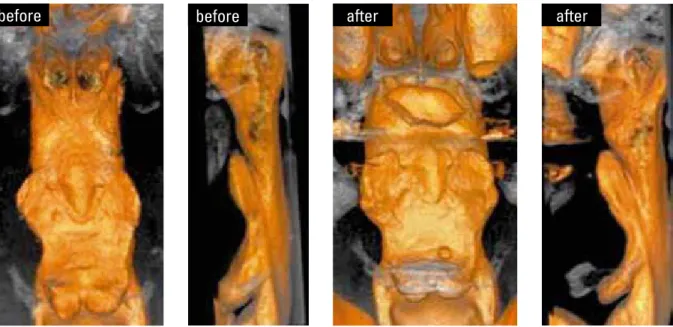
FIGURE 9 - CT images obtained with i-CAT software, illustrating the increased air space obtained using a mandibular advancement device in the treatment of OSAS.
1. McNamara JA. Inluence of respiratory pattern on craniofacial growth. Angle Orthod. 1981 Oct;51(4):269-300. 2. Vig KW. Nasal obstruction and facial growth: the strength of evidence for clinical assumptions. Am J Orthod Dentofacial Orthop. 1998 Jun;113(6):603-11.
3. Subtelny JD. Oral respiration: facial maldevelopment and corrective dentofacial orthopedics. Angle Orthod. 1980 Jul;50(3):147-64.
4. Hartgerink DV, Vig PS. Lower anterior face height and lip incompetence do not predict nasal airway obstruction. Angle Orthod. 1989 Spring;59(1):17-23.
5. Warren DW, Hairield WM, Seaton D, Morr KE, Smith LR. The relationship between nasal airway size and nasal-oral breathing. Am J Orthod Dentofacial Orthop. 1988 Apr;93(4):289-93.
6. Hinton VA, Warren DW, Hairield WM, Seaton D. The relationship between nasal cross-sectional area and nasal air volume in normal and nasally impaired adults. Am J Orthod Dentofacial Orthop. 1987 Oct;92(4):294-8.
7. Ricketts RM. Respiratory obstruction syndrome. Am J Orthod. 1968 Jul;54(7):495-507.
8. Mergen DC, Jacobs RM. The size of nasopharynx associated with normal occlusion and Class II malocclusion. Angle Orthod. 1970 Oct;40(4):342-6.
9. Tourne LP. The long face syndrome and impairment of the nasopharyngeal airway. Angle Orthod. 1990 Fall;60(3):167-76. 10. O’Ryan FS, Gallagher DM, LaBanc JP, Epker BN. The
relation between nasorespiratory function and dentofacial morphology: a review. Am J Orthod. 1982 Nov;82(5):403-10. 11. Schwab RJ, Goldberg AN. Upper airway assessment:
radiographic and other imaging techniques. Otolaryngol Clin North Am. 1998 Dec;31(6):931-68.
12. Major MP, Flores-Mir C, Major PW. Assessment of lateral cephalometric diagnosis of adenoid hypertrophy and posterior upper airway obstruction: a systematic review. Am J Orthod Dentofacial Orthop. 2006 Dec;130(6):700-8. 13. Martin O, Muelas L, Vinas MJ. Nasopharyngeal
cephalometric study of ideal occlusions. Am J Orthod Dentofacial Orthop. 2006 Oct;130(4):436 e1-9.
14. Handelman CS, Osborne G. Growth of the nasopharynx and adenoid development from one to eighteen years. Angle Orthod. 1976 Jul;46(3):243-59.
15. Poole MN, Engel GA, Chaconas SJ. Nasopharyngeal cephalometrics. Oral Surg Oral Med Oral Pathol. 1980 Mar;49(3):266-71.
16. Aboudara C, Nielsen I, Huang JC, Maki K, Miller AJ, Hatcher D. Comparison of airway space with conventional lateral headilms and 3-dimensional reconstruction from cone-beam computed tomography. Am J Orthod Dentofacial Orthop. 2009 Apr;135(4):468-79.
ReFeReNCes
17. Grauer D, Cevidanes LS, Profit WR. Working with DICOM craniofacial images. Am J Orthod Dentofacial Orthop. 2009 Sep;136(3):460-70.
18. Moshiri M, Scarfe WC, Hilgers ML, Scheetz JP, Silveira AM, Farman AG. Accuracy of linear measurements from imaging plate and lateral cephalometric images derived from cone-beam computed tomography. Am J Orthod Dentofacial Orthop. 2007 Oct;132(4):550-60.
19. Kumar V, Ludlow JB, Mol A, Cevidanes L. Comparison of conventional and cone beam CT synthesized cephalograms. Dentomaxillofac Radiol. 2007 Jul;36(5):263-9.
20. Iwasaki T, Hayasaki H, Takemoto Y, Kanomi R, Yamasaki Y. Oropharyngeal airway in children with Class III malocclusion evaluated by cone-beam computed tomography. Am J Orthod Dentofacial Orthop. 2009 Sep;136(3):318.e1-9.
21. Abramson Z, Susarla S, Troulis M, Kaban L. Age-related changes of the upper airway assessed by 3-dimensional computed tomography. J Craniofac Surg. 2009 Mar;20(Suppl 1):657-63. 22. Schellenberg JB, Maislin G, Schwab RJ. Physical indings and the
risk for obstructive sleep apnea. The importance of oropharyngeal structures. Am J Respir Crit Care Med. 2000 Aug;162(2 Pt 1):740-8. 23. Ogawa T, Enciso R, Shintaku WH, Clark GT. Evaluation of
cross-section airway coniguration of obstructive sleep apnea. Surg Oral Med Oral Pathol Oral Radiol Endod. 2007 Jan;103(1):102-8. 24. Haskell JA, McCrillis J, Haskell BS, Scheetz JP, Scarfe WC, Farman
AG. Effects of Mandibular Advancement Device (MAD) on airway dimensions assessed with cone-beam computed tomography. Semin Orthod. 2009;15(2):132-58.
25. Lowe AA, Ono T, Ferguson KA, Pae EK, Ryan CF, Fleetham JA. Cephalometric comparisons of craniofacial and upper airway structure by skeletal subtype and gender in patients with obstructive sleep apnea. Am J Orthod Dentofacial Orthop. 1996 Dec;110(6):653-64.
26. Huang J, Shen H, Takahashi M, Fukunaga T, Toga H, Takahashi K, et al. Pharyngeal cross-sectional area and pharyngeal compliance in normal males and females. Respiration. 1998;65(6):458-68.
27. Ono T, Otsuka R, Kuroda T, Honda E, Sasaki T. Effects of head and body position on two- and three-dimensional conigurations of the upper airway. J Dent Res. 2000 Nov;79(11):1879-84.
28. Lohse AK, Scarfe WC, Shaib F, Farman AG. Obstructive sleep apnea-hypopnea syndrome: Clinical applications of cone beam CT. Aust Dent Pract. 2009;Sep-Oct:122-32.
Contact address Sabrina dos Reis Zinsly Rua Atibaia, 100 - Jd Apolo CEP: São José dos Campos / SP E-mail: szinsly@hotmail.com