Faculdade de Ciências
Departamento de Química e Bioquímica
Molecular switches in CFTR processing
and trafficking: the role of the novel
interactor Rap1A
João Miguel Parente Fernandes
Dissertação
Mestrado em Bioquímica
Especialização em BioquímicaOrientador: Professor Doutor Carlos Farinha 2011/2012
Index
I Acknowledgements……….….iii II Resumo……….v III Abstract………..viii IV Abbreviations………..ix 1 Introduction ... 11.1 Cystic Fibrosis - Overview ... 1
1.2 Clinical features of Cystic Fibrosis and therapeutics ... 2
1.3 CFTR structure ... 3
1.4 CFTR function ... 4
1.5 CFTR folding ... 5
1.6 CFTR trafficking ... 6
1.7 The CFTR interactome and Rap1A ... 7
1.8 Rap1A – an overview ... 8
1.9 Rap1A post-translational modifications ... 10
1.10 RapGEFs and RapGAPs ... 11
1.11 Rap effectors and function ... 12
2 Objectives ... 14
3 Materials and Methods ... 15
3.1 Production and characterization of vectors to study the impact of Rap1A on CFTR biogenesis ... 15
3.1.1 Bacterial strain ... 15
3.1.2 Plasmid vectors and siRNAs ... 15
3.1.3 Production of competent bacteria ... 15
3.1.4 Transformation of competent bacteria ... 16
3.1.5 DNA extraction and quantification ... 16
3.1.6 DNA sequencing ... 17
3.1.7 Cloning ... 17
3.1.8 Mutagenesis ... 17
3.2 Biochemical and electrophysiological analysis of Rap1A impact on CFTR biogenesis ... 19
3.2.1 Characterization, Culture and Maintenance of cell lines ... 19
3.2.2 Transfection ... 20
3.2.4 Western blot ... 21
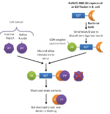
3.2.5 Rap1A activity assay ... 22
3.2.6 Immunoprecipitation ... 23
3.2.7 Immunofluorescence ... 23
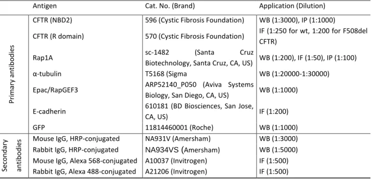
3.2.8 Antibodies ... 24
3.2.9 Ussing Chamber assays ... 24
4 Results ... 26
4.1 Rap1A expression in CFBE cells and primary cultures of human bronchial cells 26 4.2 Co-immunoprecipitation ... 28
4.3 Molecular cloning and mutagenesis ... 28
4.4 Transfection optimization ... 32
4.5 Rap1A expression and activity and its impact on steady-state CFTR... 35
4.6 Ussing chamber analysis of CFTR function upon down-regulation of Rap1A 37 5 Discussion ... 40
5.1 Validation of the central questions ... 40
5.2 Objective 1 – Producing plasmid constructs... 41
5.3 Objective 2 – Optimizing transfection and assessing Rap1A expression and activity ... 42
5.4 Objective 3 – Validating the CFTR-Rap1A interaction ... 43
5.5 Objective 4 – Effect of up and down-regulation of Rap1A on CFTR ... 43
6 Future perspectives ... 45
7 Bibliography ... 46
8 Appendices ... 50
8.1 Appendix I – pCMV6-AC-Rap1A-GFP plasmid map and cloning scheme ... 50
8.2 Appendix II – pCMV6-AN-GFP plasmid map and cloning scheme ... 51
8.3 Appendix III – Preparation of GST-RalGDS-RBD-coupled beads for Rap1A activity assays ... 52
Acknowledgements
I would like to express my gratitude first and foremost to my supervisor, Prof. Carlos Farinha, who was always very supportive of my work, especially when it didn’t go as planned. I owe to him what has become my personal motto in everything concerning life in the lab: try better, fail better. I am also grateful to Prof. Margarida Amaral, for welcoming me into her group and providing me with a very stimulating work environment in which I never lacked for intellectual challenge.
Special thanks are due to the many people in the group which helped me in the daily life in the lab. Sara was a huge support for the whole year, and kept me company everytime I went for snacks or needed to complain (which was very often); this year would have been much duller without her. Simão was always there to point the way in everything biochemical, to provide me with cheesy music and to pester me about my plans for the future. Verónica was always a constant source of laughter and support, especially when we made fun of Sara. I probably wouldn’t be presenting this thesis if it weren’t for Marisa and her help with the Ussing chambers, with planning experiments, and analyzing data. Thank you for helping me see that I actually had some results! Marta was as instrumental for my work as everyone else combined, for making all the operations in the lab run smoothly, for providing me with reagents, materials and cells whenever needed and for all the moments of fun. I also had a lot of help from Inna and Anabela, especially with the molecular biology stuff and with the more confusing and unexpected results. Finally, my days in the lab (and out of it) were made a lot more fun by the comraderie and support of Ana Luísa, Ana Margarida, Jorge, Georgina and Johnny Johnny, and lately also by Sara Afonso and Johnny Johnny Johnny.
I am also grateful to Marta Sousa Silva, who was always helpful and interested; I might still be struggling with my cloning experiments if it weren’t for the ligase and ligase buffer she kindly provided me.
Outside of the lab, my friends were indispensable. Special thanks are due to Carapeta, for being the perfect wingman, my personal jiminy cricket and an awesome friend. Carlos is an alternating source of fun and boredom, but I would have it no other way, even though he sometimes seems to doubt our friendship. Cátia has long been the most constant of friends and, even though we fight over so many little things, I know she’ll be there when I’m in a nursing home, polishing my Nobel Prize in my wheelchair with my hearing aid turned off. To Ruca (born João Tiago Vieira), I hope you know that the fact that I get green with envy every time you talk of your results, or how your thesis
is going, is actually the best compliment I could give you and not a sign of ill will towards you. And I’m sorry I keep forgetting your birthday!
I must also thank my family for supporting me in this five-year endeavor of getting a college education. To my parents; it’s been a rocky road, but I think we’re getting in the right track. Achieving this was only possible thanks to you and your dedication to my education. To my grandparents, Rita and Eduardo, who gave me an example to follow, a home and wise advice (that I should follow more often), a very big thank you. Finally, I thank my sister (and, sadly, no longer roommate), Damiana, for the camaraderie, support and hours of laughter and to my aunt, Clara, for always being there whenever needed.
I Resumo
A Fibrose Quística é a doença recessiva autossómica letal mais comum na população caucasiana. É caracterizada do ponto de vista clínico por um declínio rápido na função pulmonar, com obstrução das vias aéreas devido à ineficaz remoção de muco e consequentes infecções bacterianas recorrentes. Estas alterações estão associadas desde muito precocemente a um ambiente de hiperinflamação, que exacerba os processos de remodelação do tecido pulmonar e acaba por culminar na fibrose dos tecidos e perda da sua função. Além deste fenótipo respiratório, os pacientes apresentam ainda problemas digestivos graves, nomeadamente a nível pancreático, intestinal e hepático, e são inférteis no caso dos homens e de uma elevada percentagem das mulheres.
A fibrose quística é causada por mutações no gene CFTR (do inglês Cystic
Fibrosis Transmembrane Conductance Regulator) que codifica para uma proteína com
o mesmo nome. Neste momento, conhecem-se cerca de 1930 mutações causadoras da doença, sendo a mais comum a delecção de três nucleótidos que correspondem ao resíduo de fenilalanina na posição 508 da proteína. Esta mutação está presente em cerca de 90% dos pacientes.
Esta proteína é expressa em células epiteliais de vários tecidos, e além de funcionar como um canal de cloreto e outros aniões na membrana apical dessas células, é também responsável por regular outros canais e transportadores, tendo um impacto global no transporte iónico epitelial. A fosforilação pela proteína cinase A em resposta a aumento dos níveis de AMP cíclico é considerada o principal mecanismo responsável pela abertura do canal.
Ao ser sintetizada, a CFTR é inserida co-traducionalmente na membrana do retículo endoplasmático, onde adquire a sua conformação nativa com o auxílio de vários chaperones aí presentes e sofre uma glicosilação inicial. Do retículo endoplasmático, é transportada para o Golgi, onde os seus resíduos glicídicos são processados até atingir uma forma madura, que é transportada em vesículas para a membrana plasmática, onde exerce a sua função. Uma vez na membrana, a CFTR é endocitada e segue uma de duas vias: ou é reciclada de volta para a membrana ou é enviada para degradação no lisossoma. O balanço destes processos determina a quantidade de proteína presente na membrana, e como tal são relevantes para a compreensão do defeito molecular que origina a doença..
As interacções que a CFTR estabelece com outras proteínas ao longo deste trajecto são determinantes para que a proteína consiga exercer convenientemente a sua função. Recentemente, a pequena GTPase Rap1A foi identificada como
potencialmente interagindo com a CFTR. O objectivo deste trabalho consistiu na validação dessa interacção e no estabelecimento de um sistema experimental que permitisse avaliar o impacto da Rap1A na biogénese, tráfego e função da CFTR.
A Rap1A é uma pequena GTPase da família Ras que está activa quando ligada a GTP e inactiva quando ligada a GDP. A interconversão destas duas formas é auxiliada por proteínas reguladoras chamadas GEFs (activadores) e GAPs (inactivadores), que respondem a uma variedade de estímulos intra e extracelulares. Quando activa, a Rap1A induz alterações conformacionais em determinadas proteínas alvo, envolvidas na regulação da adesão à matriz extracelular e célula-célula, dinâmica do citoesqueleto e polarização celular, alterando a sua função.
O primeiro passo do trabalho foi avaliar a expressão da Rap1A no tecido mais relevante do ponto de vista clínico, o pulmão. Para isto, realizaram-se experiências de
Western Blot a partir de culturas primárias de células epiteliais dos brônquios de
indíviduos saudáveis e de uma linha celular do epitélio brônquico de pacientes com fibrose quística (CFBE) que sobreexpressa CFTR. Observou-se em ambos os tipos celulares expressão da proteína.
A estratégia escolhida para validar a interacção entre a CFTR e a Rap1A foi tentar co-imunoprecipitar ambas as proteínas. No entanto, não foi possível validar esta associação, tendo sido unicamente observada uma possível colocalização por ensaios de imunofluorescência.
De seguida, foram produzidos com sucesso plasmídeos para expressão da Rap1A wt em fusão com GFP, bem como de um mutante constitutivamente activo e um dominante negativo. O protocolo de transfecção de células CFBE com estes plasmídeos e com siRNAs foi optimizado, embora as eficiências de transfecção atingidas tenham sido modestas. A expressão dos constructos fluorescentes foi analisada por Western Blot e revelou um problema de folding e/ou processamento, provavelmente decorrente da fusão com a GFP. A actividade dos constructos com Rap1A foi também avaliada com um ensaio de actividade específico. Observou-se que o mutante dominante negativo apresentava uma actividade igual ou superior à do constructo wt, enquanto o constitutivamente activo apresentava uma actividade igual ou inferior, o que levou a abandonar ambos estes mutantes. Estas discrepâncias em relação à literatura devem-se provavelmente à marcação com GFP. Em nenhuma destas experiências se observaram alterações na quantidade de CFTR total ou madura. O facto de não se ter observado um efeito pode ter sido devido à baixa eficiência de transfecção obtida, à baixa quantidade de proteína com o
Resumo vii Finalmente, recorreu-se a câmaras de Ussing para analisar a função da CFTR em monocamadas polarizadas de células CFBE transfectadas com siRNA contra a Rap1A ou siRNA scrambled. Não se observaram diferenças na corrente transepitelial induzida pelo tratamento com forscolina (um composto que faz aumentar os níveis de AMP cíclico na célula, activando a CFTR) ou com genisteína (um potenciador específico da CFTR que maximiza a abertura do canal) em células tratadas com siRNA contra a Rap e tratadas com siRNA scrambled. Posteriormente, analisando por Western Blot a expressão de Rap1A nestas células, concluiu-se que a transfecção foi pouco eficiente, o que justifica a ausência de efeito.
De uma forma geral, houve dois factores limitantes neste trabalho: o facto de, apesar de um longo processo de optimização, as eficiências de transfecção continuarem bastante modestas, e o facto de o sistema de expressão utilizado ser de difícil utilização.
As observações mais significativas resultantes deste trabalho são:
- Tanto em culturas primárias de células epiteliais humanas dos brônquios e em células CFBE41o- observa-se expressão endógena de Rap1A e Epac.
- O padrão de expressão dos factores Epac em células CFBE altera-se quando as células são polarizadas.
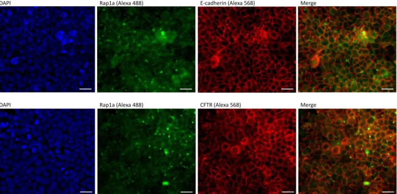
- Rap1A é expressa abundantemente nas junções célula-célula no epitélio respiratório, onde co-localiza com a E-caderina.
- A Rap1A parece co-localizar com a CFTR em células do epitélio respiratório polarizadas.
- A quantidade total de CFTR e a quantidade da forma madura (banda C) estão positivamente correlacionadas com os níveis de expressão endógena de Rap1A.
II Abstract
CFTR, the protein whose malfunction causes cystic fibrosis, is a chloride channel expressed in the apical membrane of epithelial cells. CFTR undergoes a long journey in the cell, since it is translated in association with the endoplasmic reticulum until its degradation, throughout which it interacts with numerous proteins. One of these was recently identified as Rap1A, a small GTPase that regulates cell-cell and cell-matrix adhesion, cell polarity and cytoskeleton dynamics. The aim of this work was to validate the interaction between CFTR and Rap1A and evaluate the impact of this small GTPase on CFTR biogenesis, trafficking and function.
Rap1A expression was observed by Western blot in primary cultures of human bronchial epithelial cells and in the CFBE cell line. Plasmids encoding for several variants of GFP-tagged Rap1A were generated. Transfection with these or with siRNAs was optimized, although only sub-optimal efficiencies were achieved. Expression of Rap1A constructs was analysed by Western Blot, revealing a folding and/or processing defect for most constructs. Rap1A activity assays showed that the constitutively active mutant didn’t have higher activation levels than the wt construct, whereas the dominant negative had more activity than the wt construct, suggesting that the GFP-tagged constructs were not the most appropriate for this kind of study. No effects on CFTR were observed with Rap1A upregulation, possibly because of the low transfection efficiencies.
Finally, CFTR function upon knockdown of Rap1A was analyzed in Ussing chambers. However, only a minor trend of decrease of CFTR function was observed for Rap1A siRNA transfected samples.
Although suggestive of the need of a different experimental system, the results evidenced that:
- Lung epithelial cells have endogenous expression of Rap1A.
- The band pattern for Epac changed when CFBE cells were polarized;
- Rap1A appears to be enriched in cell-cell junctions, where it co-localizes with E-cadherin;
- Rap1A and CFTR seem to overlap;
- There seems to be a positive correlation between Rap1A levels and both total CFTR expression and band C intensities.
III Abbreviations
ABC – ATP-binding cassete AF6 – afadin
AKAP – A-kinase anchoring proteins
ARAP3 – ArfGAP with RhoGAP domain, ankyrin repeat and PH domain 3 ATP – adenosine triphosphate
BHK – baby hamster kidney (cell line) BSA – bovine serum albumin
C3G – CRK SH3-binding GEF CA – constitutively active
CAAX – cystein-alyphatic-alyphatic-any aminoacid motif CaCC – calcium activated chloride channel
Calu-3 – human caucasian lung adenocarcinoma (cell line) cAMP – cyclic adenosine monophosphate
CAPRI – Ca(2+)-promoted Ras inactivator
CCM1/KRI –T-1 cavernous cerebral malformation protein 1/KREV interaction-trapped 1 CF – cystic fibrosis
CFBE41o-/CFBE – cystic fibrosis bronchial epithelial (cell line) CFTR – cystic fibrosis transmembrane conductance regulator CHO – chinese hamster ovary (cell line)
CIP – CFTR interacting protein COP-II – coating protein II
COS-7 – CV-1 (simian) in Origin, and carrying the SV40 genetic material (cell line) Crk – CT10 Regulator of Kinase
DAPI – 4',6-diamidino-2-phenylindole
DEP – disheveled, Egl-10, pleckstrin domain dsDNA – double stranded DNA
DMSO – dimethylsulfoxide DN – dominant negative E6TP1 – E6-targeted protein 1 E-cad – epithelial cadherin
EDEM – ER degradation-enhancing α-mannosidase-like protein ENaC – epithelial sodium channel
Epac/Repac – exchange factors directly activated by cAMP ER – endoplasmic reticulum
ERAD – endoplasmic reticulum-associated degradation ERM – ezrin-radixin-moesin
ERQC – endoplasmic reticulum quality control FRET – Förster resonance energy transfer GAP – GTPase activating protein
GDP – guanosine diphosphate
GEF – guanine nucleotide exchange factor GFP – green fluorescent protein
GRASP – golgi reassembly stacking protein GST – glutathione S-transferase
GTP – guanosine triphosphate
HBE – human bronchial epithelial cells
Hsc70/Hdj2 – heat shock cognate 70kDa protein/human DnaJ homolog 2 Hsp70/Hdj-1 – heat shock protein with 70kDa/human DnaJ homolog 2 Hsp90 – heat shock protein with 90kDa
HUVEC – human umbilical vein endothelial cells (cell line) IPTG – Isopropyl β-D-1-thiogalactopyranoside
LB – Luria broth LKB1 – liver kinase B1
MALDI-TOF – matrix-assisted laser desorption ionization-time of flight MAPK – mitogen-activated protein kinase
MCF-7 – Michigan Cancer Foundation-7 (cell line) MDCK – Marbin-Darby canine kidney (cell line) MEM – minimal essential medium
MS – mass spectrometry
MSD – membrane-spanning domain NBD – nucleotide binding domain
NHERF – Na+/H+ exchanger regulatory cofactor ORCC – outwardly rectifying chloride channel p130-Cas – Crk-associated antigen 130kDa protein PAGE – poliacrylamide gel electrophoresis
PBS – phosphate buffered saline
PBS-T – phosphate buffered saline supplemented with 0.1% Tween PCR – polimerase chain reaction
PDZ – post synaptic density protein (PSD95), Drosophila disc large tumor suppressor (Dlg1), and zonula occludens-1 protein (zo-1) domain
PKA – protein kinase A
PVDF – polivinylidene difluoride
qRT-PCR – quantitative real-time polimerase chain reaction R-domain – regulatory domain
RA-RhoGAP – Rap-activated Rho GTPase-activating protein RAPL – Rap ligand
RASAL – Ras GTPase-activating-like protein RBD – Rap-binding domain
RIAM – Rap1-GTP-interacting adaptor molecule ROMK – renal outer medullary potassium channel RT – room temperature
siRNA – small interfering RNA SDS – sodium dodecylsulphate
SNARE –soluble N-ethyl-maleimide sensitive factor Attachment Protein receptors Spa-1 – signal-induced proliferation-associated gene-1
SPAL – Spa-1-like protein
SPAR – surfactant protein A binding protein
TIAM1 – T-lymphoma invasion and metastasis-inducing protein 1 UGGT – UDP-glycoprotein glucosyltransferase
UPS – unfolded protein response
VASP – Vasodilator-stimulated phosphoprotein VE-cad – vascular endothelium cadherin YFP – yellow fluorescent protein
1 Introduction
1.1 Cystic Fibrosis - Overview
Cystic Fibrosis (CF) is the most common lethal autosomic recessive disorder in the Caucasian population. It affects 1 in 2500 to 6000 live births and has a carrier frequency of 1 in 25 to 40 individuals. The disease frequency is variable among different ethnic groups, being highest in Northeastern Europe and quite rare among Oriental populations (1:90,000)1.
It was first described in 1938 as a disorder of its own right by Anderson and colleagues, who also came to find that it had a recessive autossomal pattern of inheritance. At the time of its discovery as a disease in its own right, cystic fibrosis was described as a digestive disorder, since the first detectable symptoms were intestinal obstruction in newborns and malnutrition in infants, and was associated with a progressive fibrosis of the pancreatic tissue. Because infants died at a very young age, these were the only symptoms that were noticeable. As the digestive problems in newborns and children were gradually overcome with the advances in healthcare, problems in the respiratory tract became the major cause for morbidity and mortality.
In 1953, an excess of salt in the sweat of patients was identified, which was later attributed to a defect in chloride transport in the sweat glands. This defective chloride transport was also detected in the lung, pancreas and intestinal epithelium, the most affected tissues1. In 1989, the gene responsible for the disease was identified using a positional cloning strategy and cautiously named cystic fibrosis transmembrane conductance regulator (CFTR). This gene encodes for a cAMP-regulated chloride channel expressed in a number of epithelial tissues. It was found to harbour mutations in all CF patients analysed, the most common of which being a deletion of three nucleotides encoding for a phenylalanine in position 508 in the protein (∆F508, or F508del as it is most commonly termed now)2. This mutation is present in about 90% of all patients, and represents only one of the 1930 mutations in the CF gene described at the time this work was written3.
Even though CF research has gone a long way since then, therapies targeting the molecular basis are only now starting to appear and reach the patients. This has only been possible thanks to international, concerted effort in understanding the molecular mechanisms governing CFTR biogenesis and function in health and disease, which will be discussed in this Introduction.
1.2 Clinical features of Cystic Fibrosis and therapeutics
Clinical features of CF are dominated by involvement of the respiratory tract, with obstruction of the airways by thick, dehydrated airway mucus that prevents proper mucocilliary clearance. This leads to recurring bacterial infections, especially with
Pseudomonas aeruginosa and Staphylococcus aureus species. A hyperinflammation
environment is generated in the lungs of patients from a very early age, exacerbating tissue remodelling processes and fibrosis4. This degradation of the lung tissue and loss of its function is the main cause of morbidity and mortality among CF patients. The involvement of the gastrointestinal tract is also common, with 85% of the patients presenting pancreatic insufficiency as a result of the obstruction of the pancreatic ducts. 10 to 20% of newborns with CF present a form of intestinal obstruction called
meconium ileus, and 2 to 5% develop liver disease at some time during the course of
the disease. In adults with CF, infertility is almost universal in males, due to congenital bilateral absence of the vas deferens, and is also frequent in females. Patients also have elevated concentrations of sodium chloride in the sweat; in fact, the sweat test, which measures the amount of salt in the sweat of patients, is still one of the fundamental tools for the establishment of a CF diagnosis5.
Because there are so many different disease-causing mutations in CFTR, mutation-specific treatments are not a viable option. Therefore, they have been grouped in classes based on the functional defect they confer, in order to facilitate the establishment of common therapeutic strategies. Class I mutations abolish protein production. Class II mutations result in defective protein processing; this is the case of the most common F508del mutation. Class III proteins confer defects in the regulation of the channel. Class IV mutations have a defective ion conductance. Class V mutations result in decreased protein synthesis. Class VI mutations decrease the protein’s stability in the membrane.
Most of the different therapeutic approaches that have been used in the last 25 years focus mainly on the amelioration of the symptoms of CF. This includes anti-inflammatory and antibiotic drugs, as well as treatments directed towards restoring the levels of airway surface liquid, preventing mucus accumulation and overcoming the nutritional defects in patients. Currently, therapeutic focused research includes a multiplicity of approaches: search of candidate modifier proteins to identify new potential therapeutic targets and pharmacological therapy to rescue the molecular defects responsible for CF are the most important topics under development6. In January 2012, Kalydeco, the first drug targeting the molecular basis of the disease, was approved by the Food and Drugs Administration in the USA. This compound,
previously named VX-770, corrects the gating defect conferred by several mutations, and has been approved for patients over 6 years of age with the G551D mutation. Clynical phenotype of patients with the much more common F508del mutation is significantly improved by this drug; aditionally, ongoing clinical trials are exploring a combination of VX-770 with another compound, VX-809, that corrects the trafficking defect of proteins with other mutations, including F508del7.
1.3 CFTR structure
The CFTR gene (or ABCC7), is one of the longest in the human genome, approximately 250kb long, and encompasses 27 exons in total. It generates transcripts of about 6.5kb after splicing, which are translated into a protein with 1480 aminoacid residues.
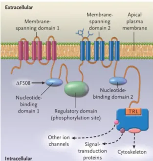
This protein was identified soon after its discovery as a member of the ATP-binding cassette (ABC) transporter family due to structure similarity. Like other ABC transporters, it has two membrane spanning domains (MSDs) with six transmembrane segments each, portions of which form the pore through which anions pass, and two nucleotide binding domains (NBD1 and NBD2). Both NBDs bind and hydrolyse ATP, which drives channel opening and closure, respectively. Recent experimental data suggest that this is linked to dimerization of these two domains8. The most common mutation in cystic fibrosis patients, F508del, is located in the NBD1. In the structure of CFTR there is also a regulatory (R) domain that is absent in all other ABC transporters which contains consensus sites for phosphorylation by various kinases, including protein kinase A (PKA). Phosphorylation of the R domain by PKA in response to cyclic AMP is regarded as the major determinant for opening of the channel. In the C-terminus, CFTR has a PSD95, Dlg1, ZO-1 (PDZ)-binding motif through which it is involved in complex PDZ-based protein interaction networks with a number of proteins9. The N-terminus is also a site for interaction with other proteins, as will be discussed in section 1.7.
Figure 1.1 - Schematic representation of the structure of CFTR. Taken from Rowe (2005)5.
1.4 CFTR function
The traditional function attributed to CFTR is that of a chloride channel. Contrary to other ABC transporters, CFTR is incapable of actively driving ion transport against a gradient; instead, it functions as a passive channel that allows bidirectional flow of ions when open.
The gating of the CFTR Cl− channel is tightly controlled by the balance of kinase and phosphatase activity in the cell and by cellular ATP levels. Activation of PKA by cAMP causes phosphorylation of multiple serine residues within the R domain. Once the R domain is phosphorylated, channel gating is regulated by a cycle of ATP hydrolysis at the NBDs. Finally, protein phosphatases dephosphorylate the R domain and return the channel to its quiescent state10.
This PKA-mediated activation of CFTR depends on the strict compartmentalization of cAMP in the cell. It has been shown that the increase in cAMP necessary for PKA to activate CFTR must be specifically localized near the membrane, in the subcortical compartment11. This compartmentalization is dependent on the integrity of the subcortical cytoskeleton and on the presence of A-kinase anchoring proteins (AKAPs), such as Na+/H+ exchange regulatory co-factors (NHERFs) and ezrin, which anchor PKA, phosphatases, phosphodiesterases and other signaling proteins near CFTR and close to the membrane12.
CFTR function is not restricted to being a chloride channel, though. It also permeates other anions, namely bicarbonate and glutathione.
It is still debated to what extent CFTR, and not the Cl-/HCO3- transporter, which is also expressed in most secretory epithelia, is responsible for bicarbonate transport in
vivo, because its permeability to this ion is low (only about 25% of that of chloride).
However, the fact is that bicarbonate transport is impaired in CF patients, and this accounts at least partially for the loss of pancreatic function13,14, so even if it doesn’t directly permeate this anion it probably regulates channels/transporters that are involved in the process.
Glutathione transport by CFTR, on the other hand, is a well established fact, and it has important effects on the extracellular redox balance, mucocilliary clearance, inflammation and immune response, processes that are impaired in the CF airways and pancreas. Defects in extracellular glutathione homeostasis have been proposed to be one of the major contributors to the exacerbation of inflammation and consequent deterioration of the lung tissue in CF15,16.
Besides acting as an ion channel itself, CFTR also regulates other channels in the cell membrane. The mechanisms underlying this are hard to pin down, because it is not easy to distinguish between effects caused by changes in CFTR itself from those that are consequences of altered chloride conductance. Regulation of the epithelial sodium channel (ENaC) by CFTR is the most studied of these phenomena. ENaC is inhibited by increased CFTR activity, even in chloride-free media. There are three main possible causes for this: an increased chloride conductance might have effects per se in ENaC; CFTR and ENaC might be part of a macromolecular complex with proteins that regulate ENaC by themselves; or CFTR and ENaC might interact directly with one another. Compelling evidence exists to support any of these three hypotheses, so no consensus has been reached so far in this matter. CFTR also regulates other ion channels, including outwardly rectifying chloride channels (ORCCs), renal outer medullary potassium channel (ROMK)17, and calcium activated chloride channels (CaCCs)18.
1.5 CFTR folding
Because CFTR is such a large membrane protein, proper folding is difficult to achieve. In fact, at least in heterologous expression systems, depending on the cell type, only 20-40% of the protein escapes the endoplasmic reticulum quality control (ERQC) system and reaches its native conformation, with the rest being tagged for degradation by the ubiquitin-proteasome pathway. Whereas the NBD1 folds mostly co-translationally, the native structure of the NBD2, and therefore of the full channel, is
only attained post-translationally. This slow, stepwise folding is facilitated by the interaction with several chaperones at the endoplasmic reticulum (ER)19.
As CFTR begins to be translated by the ribosome, an ER targeting motif is recognized by the signal recognition particle, causing it to be taken to the surface of the ER and co-translationally inserted in the ER membrane via the translocon complex. While the nascent polypeptide chain is being inserted in the ER membrane, the exposed parts in the cytosol associate with the cytosolic molecular chaperones Hsp90, Hsc70/Hdj-2, and Hsp70/Hdj-1. A branched 14-unit oligosaccharide is added at asparagine residues 894 and 900 in the fourth extracellular loop in a process called N-glycosylation or core-N-glycosylation20. Initially, this glycosidic moiety has three glucose residues that are trimmed away by glucosidases I and II. The chaperones calnexin and calreticulin recognize the monoglucosylated intermediate and shield it from the highly crowded environment of the ER, preventing unwanted interactions and aggregation with other molecules and allowing folding to progress. The protein then dissociates from calnexin/calreticulin and has its last glucose residue removed.
If the protein is folded at this point, it proceeds to the secretory pathway. If, however, the protein didn’t attain the proper folding, it is recognized by UDP-glycoprotein glucosyltransferase (UGGT), which re-glucosylates it and begins the next round of chaperone binding, deglucosylation and proofreading. If the protein undergoes too many of these rounds, eventually it becomes subject to ER-associated degradation (ERAD): it is marked by the ER degradation-enhancing α-mannosidase-like protein (EDEM) for degradation, is retrotranslocated to the cytoplasm and degraded by the ubiquitin-proteasome pathway.
Most of the CFTR bearing the F508del mutation falls victim to the ERQC system and is degraded before evading the ER into the Golgi21.
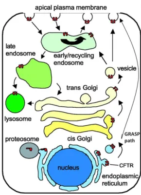
1.6 CFTR trafficking
If CFTR folds properly and is allowed to progress to the secretory pathway, it is loaded into COP II-coated vesicles that bud from the ER and traffic to the Golgi. Along the Golgi, its glycosidic moiety is further processed into a mature, fully glycosylated, transport-competent form. This form has a lower electrophoretic mobility than the core-glycosylated protein, allowing their easy distinction by Western Blotting as band C and band B, respectively. After this fine processing, CFTR is incorporated into secretory vesicles that deliver the protein into the apical membrane, where it will exert its functions22. Alternatively, some data suggests that at least a portion of CFTR may be
delivered directly from the ER to the apical membrane without going through the Golgi through a GRASP-dependent pathway23.
Not surprisingly, CFTR trafficking to the membrane in epithelial cells is dependent on their polarization state24. Cell polarization is characterized by, among other things, a selective redistribution of membrane proteins among the apical and basolateral sides, which relies on polarized sorting and trafficking of those proteins25. However, the precise molecular mechanisms governing polarization-dependent delivery of CFTR to the apical membrane have not been well studied.
After being delivered to the membrane, CFTR becomes subject to endocytosis. CFTR is internalized into early endosomes, where it can either be targeted to recycling vesicles containing Rab11, which will be returned to the apical membrane, or sent to the lysosome for degradation26.
All these processes depend on the loading, budding, docking and fusing machinery that regulate intracellular trafficking that comprise a huge number of proteins, including Rab GTPases and soluble NSF Attachment Protein receptors (SNAREs).
Figure 1.2 - Schematic representation of CFTR trafficking, starting at the endoplasmic reticulum.
Adapted from Bertrand (2003)27.
1.7 The CFTR interactome and Rap1A
Throughout its lifetime, CFTR makes a very long travel in the cell, that begins with translation in the cytoplasm, traversing the ER and Golgi to the membrane, and being
recycled or sent for degradation. Along this journey, CFTR interacts with numerous proteins that have all sorts of effects on it. Throughout the previous sections of this Introduction, a reasonably large number of proteins have been mentioned as being relevant in CFTR biogenesis, from kinases, scaffolding proteins and chaperones to trafficking small GTPases and SNAREs. The number of known CFTR-interacting proteins (CIPs) is staggering, and it keeps on growing. Understanding how CFTR interacts with all these partners and how they affect it is crucial for proper therapeutic strategies to be devised that target the molecular basis of cystic fibrosis.
Proteomic technologies are especially well suited to identify CIPs28,29. In 2010, Faria and other colleagues from our group reported having identified several CIPs with one such approach30. Using a recombinant CFTR NBD1 as affinity chromatography bait, CIPs in Calu3 (a human airway epithelial cell line) cell extracts were captured, separated by 2D gel electrophoresis and identified by MALDI-TOF. One of the proteins identified was a small GTPase called Rap1A, which will be described in the following sections.
1.8 Rap1A – an overview
Rap1A is a small GTPase belonging to the Ras superfamily of GTPases, specifically to the Rap subfamily. It was discovered in the late 1980s for its ability to revert the morphological phenotype of Ras-transformed cells. It was initially postulated that it merely competed for binding to Ras effectors without activating them, but this view has been challenged with the appearance of a myriad of Rap modulators and interactors with varying specificities. Rap1A is seen nowadays as a node in a complex signaling pathway of its own, which interacts with several other signaling pathways involved in key biological processes in the cell. The most prominent of these is control of cell adhesion, both to the extracellular matrix and to neighboring cells. It also takes part in regulating actin cytoskeleton dynamics and cell polarity (both in resting states and during cell migration), exocytosis and membrane protein recycling, and it might have a role in cell cycle regulation31.
There are 5 known members of the Rap family: Rap1A and B and Rap2A, B and C. Rap2 proteins are mainly expressed in platelets and other hematopoietic cells, whereas Rap1A and B are expressed in a broader range of human tissues, including the lung, colon and pancreas, the most affected tissues in cystic fibrosis. Rap1A, on which this work is focused, is 181 residues long and approximately 21kDa when mature.
Figure 1.3 - The Ras superfamily of GTPases, with the Rap subfamily outlined in red. Adapted from
Colicelli (2004)32.
Rap1A, like all other small GTPases, cycles between two structural conformations. When it binds GTP, Rap1A is in an active form, capable of binding to and inducing specific conformational changes in downstream effectors. When it binds GDP it shifts to an inactive form, which is incapable of interacting with those effectors. The intrinsic GTPasic activity and nucleotide release rate of Rap1A are very low, so when it binds GTP it tends to stay locked in the active conformation, and when it binds GDP it remains in the inactive conformation, thus functioning as a molecular switch. Interconversion of these two conformations depends on auxiliary regulatory proteins that either activate or inactivate Rap1A. Activators are called guanine nucleotide exchange factors (GEFs) and act by stimulating the release of the bound GDP, allowing for binding of the much more abundant GTP. GTPase activating proteins (GAPs), on the other hand, are actually Rap inactivators, because when they increase the very low GTPase activity of the protein, which is where their designation comes from, they lead to hydrolysis of the bound GTP to GDP, converting Rap1A in the inactive form. Several GEFs and GAPs with varying specificities for Rap1A have been identified and characterized, and will be adressed in section 1.10.
The localization of Rap1A is essential to its function, as it must be close to both its activators and downstream effectors in order to have an effect in the cell. Rap1A localization is defined by its post-translational modifications, which are discussed in section 1.9. While some other GTPases, from the Rho family, for instance, translocate
between the cytosol and the membrane (depending whether they bind proteins that mask the lipid anchor or not), the localization of Rap proteins doesn’t vary a lot. Instead, Rap signaling is spatially and temporally controlled by regulating the localization and activation state of RapGEFs and GAPs, respectively, as will be discussed in section 1.10.
1.9 Rap1A post-translational modifications
Rap1A is subject to four kinds of posttranslational modifications necessary for its proper function: prenylation and methylation of Cys181, proteolysis of the 3 C-terminal residues and phosphorylation of Ser180. Rap1A contains a CAAX (C – cysteine, A – any aliphatic aminoacid, X – any aminoacid) consensus motif in the very end of the C-terminus that is recognized by geranylgeranyl transferase 1. The Cys residue is geranylgeranylated, the three last residues are proteolitically removed by a CAAX protease and the cysteine, now the last residue in the C-terminus, is methylated by a methyl transferase. The X residue at the C-terminus determines what kind of lipid anchor is added to the protein; for instance, Rap1A, Rap1B and Rap2A all end with leucine and are geranylgeranylated, whereas as Rap2B, which ends with glutamine, is farnesylated33. Experimental information concerning Rap2C prenylation has not been obtained so far. Phosphorylation of Rap1A by PKA at Ser180 appears to have an effect on its function, affecting its ability to bind some downstream effectors34.
Due to its geranylgeranyl anchor, Rap1A localizes to membranes in the cell. It has been reported to be either perinuclear and co-localized with Golgi markers in COS-735 cells or in a late endocytic vesicular compartment in the fibroblast cell lines BHK, NIH3T336 and C237. In endothelial cells, namely HUVECs38, and in epithelial cells, such as MCF-739 and MDCK40 cells, Rap1A is seen at the plasma membrane and enriched at cell-cell junction sites. Two papers reported that in CHO41, which are epithelial-like, and A54942 cells, which are epithelial, Rap1A has been identified in perinuclear locations; however, in these reports, cells were cultured in suspension, which may explain why Rap1A was located differently than in other epithelial cell lines. It seems to be a general rule that in endothelial and epithelial cells, when grown in conditions that allow for cell-cell adhesion to develop, Rap1A is located at cell junctions.
Prenylation seems to be necessary for the membrane localization of Rap1A, but it doesn’t seem to affect which membranes it localizes to. Instead, an internal TAQST sequence between residues 85 and 89 seems to be responsible for its perinuclear location in COS-7 cells. It is unclear how it functions in maintaining Rap1A at the nuclear periphery in fibroblasts but not in endothelial and epithelial cells. It is likely that
this differential behavior involves interaction with other proteins differentially expressed in these distinct cell types, but this doesn’t seem to have been explored so far35
.
1.10 RapGEFs and RapGAPs
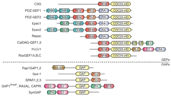
Rap GEFs are much better understood than GAPs, and great effort has been focused on understanding how they activate Rap in a spatially and temporally strict way. Most of them respond to extracellular stimuli, relayed by either second messengers or tyrosine kinase receptors. Many of them also activate other Ras-family GTPases to lesser extents43.
Figure 1.4 - Schematic representation of the multi-domain structure of RapGEFs and RapGAPs.
Taken from Gloerich (2011)44.
Epac is a 104kDa multidomain protein that activates Rap proteins in response to direct binding of cAMP, constituting a cAMP sensor in the cell that is independent of protein kinase A (PKA). Binding to cAMP has a double action on Epac: it activates it, allowing it to activate Rap in its turn, and it induces its translocation to the plasma membrane. The disheveled, Egl-10, pleckstrin domain (DEP) is necessary for this translocation and has been shown to tether Epac to phosphatydic acid in the membrane.
Epac is also tethered to the plasma membrane by phosphorylated (active) ezrin, radixin and moesin (ERM) scaffolding proteins. It is worthy of note that ezrin also interacts with CFTR (through E3KARP/NHERF2) and with PKA, forming a macromolecular complex that facilitates phosphorylation of CFTR and its activation12. Ezrin also tethers CFTR to the actin cytoskeleton, increasing its membrane stability11. Radixin has also been reported to bind Epac and PKA simultaneously, bringing these two cAMP sensors together45. All these data suggest that Epac might co-localize with
CFTR, which argues for a possible function of Rap1A in regulating CFTR function or membrane stability. Besides, Epac is activated by stimuli that result in increased subcortical cAMP, the same stimuli that induce PKA-mediated activation of CFTR, which further supports this hypothesis.
C3G is another GEF that has been shown to mediate signals received from B and T cell receptor activation, growth factors, cytokines, G protein coupled receptors and integrins. C3G is present in the cytoplasm in a complex with members of the Crk family of small adapter molecules. In response to stimuli, this complex is recruited to the cell membrane. This involves association of Crk with phosphotyrosine containing proteins like receptor tyrosine kinases, p130 Cas, insulin receptor substrate 1 and paxillin. Phosphorylation of C3G by Src-family kinases is also important in this translocation from cytosol to cell membrane. At the membrane, C3G activates downstream signaling by Rap1, Rap2 and R-Ras.
Other GEFs include PDZ-GEFs, which are involved in cadherin-mediated cell-adhesion and polarization and localize active Rap1 to the cortical cytoskeleton near the plasma membrane, and CalDAG-GEFs, which activate Rap1 upon binding to diacylglycerol, another important second messenger.
Rap GAPs include RapGAPs I and II, the SPA-1 family (SPA-1, SPAR, SPAL, and E6TP1), and tuberin. These are specific to Rap proteins, but even though the catalytic mechanisms for Rap and Ras deactivation are different, some GAPs exist that have dual activity, namely GAP1IP4BP, GAP1m, CAPRI, and RASAL46. Most research on these regulators has focused on catalytic mechanisms and on the role of RapGAPs in tumorigenesis rather than in elucidating how they regulate Rap signaling.
1.11 Rap effectors and function
Rap signaling has been implicated in a growing number of cellular processes, of which the most prominent is cell adhesion, both between cells and between the cell and the extracellular matrix. The involvement of Rap in the establishment of cell polarity will also be discussed. Its role in cell migration, cell cycle regulation, lymphocyte-specific (reactive oxygen species formation, B- and T-cell receptor signaling) and neuron-specific functions (neurite outgrowth, synaptic plasticity) are beyond the scope of this work.
All these functions are implicit in the set of proteins whose activity or localization Rap modulates. By interacting with VASP (an actin polymerizing protein)47, the RhoGEFs TIAM, Vav248 and FRG49 and the RhoGAPs ARAP350 and RA-RhoGAP51, Rap signaling causes sometimes drastic changes in cytoskeleton dynamics. Interaction
with Afadin/AF6 and p120-catenin is necessary for the regulation of E-cadherin stability at nascent adherens junctions in epithelial cells52, and interaction with CCM1/KRIT provides a similar role in VE-cadherin-based adherens junctions in the endothelium53. By activating the effectors RAPL and RIAM, Rap proteins are involved in the activation and clustering of various integrins and consequently in adhesion to several extracellular matrix proteins, including fibronectin, fibrinogen, collagen and laminin31. Adhesion to the extracellular matrix doesn’t seem to affect Rap signaling, though, so this phenomenon seems to be unilateral. Depending on the cell type and mode of activation, Rap1 can also activate B-Raf, a serine/threonine kinase that regulates the MAPK signaling pathway54.
The regulation of cadherin stability at adherens junction by Rap signaling is one of the subjects of major focus in Rap-related research. Stabilization of newly-recruited E-cadherin in nascent adherens junctions is dependent on Rap1 and is mediated by p120-catenin52.Although, as stated above (see section 1.9), Rap1 in epithelial cells is located mainly at the plasma membrane, upon the loss of adherens junctions it translocates to recycling vesicles that also contain endocytosed E-cadherin and Rab11. Furthermore, disengagement of adherens junctions results in activation of Rap1 via C3G, which facilitates E-cadherin recycling to the membrane and allows adherens junctions to be reformed40. It is interesting to note that recycling of CFTR also involves sorting to Rab11-positive vesicles26, making this a possible meeting point between both proteins.
Rap proteins have a well described role in the establishment of cell polarity in neurons, where they define which out of a number of seemingly equal dendrites becomes the axon. In yeasts, budding is also spatially controlled in a similar fashion55. However, information is still scarce in what concerns the role of Rap signaling in the establishment of epithelial cell polarity. It has been shown that it is necessary for Arg kinase-dependent polarization in 3D cultures of MDCK cells56. It is also known that, in hepatocytes, the bile acid taurocholate increases cAMP levels in the cytoplasm, which are relayed to Rap1 by Epac and eventually result in the activation of the kinase LKB1, a master regulator of polarization in many cell types54. Although a role for LKB1 in the polarization of lung epithelial cells has only been shown so far in adenocarcinomas, it is plausible to think that this signaling module might be relevant for polarization of these cells in normal conditions as well. Whether this is the case or not, an effect of Rap1 in polarization can have an impact on CFTR biogenesis, as the delivery of CFTR to the membrane is dependent on proper cell polarization.
2 Objectives
The global aims of this work were to validate the previously reported interaction between Rap1A and CFTR and to assess the impact of up- and down-regulation of this small GTPase on CFTR biogenesis. In order to achieve this, several milestones were established, namely:
1 – To validate the interaction between Rap1A and CFTR through co-immunoprecipitation and immunofluorescence;
2 – To produce plasmid constructs suitable for expression of GFP-tagged wt, constitutively active and dominant negative Rap1A;
3 – To optimize transfection conditions with plasmids and siRNA and assess Rap1A expression and activity in transfected cells;
4 – To evaluate the effect of up- and down-regulation of Rap1A on CFTR expression, trafficking and function.
3
Materials and Methods
3.1 Production and characterization of vectors to study the impact of
Rap1A on CFTR biogenesis
3.1.1 Bacterial strain
The bacterial strain used for cloning and DNA amplification was XL1-Blue (Stratagene, La Jolla, CA, USA), which is tetracycline resistant. XL1-Blue cells are endonuclease (endA) deficient, which greatly improves the quality of miniprep DNA, and are recombination (recA) deficient, improving insert stability. The hsdR mutation prevents the cleavage of cloned DNA by the EcoK endonuclease system. The lacIqZΔ M15 gene on the F´ episome allows blue-white color screening.
XL1-Blue Genotype: recA1 endA1 gyrA96 thi-1 hsdR17 supE44 relA1 lac [F´ proAB lacIqZΔM15 Tn10 (Tetr)]. (Genes listed signify mutant alleles. Genes on the F´ episome, however, are wild-type unless indicated otherwise).
3.1.2 Plasmid vectors and siRNAs
pCMV6-AC-Rap1A-GFP (RG215248, Origene, Rockvile, MD, US), which encodes for a Rap1A-GFP fusion protein (GFP tag at the C-terminus of Rap1A), and pCMV6-AN-GFP (PS100048, Origene), a destination vector for expression of proteins with a GFP tag in the N-terminus, were used. Maps and cloning schemes for both vectors are available in Appendices 1 and 2. Both plasmids contain the ampicillin resistance gene, which was used for selection of transformed bacteria.
The fragment containing the Rap1A open reading frame was cloned from pCMV6-AC-Rap1A-GFP into pCMV6-AN-GFP, yielding the pCMV6-AN-GFP-Rap1A plasmid, as described in section 3.1.7. Variants of pCMV6-AN-GFP-Rap1A encoding for constitutively active (G12V) and dominant negative (S17A) mutants of Rap1A were obtained by site-directed mutagenesis, as described in section3.1.8.
siRNAs used were Silencer Select Negative control #2 (4390846), Rap1A siRNA (s11781) and CFTR siRNA (52945) (all from Applied Biosystems, Foster City, CA, USA).
3.1.3 Production of competent bacteria
Bacteria were plated in LB-agar medium supplemented with tetracycline and a single colony was used to inoculate a small volume of LB medium overnight at 37°C with vigorous shaking (220 rpm). This pre-inoculum was then used to inoculate at dilution 1/100 a larger volume of LB medium, typically 100 ml, which was also grown at
37°C (220 rpm) to final concentration of 5 x 10⁷ bacteria/ml (corresponding to an absorbance of 0.3 at 600 nm). Bacteria were transferred to ice and pelleted by centrifugation (1000 g for 15 min at 4°C). The bacterial pellet was then ressuspended, incubated on ice for 15 min in 33mL RF1 buffer (100 mM RbCl, 50mM MnCl2, 30 mM KCH3COO pH 7.5, 10 mM CaCl2, 15% (w/v) glycerol, pH 5.8; all from Sigma-Aldrich, St. Louis, MO, USA) and re-pelleted by centrifugation (1000 g for 15 min at 4°C). This second pellet was ressuspended and incubated on ice for 15 min in 8.3mL RF2 buffer (10 mM MOPS, 10mM RbCl, 75mM CaCl2, 15% (w/v) glycerol, pH 6.8; all from SigmaAldrich). 200 μl aliquots were then rapidly frozen with liquid nitrogen and stored at -80°C.
3.1.4 Transformation of competent bacteria
Bacteria were transformed by incubating a 200 μl aliquot of competent cells with DNA (100-200ng of ligation products; 0.1-10ng of plasmid DNA) for 30 min on ice, followed by heat-shock (2min at 42°C), incubation for 3min on ice, and then allowing antibiotic resistance to be expressed by growth in antibiotic-free LB medium for 45 min at 37°C at 220rpm. Bacteria were then pelleted (5000g for 2min), the supernatant was discarded and the pellet was ressuspended in the remaining supernatant medium. This suspension was then plated into LB-agar supplemented with selection antibiotic (100 μg/ml ampicillin, Sigma-Aldrich) and left to grow overnight.
Transformed bacterial colonies were grown in LB medium supplemented with 100μg/mL ampicillin and used to extract plasmid DNA. Clones were stored in liquid LB medium supplemented with 15 % (w/v) glycerol (Sigma-Aldrich) at -20°C.
3.1.5 DNA extraction and quantification
Small scale plasmid DNA was purified with the PeqGOLD Plasmid Miniprep Kit (PeqLab, Erlangen, Germany). This protocol is based on an alkaline lysis of the bacterial cells in the presence of SDS, to denature bacterial proteins, followed by a centrifugation step to remove cellular debris, genomic DNA and denatured proteins, and adsorption of the plasmid DNA in the supernatant to an anionic exchange matrix in the presence of high saline concentrations. After adsorption, the DNA is washed and eluted in water or TE buffer (10mM Tris/HCl, EDTA 1mM, pH8). Sequence of plasmid DNA preparations was confirmed by automatic DNA sequencing at least once for every plasmid.
DNA concentration was determined by measurement of absorbance at 260nm (one absorbance unit corresponding to 50μg/ml of dsDNA) using a Nanodrop 2000 spectrophotometer (Thermo Scientific, Waltham, MA, USA) and its purity was
evaluated by assessment of the ratio A260/A280. Only DNA with a ratio above 1.8 was considered pure enough for further use.
3.1.6 DNA sequencing
Plasmid DNAs were purified as described in section 3.1.5. The sequencing reactions were performed using the ABI Prism BigDye Terminator Cycle Sequencing Kit (Applied Biosystems) according to the manufacturer’s instructions. The products were analyzed in the automatic sequencer 3130 XL Genetic Analyzer (Applied Biosystems). Alternatively, the sequencing reactions were outsourced to StabVida, using BigDye Terminator 3 (Applied Biosystems) and analysed in a ABI 3730XL sequencer (Applied Biosystems). Sequencing primers used were VP1.5 (forward primer, sequence 5’-GGACTTTCCAAAATGTCG-3’) and XL39 (reverse primer, sequence 5’-ATTAGGACAAGGCTGGTGGG-3’) (obtained through Thermo Scientific).
For sequence analysis, the sequences obtained were analysed through comparison with the reference human Rap1A sequence (NCBI reference sequence
NM_002884.2) using the software Bioedit
(http://www.mbio.ncsu.edu/BioEdit/bioedit.html).
3.1.7 Cloning
The fragment containing the Rap1A open reading frame was cloned from pCMV6-AC-Rap1A-GFP into pCMV6-AN-GFP. Briefly, the two plasmids were separately hydrolysed with MluI and AsiAI (an SgfI isoschizomer) (Fermentas, Burlington, Canada) and the resulting fragments were separated by low-melting agarose gel electrophoresis. The bands containing the fragments of interest were excised from the gel and melted at 70ºC. The melted portions of gel containing insert and empty vector were combined at a ratio of 4:1, respectively, and incubated with ligase and ligase buffer (Roche) for 72h at 4ºC. Bacteria were then transformed with the ligation product, plated in LB-Agar plates supplemented with ampicillin 5ug/mL and allowed to grow overnight at 37ºC. Single colonies were picked to LB medium with ampicillin 5ug/mL and grown for plasmid extraction and sequencing. One clone containing the correct recombinant plasmid, pCMV6-AN-GFP-Rap1A, was chosen for further use.
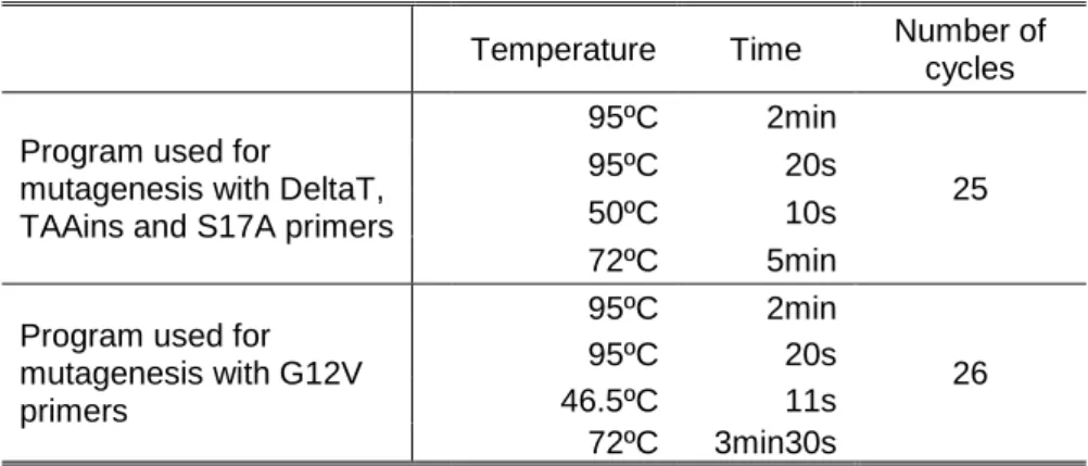
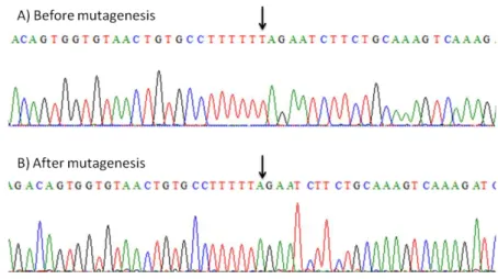
3.1.8 Mutagenesis
Mutations were introduced into pCMV6-AN-GFP-Rap1A using the KOD Hot Start Kit (Novagene, Darmstadt, Germany) with complementary pairs of the custom designed HPLC-purified mutagenic primers described in Table 3.2 (Thermo Electron Corporation, Waltham, MA, USA). Primers were designed with the software PrimerX (http://www.bioinformatics.org/primerx).
The mutagenesis reaction is a regular PCR reaction, with the peculiarity being that the primers are not completely complementary to the template sequence; instead, they contain the desired point mutations. The PCR programs used are displayed in Table 3.1. The result of this mutagenesis reaction is a mixture of the template plasmid and a new nicked plasmid containing the desired mutations. After confirming the amplification by agarose gel electrophoresis, the PCR products were incubated with DpnI (Invitrogen, Carlsbad, CA, USA), a restriction enzyme that specifically hydrolyzes methylated and hemi-methylated DNA. This results in complete degradation of the template DNA, which was produced in bacteria and, thus, is heavily methylated. The mutagenized plasmid, however, was synthesized in vitro and was not methylated, therefore resisting to hydrolysis by DpnI.
Table 3.1- PCR programs used for the mutagenesis reactions
Temperature Time Number of cycles Program used for
mutagenesis with DeltaT, TAAins and S17A primers
95ºC 2min
25
95ºC 20s
50ºC 10s
72ºC 5min
Program used for mutagenesis with G12V primers 95ºC 2min 26 95ºC 20s 46.5ºC 11s 72ºC 3min30s
After the hydrolysis step, bacteria were transformed with the PCR products (section 3.1.4 from this Chapter). Bacteria not only amplify the plasmid DNA but also repair the nicks left by the polymerase. Following transformation, plasmid DNA was extracted (section 3.1.5 from this Chapter) and the presence of each mutation was confirmed by automatic DNA sequencing (section 3.1.6from this Chapter).
Table 3.2 - Primers for mutagenesis reaction. The table presents only the forward primers. For each, a complementary reverse primer was also used.
Name Sequence G12V 5’- GGTCCTTGGTTCAGTGGGCGTTGGGAAGTC -3’ G15D 5’- GTTCAGGAGGCGTTGACAAGTCTGCTCTGAC -3’ S17A 5’- GTTCAGGAGGCGTTGGGAAGGCCGCTCTGACAGTTCAGTTTG -3’ DeltaT 5’- GGTGTAACTGTGCCTTTTTAGAATCTTCTGCAAAGTC -3’ TAAins 5’- CATGTCTGCTGCTCTAAACGCGTTAAGCGGC -3’
3.2 Biochemical and electrophysiological analysis of Rap1A impact
on CFTR biogenesis
3.2.1 Characterization, Culture and Maintenance of cell lines
The majority of the experiments were performed in CFBE41o- cells57 (cystic fibrosis bronchial epithelial, further referred to simply as CFBE) stably overexpressing wt CFTR. This cell line is one of the most commonly used cell models for the study of CFTR in a lung context. Because they are able to polarize, these cells mimic what happens in the lung epithelium in physiological conditions.
Cells were cultured in Minimal Essential Medium supplemented with L-alanyl-L-glutamine (MEM+Glutamax) (Gibco, Paisley, UK), 10% (v/v) fetal bovine serum and 100U/mL penicillin and 100mg/mL streptomycin (Invitrogen) in plastic flasks or plates coated with LHC basal medium (Invitrogen), bovine serum albumin, fraction V 0.1mg/mL, collagen from rat tail 29μg/mL and fibronectin 10μg/mL (all from Roche). For polarization-dependent assays, cells were seeded in either Snapwell (Sigma-Aldrich) or Millicell (Millipore, Billerica, MA, USA) filters at a density of 2x10⁵ cells/filter coated with collagen IV (Sigma-Aldrich) and cultured with MEM+Glutamax supplemented with 5% fetal bovine serum (FBS) and 100U/mL penicillin and 100mg/mL streptomycin on both apical and basolateral compartments. Transepithelial resistance was measured every other day with an STX2 Chopstick Electrode (World Precision Instruments, Inc., Sarasota, FL, USA), in order to monitor monolayer integrity, and experiments were performed when it exceeded 750Ω.cm2.
Primary cultures of human bronchial cells (HBE) from 3 healthy donors were prepared by Marta Palma in our unit. The access to explanted lungs was established through a collaborative project between the Faculty of Sciences of the
University of Lisboa and the Cardio-Thoracic Surgery Department of the Hospital de Santa Marta (Lisboa), which received approval from the hospital’s Ethics Committee, and primary HBE cells were isolated as described in reference 58.
Continuous growth was made possible by pre-confluence enzymatic dissociation with trypsin (Invitrogen). After dissociation, cells were resuspended and redistributed in new flasks or plates. Cell lines were stored in liquid nitrogen in aliquots in 40% (v/v) Minimal Essential Medium (Gibco), 50 % (v/v) FBS (Invitrogen) and 10 % (v/v) DMSO (Sigma-Aldrich), a cryoprotectant that prevents the formation of ice crystals during the freezing process. Freezing was performed in such a way that the cooling speed was about 1ºC/min, in order to minimize damage to the cells. Thawing was done by quickly submerging the vials in water at RT, washing the cells with F12 Medium (Invitrogen) and seeding in the medium described above.
Cultures were maintained at 37ºC in a humidified atmosphere of 5% (v/v) CO2.
3.2.2 Transfection
CFBE cells were transfected using either cationic liposomes or electroporation.
Lipofection is based on the ability of cationic lipids to form unilamelar liposomes that adsorb nucleic acid molecules to their surface and are internalized by the cells. The delivery mechanisms of the nucleic acid to the cytoplasm and nucleus are poorly understood, but the practical result is protein expression (plasmids) or down-regulation of the target genes (siRNAs)59. In this work, several such liposomal formulations were used, namely Lipofectamine 2000 (Invitrogen), Fugene HD (Promega, Fitchburg, WI, US) and Hiperfect (Qiagen, Venlo, Netherlands). Liposomal solutions and DNA or siRNA were separately incubated for 5 min in OPTIMEM (Invitrogen), mixed together and allowed to incubate for 15 min at room temperature (RT). They were then added dropwise to either 70-80% confluent cells or freshly trypsinized cells. Medium was changed after 24h and the experiments were performed 48-72h post-transfection.
Another way of transfecting cells is by delivering the plasmid DNA or siRNA through an electrical shock. This causes transient, reversible changes in the organization of the lipid bilayer in the membrane, resulting in the opening of pores big enough for nucleic acids to enter60. A Microporator MP100 (Digital Bio, Seoul, South Korea) was used for electroporation experiments. Briefly, 1x10⁵ cells were mixed with 1.5μg of the appropriate DNA or siRNA, pipetted into a gold-covered tip and delivered a certain number of electrical pulses. Pulse voltage ranged from 1075-1500V, pulse width from 10 to 25ms and the number of pulses was 2-3. Cells were then seeded in
12-well plates in the medium described in section 3.2.1. Medium was changed after 24h and the experiments were performed 48-72h post-transfection.
For transfection with plasmids, which encode for GFP or GFP fusion proteins, transfection efficiency was estimated by direct counting of fluorescent vs. non fluorescent cells under an epifluorescence microscope (check brand). This was only feasible when the number of attached cells at 24h post-transfection was relatively small. When there was a great number of attached cells, and for siRNAs, transfection efficiency could only be evaluated by Western Blot.
3.2.3 Preparation of total protein extracts
For Western blot (WB), protein extracts were prepared by cell lysis with sample buffer (1.5 % (w/v) SDS; 5 % (v/v) glycerol; 0.01 % (w/v) bromophenol blue; 0.05 mM dithiotreitol; 0.095 M Tris pH 6.8) and DNA was sheared by passing the sample first through a 22G and then a 27G needle until viscosity decreased or sheared by enzymatic action of 5-25U/mL of benzonase (Sigma-Aldrich) in the presence of MgCl2 2.5mM.
Total protein concentration in different samples was assessed by a modified Lowry protein assay. Proteins were solubilized with 0.015% (w/v) sodium deoxycholate for 10 min at RT, precipitated with 6% (w/v) trichloroacetic acid and then centrifuged at 14000 g for 5 min. The supernatant was removed and the pellet was resuspended in equal volumes of H2O and Reagent A (0.1 g/l CuSO4.5H20; 0.2 g/l potassium tartarate; 10 g/l Na2CO3; 2.5% (w/v) SDS; 0.2 M NaOH) followed by 10 min incubation at RT. Finally, Reagent B (Folin-Ciocalteau Reagent diluted 5-fold in water) was added, followed by incubation for 30 min. Protein concentration was determined by measurement of A750 and comparison with a regression line obtained for BSA (BioRad, Hercules, CA, USA) standards of increasing concentrations.
3.2.4 Western blot
After quantification of total protein (see section 3.2.3), protein extracts were separated by SDS-polyacrylamide gel electrophoresis (PAGE) on 7, 10 or 12.5% (w/v) minigels, followed by transfer onto Immobilon polivinylidene difluoride (PVDF) -membranes (Millipore). After blocking with 5 % (w/v) skimmed milk in phosphate-buffered saline (PBS, NaCl 137mM; KCl 2.7mM; KH2PO4 1.5mM; Na2HPO4 6,5mM, pH 7.4) containing 0.1% (v/v) Tween (PBS-T) for 2 h, the membranes were probed for 2 h at RT with primary antibodies diluted in 5% (w/v) milk in PBS-T, washed 3x10min with PBS-T, followed by incubation for 1 h at RT with horseradish peroxidase-conjugated secondary antibodies in 5% (w/v) milk-PBS-T. Blots were developed either by