i
Terminal Sterilization Using
Supercritical Carbon Dioxide (
SC
CO
2
):
Influence of Several Processing
Parameters
by
Fábio André Teixeira Pinheiro
i
Terminal Sterilization Using
Supercritical Carbon Dioxide (
SC
CO
2
):
Influence of Several Processing
Parameters
Mestrado em Engenharia Biomédica
Dissertação de Mestrado:
Orientador da ESB-UCP: Prof. Ana Leite Oliveira
Co-Orientadores da ESB-UCP: Nilza Ribeiro, Gonçalo Soares
by
Fábio André Teixeira Pinheiro
iii
Acknowledgements
No decorrer desta etapa académica devo o meu especial agradecimento à Escola Superior de Biotecnologia da Universidade Católica Portuguesa e a todos os professores e colaboradores desta grande instituição.
Primeiramente devo agradecer à minha equipa especial de supercríticos, a “CriticalTeam” que foram essenciais no conhecimento nesta área de esterilização e que me ajudaram a desenvolver o trabalho realizado.
Devo um agradecimento especial à professora Ana Oliveira que me ajudou muito e esteve sempre disponível, mesmo quando a sua agenda estava lotada. Um obrigado também por todos os puxões de orelhas e pelas viagens e congressos que disfrutei.
A Nilza Ribeiro e o Gonçalo Soares, meus co-orientadores e colegas de laboratório, agradeço-lhes por toda a ajuda, conhecimento e paciência. Sem eles seria muito mais difícil.
Devo um agradecimento especial também à minha família, pais, irmã e avós. Ajudaram a seguir sempre em frente neste percurso académico. Os meus amigos também não podem ficar esquecidos e também lhes dedico este trabalho a eles. A minha namorada, Joana Barbosa, que me ajudou sempre desde o 1º ano de faculdade até aos dias de hoje. Aos colegas de laboratório do 2º piso que se mostraram sempre disponíveis para ajudar.
v
Resumo
Dada a expansão do mercado de biomateriais e sofisticação dos dispositivos médicos, os processos de esterilização terão necessariamente de ser reformulados e melhorados para responderem eficazmente. Algumas das técnicas de esterilização utilizadas atualmente não conseguem executar a sua função de uma forma eficaz. Em muitos casos o material é danificado durante o processo ou a esterilização não é realizada eficientemente. Assim sendo, a esterilização por dióxido de carbono em estado supercrítico (SCCO2) responde eficazmente ao esterilizar diversos materiais biológicos e polímeros sensíveis, que antes não eram esterilizados por outras técnicas. A par desta vertente, o custo de operação, a sua segurança e rapidez processual são características positivas na esterilização por SCCO2.
O trabalho realizado no âmbito da presente tese teve como objetivo estudar e analisar as variáveis de esterilização em tiras de esporos bacterianos de três estirpes diferentes: Bacillus
atrophaeus, Bacillus pumilus e Bacillus stearothermophilus. Com este estudo, as variáveis
como o tempo de esterilização efetiva, pressão e “Shelf life” foram aprimorados, aumentando assim a eficiência do processo. Em paralelo a este estudo, foram também estudadas amostras de placenta humana antes e depois do processo de esterilização de forma a verificar se a esterilização por SCCO2 poderá danificar ou modificar as propriedades físicas ou químicas deste material biológico.
Conclui-se que para esterilizar eficazmente as tiras de esporos bacterianos é recomendável um tempo de esterilização efetiva no mínimo de 3 horas com um shelf life mínimo de 1 semana. De forma a aumentar a viabilidade do processo, demonstrou-se que é possível a utilização dos materiais esterilizados logo após o processo sem reativação dos esporos, quando estes estão em contacto com o SCCO2 no mínimo 3 horas. No reator os valores recomendados são: pressão a 140 bar, 600 rpm, 40ºC e com a adição de 300 ppm H2O2. As análises de High-performance liquid chromatography (HPLC) e Fourier-transform infrared
spectroscopy (FTIR) demonstraram que as amostras de placenta após esterilização não sofrem
alterações químicas significativas. Contrariamente, os testes mecânicos e análises de
Differential scanning calorimetry (DSC) revelaram mudanças físicas e químicas significativas
nas amostras. A esterilização por SCCO2 conferiu um aumento de rigidez nas amostras biológicas estudadas, assim como um aumento na sua resistência térmica. O estudo da eficácia desta técnica de esterilização (SCCO2) em amostras biológicas deve ser continuado para que se
vi possa reunir mais informação que permita validar a sua aplicabilidade na área dos materiais médicos e biomateriais.
Palavras-chave: Esterilização, CO2 supercrítico, Esporos bacterianos, placenta humana descelularizada.
vii
Abstract
Due the expanding market for biomaterials and the sophistication of medical devices, sterilization processes need to be refined and reinvented to respond effectively to this demand. Nowadays the current sterilization techniques, cannot perform their function effectively without damaging the material during the process. Thus, supercritical carbon dioxide (SCCO2) sterilization can be an alternative solution for sterilizing various biological materials and complex/sensitive polymers. The great advantages of SCCO2 sterilization englobes the price of operation, its safety and procedural speed among others.
The main goal of this thesis project was to study and analyse different variables, like effective sterilization time or pressure in the effective sterilization process of three different bacterial spore strains: Bacillus atrophaeus, Bacillus pumilus and Bacillus stearothermophilus. In this study, the effect of the time interval between SCCO2 sterilization and the microbiological validation was also evaluated, despite of being a parameter that is not taken into account in the works described in literature to date. Parallel to this work, human placenta samples were analysed before and after the sterilization process to verify whether SCCO2 sterilization could damage or modify the physical or chemical properties of this biological material.
Regarding the results obtained of the sterilization of the bacterial spore strips, the mildest conditions optimized were: 140 bar, 600 rpm, 40ºC, 300 ppm H2O2 and 4 hours of effective sterilization. Interestingly, the effectiveness of terminal sterilization of these biological indicators was dependent on the time period after scCO2 treatment. In addition, the minimum “shelf life” required is at least 1 week.
In the second part of the work, the placenta samples sterilized by scCO2 did not undergo significant chemical changes as demonstrated by high-performance liquid chromatography (HPLC) and Fourier-transform infrared spectroscopy (FTIR) analyses that. In contrast, the mechanical tests and analyses of Differential scanning calorimetry (DSC) demonstrated significant physical and chemical changes in the samples. SCCO2 sterilization conferred rigidity to the biological sample and a different behaviour of the samples were detected when exposed to a constant increase of temperatures.
The study of the innovative sterilization technique (SCCO2) should be continued to increase its applicability in the area of medical materials and biomaterials and their efficiency.
viii
ix
Contents
Acknowledgements ... iii Resumo ... v Abstract ... vii List of Figures ... xiList of Tables ... xiii
List of Abreviations ... xv
1. Introduction ... 1
1.1 Sterilization History in Medicine ... 1
1.2 Main Sterilization Techniques in Medicine ... 3
1.3 Supercritical Carbon Dioxide (SCCO2) as a Sterilization Method ... 10
1.3.1 SCCO2 Technology ... 10
1.3.2 Principles of SCCO2 Sterilization Technique ... 12
1.3.3 Current Sterilization Technology vs SCCO2 ... 15
1.3.4 Sterilization of Biological Material by using SCCO2 ... 15
2. Material and Methods ... 20
2.1 Biological Samples ... 20
2.2 SCCO2 Sterilization ... 22
2.2.1 Optimization of the Mildest Conditions for Effective SCCO2 Sterilization ... 24
2.2.2 Influence of the Time Interval between the SCCO2 Sterilization and Microbiological Validation ... 28
2.3 Sterilization of Decellularized Placenta and further Evaluation of its Physicochemical Properties ... 30
3. Results ... 34
3.1 Optimization of the Mildest Conditions for Effective SCCO2 Sterilization ... 34
3.2 Influence of the Time between SCCO2 Sterilization and Microbiological Validation: Shelf Life ... 36
x 3.3 Physical, Morphological and Chemical Characterization of the Decellularized Placentas
... 39
3.3.1 Fourier Transform Infrared (FTIR) ... 39
3.3.2 High-performance Liquid Chromatography (HPLC) ... 40
3.3.3 Differential Scanning Calorimetry (DSC) ... 44
3.3.4 Mechanical Tests ... 45
4. Discussion ... 48
4.1. Different Processing Parameters on SCCO2 Sterilization Efficiency ... 48
4.2. Effective Sterilization of placenta by SCCO2 ... 51
5. General Conclusions ... 54
6. Ongoing Projects ... 56
xi
List of Figures
Figure 1.1–Pressure steam sterilizer built in 1884 by Charles Chamberland (Skellie, 2010). ... 2
Figure 1.2– Scheme representing the main methods used for sterilization nowadays. Adapted from (National Institute of Open Schooling (NIOS), 1998) ... 3
Figure 1.3 –Recent model of an autoclave machine (STE Class B, IcanClave). ... 4
Figure 1.4a –Example of a recent model of EtO sterilizer machine (STI-500 Sterilizer, Treated Technologies). 1.4b – Sterilization facility floor layout (Sterijet, North Carolina, USA). 6 Figure 1.5 –Gamma radiation used during surgical procedures in hospitals to maintain patient and practitioner safety in the healthcare industry (Pacific Integrated Manufacturing, 2018). ... 8
Figure 1.6 –Hydrogen peroxide sterilizer (Sterrad 100S, Advanced Sterilization Products (ASP)). ... 9
Figure 1.7 –A 1680 version of Papin’s steam digester. Adapt from Thurston (1878). ... 10
Figure 1.8 –Examples of several applications of SCCO2. Adapted from:[1] (Lewa, 2017) ; [2](Zhang et al., 2014) and [3](Rawson et al., 2012). ... 11
Figure 1.9 –Schematic p-T phase diagram of CO2. From Budisa & Schulze-Makuch (2014). 12 Figure 1.10 – Comparison of the standard sterilization techniques with the SCCO2 process. ++: High probability that the sterilization method does not interfere with the properties of the material; ‒: Low: probability that the sterilization method does not interfere with the properties of the material. ... 15
Figure 1.11 – Decellularized heart valve and the results of sterilization for the different methods. Adapted from Hennessy et al (2017). ... 16
Figure 2.1– Decellularized human placenta after lyophilization. ... 20
Figure 2.2– B. stearothermophilus, B. atrophaeus and B. pumilus spore strips. ... 21
Figure 2.3– SCCO2 sterilization equipment. ... 22
Figure 2.4– Operating system created to the sterilization system. ... 23
Figure 2.5a – Biological indicators packed and sealed in Tyvek pouches. 2.5b– Decellularized and lyophilized placenta packaged in Tyvek pouches. 2.5c – placed in the reactor for the sterilization process. ... 24
Figure 2.6– An example of a complete sterilization process. ... 26
Figure 2.7–Interpretation of results in a broth medium inoculation... 27
Figure 2.8–Comparation between a turbid medium (left test tube) and clear medium (right test tube) containing spores strips before and after the SCCO2 treatment, respectively. ... 27
xii Figure 2.9– Pressure limits and programmed flow from the HPLC system. ... 31 Figure 3.1– Superimposed FTIR spectra of the lyophilized and decellularized placenta
samples. Blue: Before sterilization; Orange: After sterilization. ... 39 Figure 3.2a – HPLC chromatogram of untreated placenta sample. 3.2b – Chromatograms of
the 4 separated peaks from the untreated placenta. 3.2c – HPLC chromatogram of treated placenta sample. 3.2d – Chromatograms of the 3 separated peaks from the treated
placenta sample. ... 41 Figure 3.3a – HPLC chromatogram of untreated placenta sample. 3.2b – Chromatograms of
the 6 separated peaks from the untreated placenta. 3.2c – HPLC chromatogram of treated placenta sample. 3.2d – Chromatograms of the 5 separated peaks from the treated
placenta sample. ... 42 Figure 3.4– Comparison of DSC thermograms of untreated and treated placenta. ... 44 Figure 3.5– Representative stress-strain curves of untreated and treated placenta samples. ... 45
xiii
List of Tables
Table 1.1– Overview on publications describing the potential of SCCO2 sterilization with effective additives. Adapted from Bernhardt et al (2015). ... 14 Table 2.1– Sterilization parameters for optimizing of the mildest conditions for effective
SCCO2 sterilization. ... 25 Table 2.2 – Defined sterilization conditions to evaluate the effectiveness of terminal
sterilization. ... 29 Table 3.1– Microbiological validation: results of microbiological growth in TBS medium ... 35 Table 3.2– Microbiological validation: results of microbiological growth in TBS medium. .. 37 Table 3.3 – Comparison between the calculated retention time values, in minutes, and the
maximum absorbance, in absorbance units, of the peaks of untreated and treated
placenta. ... 41 Table 3.4– Comparison between the calculated retention time values, in minutes, and the
maximum absorbance, in absorbance units, of the peaks of untreated and treated
placenta. ... 43 Table 3.5–DSC thermograms peaks and respective enthalpies. ... 44 Table 3. 6 – Calculated values of Young´s modulus, stress and strain at burst. ... 46
xv
List of Abreviations
SCCO2 60Co
Supercritical carbon dioxide Cobalt-60 Ar Argon AU Maximum absorbance BI Biological indicators BPR Back-pressure regulator C2H4O2 CFC-12 Acetic acid Dichlorodifluoromethane CF3COOH Trifluoroacetic acid CH3CN Acetonitrile
CO2 Carbon dioxide CO32- Carbonate ion
DNA Deoxyribonucleic acid
DSC Differential scanning calorimetry ECM Extracellular matrix
EtO Ethylene oxide FSR Force-sensing resistor
FTIR Fourier-transform infrared spectroscopy
H+ Hydron
H2CO3 Carbonic acid
H2O Water
H2O2 Hidrogen peroxide HCO3- Bicarbonate ion
HPLC High-performance liquid chromatography ISO International Organization for Standardization
K Kelvin Kpa Kilopascal L Liters M Molar mg Milligrams min Minute ml Milliliters
xvi mm Millimeters
MPa Millipascal
MΩ Megaohm
N2 Nitrogen
N2O Nitrous oxide
nm Nanometers
PAA Paracetic Acid
PC Computer
PDA Photodiode array pH Potential of hydrogen RNA Ribonucleic acid rpm Rotations per minute SAL
SFC SFR
Sterilization Assurance Level Supercritical Fluid Chromatography Supercritical Fluid Reaction
TSB Trypto-casein soy broth wt % Weight percent
1
1. Introduction
1.1 Sterilization History in Medicine
The enormous evolution and innovation of surgical techniques and medical devices has lead to the development of more and more complex and sophisticated sterilization procedures and machinery. In fact, despite the advances in medical surgery, the lack of knowledge in sterilization and asepsis procedures, has made of medical rooms and hospital materials lethal weapons of contamination with infectious diseases during surgical interventions.
During the nineteenth century, the first attempts to effectively sterilize operating rooms and surgical materials were taken by scientists and doctors dealing with high rates of postpartum mortality and infectious diseases. The father of modern microbiology, Louis Pasteur proved that microorganisms, such as bacteria and parasites, were a principal cause of disease in humans (Dubos, 1951). Besides that, he also demonstrated the high potential of bacterial propagation. These microorganisms could live in dead tissues and spread through the air as spores. The British surgeon Joseph Lister pioneered modern antisepsis by introducing the washing of exposed wounds with liquid solutions and aerosols with carbolic acid (phenol)(Lister, 1867). This invention, considered one of the great evolutions in medicine, proved to be quite efficient, since there was a reduction in deaths from postoperative infections. Besides, Lister suggested the use of phenol for cleaning medical instruments, surgical sites, among others, since this agent acts by destroying and preventing the growth of microorganisms in living tissue. Prior to his studies, the Hungarian Ignaz Semmelweis demonstrated the importance of the simple act of hand washing of doctors and nurses before contact with the patient (Semmelweis, 1861). The correct hand washing, by a chlorinated solution, could prevent infections and deaths in childbirths and operations (Mcdonnell et al., 1999). These procedures attempted to create a germ-free environment in the operation room and became a frequent practice before medical observations, after child-births and autopsies.
Furthermore, Earle Spaulding sought to find a rational approach to disinfection and sterilization of medical devices and surgical tools. Accordingly, “Spaulding classification”, presented to the medical community in 1939, the materials were divided into three distinct categories (Spaulding, 1939). Briefly, the noncritical materials where those contacting only with the outside of the skin, the semicritical, contacting with the mucous membranes, and the critical materials where the tools that contacted the inside of the body. Spaulding further
2 improved his classification with the type of treatment that should be implemented, based in the use of each material/tool. The disinfectant´s strength should be related to the type of decontamination requirements of a medical device or area (From Ignaz Semmelweis to the
present: Crucial problems of hospital hygiene, n.d).
The first machines of sterilization began to be produced by Charles Chamberland in 1879. These pioneering machines, called autoclaves, were capable of sterilizing surgical materials with the use of water vapor at a temperature of 140 ºC, similar to nowadays’ autoclaves. Over the years, these sterilization machines have undergone improvements and began to be implemented in hospitals with the purpose of eliminating all forms of live and other biological agents in the materials (Harvey, 2011).
Figure 1.1–Pressure steam sterilizer built in 1884 by Charles Chamberland (Skellie, 2010).
The sterilization process has been following the advances of medicine and new methods using radiation and chemicals have been implemented. Therefore, the concept of sterilization has evolved, being defined by the destruction of all forms of life by heat, irradiation, gas or chemical treatment. Considering the several techniques and innovations in sterilization equipment, this field should contribute to a higher safety with obvious consequences in the quality care to the patients.
3
1.2 Main Sterilization Techniques in Medicine
There are several ways to perform sterilization efficiently. Figure1.2 lists the various sterilization techniques performed in various industrial sectors (National Institute of Open Schooling (NIOS), 1998).
Figure 1.2– Scheme representing the main methods used for sterilization nowadays. Adapted from (National Institute of Open Schooling (NIOS), 1998)
The techniques most commonly found in hospital facilities, which are more suitable for medical devices, are physical sterilization using heat and ionizing radiation and chemical sterilization using gas plasma and ethylene oxide (EtO) (Hurrell, 1998). Autoclave is a physical method which uses moist heat, and the most used in any hospital for many years. Other methods are rarely adopted, due to the lack of studies demonstrating its efficiency or to the low benefits of implementing a new technique in a hospital.
Along with the evolution of medicine and material’s industry, medical materials and devices have diversified and undergone changes in their composition. The need to keep the products treated implied the creation of appropriate methods for each type of product. On the other hand, the sterilization procedures for these devices has evolved and presently it must be carried out in accordance with an established Sterilization Assurance Level (SAL) which has
4 been standardized under ISO 14937-2009, by the directive EN 556-1. SAL is used to determine the effectiveness of a sterilization process, and expresses the likelihood that a specific product, from a batch of products submitted to this process, has not been sterilized. The value accepted for medical applications corresponds to SAL6, meaning the likelihood of 1 product being contaminated within a 1,000,000 batch of sterilized products (ISO, 2014).
Autoclave
As mentioned before, autoclave is widely used to sterilize medical devices. Created in the nineteenth century, the autoclave machine provides a physical method for disinfection and sterilization (Sultana, 2007). It uses a combination of heat with humidity and pressure during a stipulated time (Figure 1.3). Basically, the high-temperature steam is forced in under high pressure, thereby displacing air. Inside of the autoclave´s chamber, moist heat destroys all microorganisms by denaturation of enzymes and/or structural proteins (Ravikrishna, 2013). The two variables, time and temperature, may vary according to the type of microorganisms to be inactivated, packaging material and the material nature. There are two basic types of steam sterilization, which are gravity displacement autoclave and high-speed pre-vacuum sterilizer. In the first type, steam displaces air in the chamber by gravity without mechanical procedures. The second, the air is mechanically removed and load through a series of vacuum and pressure pulses. By this procedure, the steam penetrates the porous areas of the material which couldn’t otherwise be reached with other types of steam sterilization. Heat sterilization processes are indicated to heat resistant materials, like metallic surgical instruments and stainless-steel sutures (“The Chamber Blog,” 2016).
5 This sterilization procedure presents a low-cost machinery, a fast sterilization cycle, it is easy to control and non-toxic to the staff and the environment. However, the autoclaving process can damage materials which are sensitive to high temperatures. On the other hand, when considering metallic based devices and instruments, there is the possibility of corrosion due to the humid environment inside of the chamber (Tankeshwar, 2013).
Ethylene Oxide (EtO)
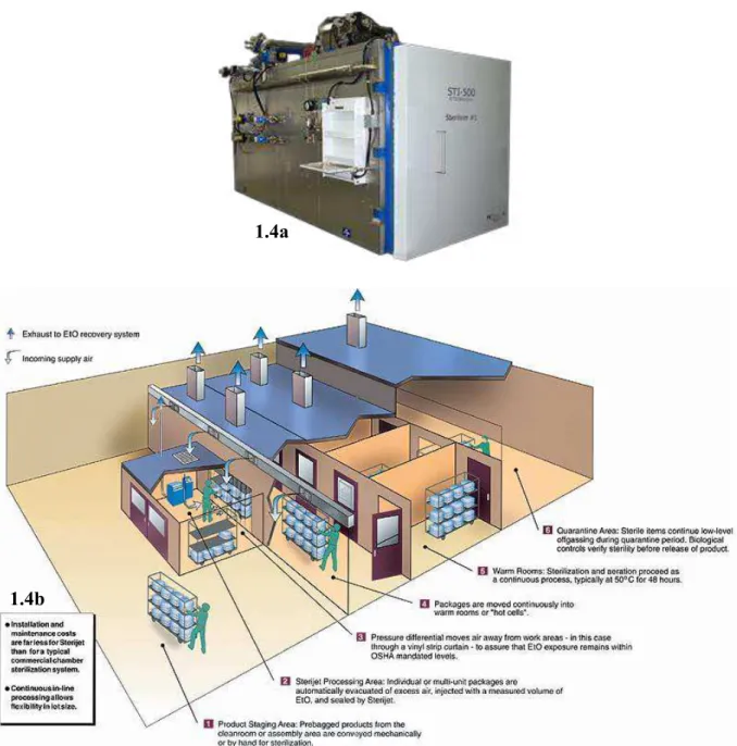
Sterilization can also be performed using chemical substances like EtO commonly used for sterilization of medical devices (Figure 1.4a, b).
6
Figure 1.4a –Example of a recent model of EtO sterilizer machine (STI-500 Sterilizer, Treated Technologies). 1.4b – Sterilization facility floor layout (Sterijet, North Carolina, USA).
EtO was developed in 1859 but only implemented in the industry and medicine in 1900s (Wurtz, 1859). This method can work at low temperatures and so, it can be applied to a vast variety of compatible materials, as compared to autoclave. The EtO sterilization cycle is divided in three stages: preconditioning, sterilization and aeration. This cycle begins with the preparation of the chamber environment to meet the ideal sterilization conditions for temperature, pressure and humidity. Then the EtO enters in the chamber via evaporation and sterilizes the material. When the sterilization process is finished, the material is exposed to an excessive aeration stage to remove any remaining EtO gas and to allow absorbed gas to
1.4a
7 evaporate again from the sterilized items (according to Figure 1.4b, performed, typically at 50 ºC for 2 hours, in room 5 “warm rooms”). If the material does not pass through this stage, materials, like plastics and rubbers, absorbed the gas and when applied to patients, the toxic gas could damage their body tissue (Henk & Finkiel, 2013). It is an alternative to medical materials that cannot support conventional steam sterilization with high temperatures. Yet, it is necessary to ensure the item to be sterilized is compatible with the sterilant. EtO gas is considered toxic, carcinogenic, flammable and explosive. Hence, pure EtO should be handled in explosion proof equipment. In addition, it is common to use EtO mixed with other components, like Chlorofluorocarbon (CFC-12) to become non-flammable sterilant. Efficiency depends heavily on the concentration of the gas, time of exposure, temperature, humidity and the nature of the material. EtO sterilization is applied to a wide range of medical material, like heart valves and stents coated with bioactive compounds (Govindaraj & Muthuraman, 2015)(Rutala & Weber, 1999).
The evident weakness of this technology is the time it takes for the sterilized material to become operational. After the sterilization process, the sterilized material is required to be placed in aeration for a minimum of 6 hours. In its entirety, the cycle has a minimum duration of 12 hours. Also, it requires special room conditions, safety equipment and separate ventilation system. EtO entails safety concerns due to its carcinogenic and flammable characteristics. Another obvious disadvantage, but less likely to occur, is the possibility of the presence of toxic EtO residues in surgical instruments and medical devices after sterilization. Ventilation may not always be sufficient to remove all residues of sterilized materials. This gas is not able to be recovered, thus it is not recyclable. Nevertheless, EtO operates under low temperature, which allows its use in thermo-sensitive materials while it is non-corrosive to plastic, rubber and metal (Henk & Finkiel, 2013).
Ionizing Radiation
E-beam, x-ray and gamma radiation are the three common ionizing radiation types used in sterilization procedures (Silindir & Özer, 2009). The first gamma irradiator was created in 1973 to sterilize medical material. One of the first industrial sterilization facilities was raised to sterilize surgical sutures with cobalt-60 (60Co) (Nahm, 2010). The innovation of this method has never ceased, and currently gamma radiation with 60Co can sterilize a larger range of materials and medical devices than EtO and autoclave. 60Co, which emits gamma rays during
8 radioactive decay, destroys microorganisms by breaking the covalent bonds of bacterial DNA. Despite that, the process presents a large amount of wasted energy, because rays are emitted in all directions beyond the desired one. Therefore, it must be carried out with safety concerns as ionizing radiation is harmful to human. In the case of 60Co, microorganisms are indirectly inactivated by the reaction of the free radicals produced in the cellular fluid. Radiation is considered one of the most effective methods for sterilization of medical devices and hospital material, despite one of the most expensive current methods (Govindaraj & Muthuraman, 2015). This technology is often found in hospitals, Figure 1.5, and in radiation sterilization plants.
Figure 1.5 –Gamma radiation used during surgical procedures in hospitals to maintain patient and practitioner safety in the healthcare industry (Pacific Integrated Manufacturing, 2018).
Despite the high sterilization efficiency rate of the technology, gamma radiation can change structural characteristics in medical devices that have been sterilized several times. These structural changes are more frequent in tissue allografts and polymer medical devices and negatively affect their clinical use. Moreover, according to studies, gamma radiation generates free hydroxyl radicals and other radiotoxins that induces toxigenic and mutagenic effects which in turn to potentially promotes cancer. Withal, sterilization by radiation demands machinery of high monetary and maintenance cost (Harrell et al., 2018).
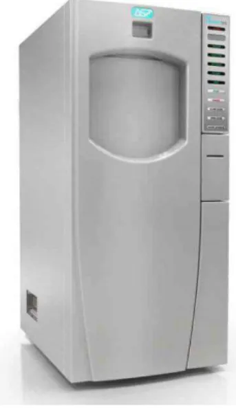
Gas Plasma: Hydrogen Peroxide (H2O2)
In the late 1980’s, the first sterilization system using hydrogen peroxide gas plasma was developed (Centers for Disease Control and Prevention, 2008). Ten years later, the company
9 Sterrad (ASP) (Irvine, USA) began the commercialization of gas plasma sterilization (Jacobs & Lin, 1996). In Figure 1.6 is represented one of the most recent models of this company.
Figure 1.6 –Hydrogen peroxide sterilizer (Sterrad 100S, Advanced Sterilization Products (ASP)).
The sterilization process begins when a strictly calculated dose of an aqueous solution of H2O2 is inserted in the plasma machine vacuum chamber where its evaporation and dispersion occur. Once the pressure inside the chamber drops, the particles become excited enough to ionize and the H2O2 turns into plasma. In this stage, the plasma breaks the genetic material of the microorganism into smaller molecules. The interaction between the free radicals generated by plasma and the cellular components such as enzymes, phospholipids, DNA or RNA, preventing normal cellular metabolism, eliminates the possibility of harmful reactions in the materials (Boiano & Steege, 2015) (Thierry Corporation, 2017).
This method presents profitable values in sterilization efficiency rates, fast sterilization cycles and a high compatibility with a lot of different medical devices and biological materials. Like EtO and radiation technologies, this entails a high price of equipment and specialized personnel (Govindaraj & Muthuraman, 2015). Compared with EtO, the H2O2 sterilization presents a better cost-efficiency ratio and begins to gain a more solid and attractive position in the market (TSO3, 2016).
10
1.3 Supercritical Carbon Dioxide (
SCCO
2) as a Sterilization Method
1.3.1
SCCO
2Technology
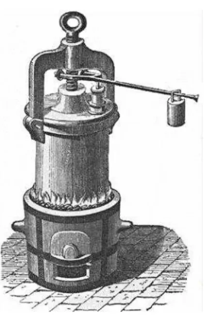
Charles Cagniard de La Tour is the man behind the discover of the critical point and, in consequence, supercritical fluids (de la Tour, 1822). The French used Papin´s digester (Figure 1.7) filled with liquid and with a silex ball inside, to study experiments in acoustic. When rotating the equipment, a noise was produced whenever the ball penetrated the liquid-vapor interface. However, when the system reaches a temperature way beyond the boiling point of the liquid, the expected noise of the stone falling into the liquid disappeared. This experiment marks the discover of the supercritical fluid phase (Berche et al ., 2009) (de la Tour, 1822).
Figure 1.7 –A 1680 version of Papin’s steam digester. Adapt from Thurston (1878).
A supercritical fluid consists of any substance whose temperature and pressure values exceed its critical values. The physicochemical properties of these fluids assume intermediate values relative to those presented in liquids and gases. So, these cannot be classified as being liquids, nor gases, and can be described as an intermediate between these two states. Under these conditions, they will have the solvability and density comparable to that of a liquid, equivalent values of viscosity compared to a gas, and values of diffusion coefficients intermediate to a liquid and a gas, thus presenting a considerable capacity of penetration into a
11 solid matrix facilitated by its carrying capacity. Furthermore, the ability to change the dissolving power of a supercritical fluid, in a continuous manner without the occurrence of a phase transition, only by adjusting the pressure and temperature values, represents an advantage which must be emphasized (Nakayma & Boucher, 1999). This combination of properties allows the application of these fluids in several areas of interest. In case of CO2, when in the supercritical state, it can be used in several applications as described in Figure 1.8.
Figure 1.8 –Examples of several applications of SCCO2. Adapted from:[1] (Lewa, 2017) ; [2](Zhang et
al., 2014) and [3](Rawson et al., 2012).
Supercritical Carbon Dioxide (SCCO2)
According to pressure-temperature (p-T) phase diagram of CO2 presented in Figure 1.9, CO2 transits to supercritical phase at the critical points of 7.38 MPa, and temperature of 31.1°C (304 K). This supercritical fluid is chemically inert, toxic, inflammable and non-polluting. It has desirable properties such as high density, low viscosity and high diffusivity that make it suiTable for use as a solvent in supercritical fluid extraction method (Lozowski, 2010).
12
Figure 1.9 –Schematic p-T phase diagram of CO2. From Budisa & Schulze-Makuch (2014).
1.3.2 Principles of
SCCO
2Sterilization Technique
The results presented by the scientist Fraser in the middle of the 20th century (Fraser, 1951), triggered the interest of using carbon dioxide (CO2) in sterilization processes. The experiment was based on blasting off Escherichia coli bacteria with the use of pressurized gases (Ar, N2, N2O, and CO2). It was noted that the rates of deactivation of E. coli between 95 and 99% were achieved with 3.4 MPa of CO2 (Enomoto et al., 1997). Some years later, in response to the strict regulations implemented on EtO and radiation sterilization by the Japanese Government, experiments were initiated using high pressured gases to treat various materials. Based on previous knowledge about the potentialities of CO2, the efficiency of gas with residual amounts of additives such as 0.5 wt.% of acetic acid or 2 wt.% of ethanol was tested in endospores of Bacillus subtilis and Geobacillus stearothermophilus. The conclusive results demonstrated that bacterial endospores were resistant to this type of treatment (Takahashi, 2004).
Kamihira et al. (1987) stated that the use of compressed CO2 caused the death of microbial cells by inactivation of important enzymes, pH decreases and/or by the extraction of intracellular substances. It was also demonstrated that CO2 is a safe sterilizing reagent to be applied to heat-sensitive substances. There is a high interest in understanding the mechanism
13 behind the bactericidal effect of SCCO2. There are two proposed mechanisms which aim to explain how this inactivation occurs. Mechanical, cell rupture is one of the accepted explanations. It is assumed that fast depressurising of CO2 at elevated pressures leads to an uncontrolled increase in cell volume, and consequently the cell lysis (Fraser, 1951).
On the other hand, it is suggested that the sudden pressure drop may not cause a significant role in microbial inactivation (Enomoto et al., 1997). Published work by Haas et al (1989) and Wei
et al (1991) demonstrated that during pressurization with CO2, the gas causes the inactivation of key enzymes essential to the metabolic process of microorganisms most likely caused by the decreased pH value inside the cells and/or the solubilization of intracellular substances. According to experimental studies focused on the inactivation of microorganisms in food, the physiological deactivation was described as a complex mechanism, divided into several stages occurring simultaneously(Garcia-Gonzalez et al., 2007). In an initial phase a pressurized CO2 solubilization occurs in the external liquid phase. It occurs the formation and dissociation of carbonic acid (H2CO3), triggered by the contact of pressurized CO2 with water. This dissociation results in hydrogen cations (H+), responsible for the acidification of the medium. The use of a more acidic medium may decrease microbial resistance to inactivation due to an increase in energy consumption to achieve stable conditions that allow cell survival (Hutkins & Nannen, 1993) (Hong & Pyun, 1999).
The acid medium may also facilitate the penetration of CO2 into microbial cells, due to an increase in cellular permeability (Ho-mu et al., 1993) (Lin et al., 1994). The next step sums up to cell membrane modifications, due to the high affinity of CO2 with the plasma membrane. The CO2 in the aqueous state may have an ability to diffuse into the cell membrane and accumulate in its inner layer, causing functional and structural damages. The increase in permeability of the membrane to the passage of pressurized CO2 will cause its accumulation in the cytoplasm, causing a decrease in intracellular pH, inactivating essential enzymes for cellular metabolism. The presence of CO2 and HCO3-, in the cytoplasm, will interfere and deregulate the carboxylation and decarboxylation reactions, translating into an inhibitory effect on microbial metabolism. This accumulation of CO2 also causes an increase in the pressure which, when released, converts HCO3- into CO32-, interfering with the electrolyte balance and damaging the cell's biological system. The high solvability of SCCO2 can also cause disturbances, with the removal of intracellular constituents (Garcia-Gonzalez et al., 2007).
14 The SCCO2 sterilization must also ensure efficient elimination/inactivation of bacterial spores. Bacterial spores (endospores) are structures created by bacteria themselves when subjected to unfavourable environmental conditions. They are quite resistant to chemical and heat sterilization and are also responsible for bacterial multiplication and propagation. Nevertheless, some experimental studies revealed that CO2 at high pressure induced the inactivation of these forms by affecting the membrane’s structure of the spores as well as inhibiting the role of important proteins associated with spore germination (Rao et al., 2016). To increase the effectiveness of sterilization, several studies demonstrated the potential of SCCO2 sterilization combined with low amounts of additives. The following table lists several publications where different combinations of additives have been used to achieve the required SAL of 10-6 for the tested bacterial spores.
Table 1.1– Overview on publications describing the potential of SCCO2 sterilization with effective
additives. Adapted from Bernhardt et al (2015).
Author (year) Bacterial spores tested
Effective additives (log reduction >6) White (2006) G. stearothermophilus B. subtilis PAA (0.002%), H2O (0.15%); TFA
Zhang (2006) B. pumilus H2O2 (0.0002%)
Hemmer (2007) G. stearothermophilus B. atrophaeus H2O2 (0.6%) Shieh (2009) B. pumilus H2O/methanol/formic acid (3.3% / 0.33 % / 0.033%), H2O/methanol/formic acid/H2O2 (3.3% / 0.33 % / 0.033% / 0.11%) (log reduction 5)
Checinska (2011) B. pumilus H2O / H2O2 (3.3% / 0.1%)
Donati (2012) G. stearothermophilus H2O2 (0.0002% - 0.0006%)
Park (2013) B. cereus Ethanol (2%)
More recent work has established that SCCO2 sterilization should occur generally under addition of 0.25% water, 0.15% hydrogen peroxide and 0.5% acetic anhydride. This
15 combination was successfully tested for the inactivation of a wide range of microorganisms including endospores of different bacterial species (Bernhardt et al., 2015).
1.3.3 Current Sterilization Technology vs
SCCO
2SCCO2 technology compared to the sterilization methods referred to in section 1.2 “Main Sterilization Techniques in Medicine”, stands out because it uses very small amounts of non-toxic sterilant, has a high penetration ability in complex structures, enables short cycle of sterilization, maintaining the intrinsic properties of materials. Figure 1.10 shows a brief comparison between technologies.
Figure 1.10 – Comparison of the standard sterilization techniques with the SCCO2 process. ++: High
probability that the sterilization method does not interfere with the properties of the material; ‒: Low:
probability that the sterilization method does not interfere with the properties of the material.
1.3.4 Sterilization of Biological Material by using
SCCO
2New emerging biomaterials, based on natural polymers, require the development of advanced methods of sterilization. During the procedures of collection and processing of the
16 biomaterials and tissue grafts, these can be easily contaminated with multiple organisms. This explains the importance of mild sterilization technologies such as SCCO2.
Several works have demonstrated the effectiveness of SCCO2 in the sterilization of various biological materials. Examples are tissue grafts and engineered tissues which have been extensively studied and tested for valve replacement in humans inflicted with vascular failure or disease. In case of the sterilization of decellularized heart valves, SCCO2 sterilization technology has been demonstrating to be superior to other tested techniques, according to histology, microbiological culture and electron microscopy results (Figure 1.11) (Hennessy et al., 2017). In fact, the standard technologies do not sterilize efficiently without impacting the structure of the biological material reflecting in several ways such as a cross-linking effect, molecular fragmentation or protein denaturation. In particular, gamma radiation provides an amount of radiation that could destroy and damage the tissue matrix of the valve cusps (Somers et al., 2009). Others, such as sterilization techniques using electrolyzed water and H2O2, proved to be inefficient to sterilize heart valves due to microbial remnants (Hennessy et al., 2017).
Figure 1.11 – Decellularized heart valve and the results of sterilization for the different methods. Adapted from Hennessy et al (2017).
Other studies were conducted on decellularized human tendons and cortical bones using different sterilization methods and compared to SCCO2 sterilization. For example the biomechanical properties of these two grafts sterilized with SCCO2 were better reinforced and maintained when compared to those sterilized by gamma irradiation (Nichols et al., 2009). Russell et al. (2013) has also demonstrated identical results in terms of mechanical properties for cortical bones from rabbits when compared to gamma radiation. Baldini et al. (2016) studied the strength and stiffness of soft tissue allografts comparing unprocessed grafts, irradiated grafts and grafts treated (sterilized) with SCCO2. As a result, the two tested methods of sterilization do
17 not affect allograft strength. The stiffness of the treated allografts was significantly lower when used SCCO2 sterilization compared to gamma radiation.
Currently, in pulmonary tissue engineering peracetic acid (PAA) is being used at high concentrations to sterilize lung tissue which negatively interferes with the extracellular matrix (ECM) (Balestrini et al., 2016). SCCO2 sterilization, compared to PAA treatment, does not significantly alter the physical or chemical properties of the lung. According to Balestrini et al (2016), PAA and SCCO2 sterilization strategies were effective in removing bioburden in decellularized lungs. Nevertheless, the histological, biochemical and mechanical assays revealed different results for both sterilization techniques. Namely, the acellular lung tissue treated with SCCO2 retained its structural and mechanical integrity and protein content like collagen, or elastin molecules, in contrast, with PAA sterilization treatment.
Qiu et al (2009) studied the effect of SCCO2 sterilization on acellular dermal matrices using PAA as additive. The mechanical parameters like maximum load, maximum stress, elasticity, and tear strength were assessed and compared between SCCO2 sterilized samples and non-sterilized samples (control group). Only differences in terms of elasticity were found, in which sterilized samples presented lower elasticity values than the control group. Regarding the biochemical and biomechanical properties of the acellular dermal matrix the changes were almost minimal. These results were consistent with other analyses which demonstrated that sterilization did not cause significant denaturation or cross-linking. However, this change can be indicative of a modification in the structure of the collagen which may affect the response in vivo.
In another work carried out by Meyer et al (2015) a set of collagen-based materials was sterilized using SCCO2 with H2O2 as an additive while other was sterilized by gamma-irradiation sterilization. Once again, the measured parameters, as structure, tensile strength and tear resistance of the SCCO2 treated samples were improved, in this case the mechanical properties, or kept as desired when compared with the standard treatment (gamma irradiation).
There is a need to reinvent and create new sterilization techniques to respond to the modernization and complexity of areas such as biomaterials, tissue engineering and tissue grafting. So far, SCCO2 sterilization has proven to be effective on a large diversity of biological materials. However, there is still a long way to go when considering the regulatory barriers of launching a new sterilization method to the market. In this sense, the present work aims at
18 investigating the ideal process parameters of scCO2 sterilization and further analysis of the impact on the physical and chemical properties of a biological samples of placenta.
20
2. Material and Methods
2.1 Biological Samples
Placenta
Placenta samples were obtained from 4 women with normal pregnancies undergoing a term (38–40 weeks of gestation) scheduled cesarean section. Samples were obtained from Hospital São João (Porto, Portugal), with the informed consent from patients and after ethical approval from Ethics Committee for Health from the referred hospital. Decellularization was performed at the Biomaterials for Multistage Drug and Cell Delivery group at i3s (Instituto de Investigação e Inovação em Saúde, Porto, Portugal) by adapting the protocol of Choi et al (2013), followed by lyophilisation performed according to the parameters of i3s. After obtaining the decellularized and lyophilized tissue, a set of samples were submitted to sterilization. Two of them of 60 mg and 135 mg were selected for structural/chemical analysis.
21
Biological Indicators
Spore strips of the species B. stearothermophilus, B. atrophaeus and B. pumilus containing 106 spores each one, were used as biological indicators (BI) to test the efficiency of the sterilization process (control samples). Namely, B. stearothermophilus is a BI for steam sterilization, B. atrophaeus for ethylene oxide and dry heat sterilization and B. pumilus for sterilization by gamma radiation (Figure 2.2).
22
2.2
SCCO
2Sterilization
The SCCO2 equipment used for the sterilization of the previously described biological samples is illustrated in Figure 2.3.
Figure 2.3– SCCO2 sterilization equipment.
Herein, the sterilization system is composed of several connected components. The CO2 supplied to the system is stored and ready for use in a cylinder with a capacity of 50 L of premium CO2 Liquid Premier with 99.995 % of purity provided by Gasin Air Products (Porto, Portugal). The CO2 will pass through a cryostat, where it liquefies before it reaches the injection pump. The CO2 will be transported to the reactor through a system of adapted pipes and valves. The correct heating and pressurizing parameters allow CO2 to reach the supercritical phase. The reactor present in the system is a Parr Instruments series 4540 high pressure reactor (Parr Instrument Company, Illinois, USA) with a capacity of 1.2 L, with a workable volume estimated in 1 L, due to the existence of the agitator and the refrigeration circuit inside it. The samples and materials that are intended to be sterilized are placed inside the reactor before closing. After processing, the SCCO2 is transported from the reactor to the atmosphere, passing through a back-pressure regulator (BPR) valve, allowing the continuous flow of SCCO2, and a trap, intended for
23 the precipitation of relevant compounds dissolved in the SCCO2, although this item is not important for sterilization. The system is composed of several resistors before and after the reactor to prevent CO2 from freezing and clogging the pipes during the depressurization. All the remaining equipment was supplied and assembled by Paralab (Porto, Portugal).
To control the conditions under which the process will take place, a customized computer program (Universidade Católica Portuguesa SC) from Paralab is used (Figure 2.4). This operating system controls the flow rate of CO2, inlet/outlet temperature (in this case 29 and 32 ºC, respectively), BPR and Trap temperatures (84 ºC and 82 ºC, respectively) and the pressure and temperature inside of the chamber (31.9 ºC and 51.5 bar). These parameters are controlled in real time.
Figure 2.4– Operating system created to the sterilization system.
Placenta samples and biological indicators were individually packaged in Tyvek pouches (Palex, Barcelona, Spain), as shown in Figure 2.5a,b , and heat-sealed before exposure to the scCO2 sterilization treatment.
24
Figure 2.5a – Biological indicators packed and sealed in Tyvek pouches. 2.5b– Decellularized and lyophilized placenta packaged in Tyvek pouches. 2.5c – placed in the reactor for the sterilization
process.
2.2.1 Optimization of the Mildest Conditions for Effective
SCCO
2Sterilization
Different approaches of the SCCO2 sterilization methodology were tested in order to determine the ideal pressure and time for terminal sterilization of B. pumilus, B. atrophaeus and
B. stearothermophilus spores. Namely, 12 different sterilization conditions were performed at
2 distinct pressures (140 bar and 245 bar) and 6 different cycle periods (1, 2, 3, 4, 5 and 6 hours of effective sterilization), all of them at 40 °C, 600 rpm and with 300 ppm H2O2 30% (Merck, Darmstadt, Germany), as presented in Table 1.1.
A
B
25
Table 2.1– Sterilization parameters for optimizing of the mildest conditions for effective SCCO2
sterilization.
(1) Quantity of Biological indicator (B.I.): 2 strips of spore of each species, namely B. pumilus,
B. atrophaeus and B. stearothermophilus.
Each sterilization cycle presents one pressurizing stage, one depressurising phase and the effective sterilization phase, also called plateau phase (Figure 2.6). The pressuring and depressurizing intervals are not considered for the effective sterilization time but are both monitored in all sterilizations.
1
6 x strips
140
1
2
6 x strips
140
2
3
6 x strips
140
3
4
6 x strips
140
4
5
6 x strips
140
5
6
6 x strips
140
6
7
6 x strips
245
1
8
6 x strips
245
2
9
6 x strips
245
3
10
6 x strips
245
4
11
6 x strips
245
5
12
6 x strips
245
6
Sterilization
Time (h) # B.I. Quantity (1)Conditions
Pressure (bar)26
Figure 2.6– An example of a complete sterilization process.
The effectiveness of the sterilization process was verified by turbidity tests carried out on liquid suspensions of trypto-casein soy broth (TSB) (Biokar, Pantin, France) containing the spores strips after SCCO2 treatment. The analysis was performed as shown in “Effective Validation Sterilization”, Figures 2.7 and 2.8. The growth was verified 15 days after the incubation start.
27
Effective Validation of Sterilization
Figure 2.7–Interpretation of results in a broth medium inoculation.
Figure 2.8–Comparation between a turbid medium (left test tube) and clear medium (right test tube) containing spores strips before and after the SCCO2 treatment, respectively.
Existance of microorganism growth. • Note: Unefficient sterilization
process.
No microorganism growth.
• Note: Efficient sterilization process.
28
2.2.2 Influence of the Time Interval between the
SCCO
2Sterilization and
Microbiological Validation
The terminal sterilization of Bacillus pumilus and Bacillus atrophaeus spores using the supercritical carbon dioxide as function of the time interval between the sterilization cycle and the start of the turbidity tests was studied. Herein it was considered one scCO2 effective sterilization time of 2 hours and another of 3 hours, both at 140 bar, 40 °C and 600 rpm, with 300 ppm H2O2 30 % (Merck, Darmstadt, Germany), as shown in Table 2.2. 3 independent sterilization processes were performed for each condition and BI. Turbidity tests were carried out on the liquid suspensions containing the spore strips selected after scCO2 treatment. The incubation of the 3 replicates of each spore species was performed at specific time points after scCO2 sterilization: same day (right after sterilization), 24 hours, 7 days and 1 month after scCO2 treatment. The growth was verified 15 days after the incubation start.
29
Table 2.2 – Defined sterilization conditions to evaluate the effectiveness of terminal sterilization.
t: Time of effective sterilization;
0 Day 1 Day 7 Days 30 Days 0 Day 1 Day 7 Days 30 Days 0 Day 1 Day 7 Days 30 Days 0 Day 1 Day 15 Days 30 Days 0 Day 1 Day 7 Days 30 Days 0 Day 1 Day 7 Days 30 Days 18 12 x strips of B.atrophaeus 2 16 12 x strips of B.atrophaeus 2 17 12 x strips of B.atrophaeus 2 14 15 12 x strips of B.atrophaeus 3
# B.I. Quantity t Incubation Start Date
13 10 x strips of B. atrophaeus 3 3 10 x strips of B.atrophaeus 0 Day 1 Day 7 Days 30 Days 0 Day 1 Day 7 Days 30 Days 0 Day 1 Day 7 Days 30 Days 0 Day 1 Day 7 Days 30 Days 0 Day 1 Day 7 Days 30 Days 0 Day 1 Day 7 Days 30 Days 2 24 12 x strips of B.pumilus 2
Incubation Start Date
19 12 x strips of B.pumilus 3 20 12 x strips of B.pumilus 3 t 3 12 x strips of B.pumilus 2 23 12 x strips of B.pumilus # B.I. Quantity 21 12 x strips of B.pumilus 22
30
2.3 Sterilization of Decellularized Placenta and further Evaluation of its
Physicochemical Properties
Two human decellularized and lyophilized placenta samples of 60 mg and 135 mg were sterilized with the following conditions: 140 bar, 40 ºC, 600 rpm, with 300 ppm of H2O2 (Durox, Rio Claro, Brazil) for 6 hours. The samples of each condition before and after sterilization were analyzed by Fourier transform infrared) spectroscopy (FTIR), High-performance Liquid Chromatography (HPLC), Differential Scanning Calorimetry (DSC), and mechanical tests using a texture analyser. For the sample’s treatment glacial Acetic Acid (C2H4O2) 1M (Merck, Darmstadt, Germany) was used as received.
Fourier Transform Infrared (FTIR)
The characterization of the sample’s chemical structure was performed using a Perkin Elmer (Wellesley, U.S.A.) Spectrum 100 FT-IR spectrometer. The analysis was performed before and after sterilization. The characterization was done at a spectral resolution of 4 cm-1 on a frequency region to 450 to 4000 cm-1. One hundred scans were accumulated per sample.
High-Performance Liquid Chromatography (HPLC)
HPLC analysis was performed using a Waters 2690 separations module connected to a Waters 2998 (PDA) photodiode array detector (Waters, Milford, USA). Absorption spectra were recorded between 270 and 550 nm. The system is connected to a PC for data acquisition and analysis using the software package (ChromQuest). The system is also composed by a Beckman 126 Pump (Analytical Instruments) and a Beckman 168 DAD Detector. The HPLC column used was C18 250 x 4.6 mm I.D. (Phenomenex, California, USA) and was packed with 5µm Kromasil. For HPLC detection, the mobile phase A consisted of Acetonitrile gradient grade 0.2% (CH3CN) (Merck), Trifluoroacetic acid (TFA) (CF3COOH) (Sigma-Aldrich, St. Louis, MO, EUA) and the mobile phase B Acetonitrile 0.2% TFA and ultra-pure H2O with 18.2 MΩ.cm resistivity (5:95). The flow rate was set at 2.0 mL . min− 1 and the signal was monitored
at 220 nm. Two solutions were made to dissolve placental samples for further HPLC analysis, as follows: 8.0 mg of treated placenta in 10 ml of acetic acid solution 1.5 M and 17.1 mg of untreated placenta in 40 ml of acetic acid solution 1.5 M. There were some difficulties in
31 dissolving the untreated sample, so a larger amount of solution was added. The injections were executed as mentioned in the programmed flow (Figure 2.9). The HPLC tests served to compare the chemical composition of treated and untreated human placenta.
Figure 2.9– Pressure limits and programmed flow from the HPLC system.
Differential Scanning Calorimetry (DSC)
Differential Scanning Calorimetry (DSC) was employed for thermal analysis of human decellularized placenta. It was used a Micro DSC III Analyzer from Setaram instrumentation (Caluire, France). The placenta samples were closed inside aluminium pans and was used a flow rate of 40 ml/min of nitrogen to keep an inert atmosphere. The samples were heated from 10ºC to 150ºC with a scan rate of 1ºC/min to detect the thermal properties. The system is connected to a PC for data acquisition and analysis using the software Thermal Analysis Software Calisto.
32
Mechanical Tests
The mechanical properties of the placenta samples were assessed by uniaxial tensile testing using the texture analyser TA. XT.Plus® (Stable Micro Systems, Surrey, UK), based on ASTMD882-02 methods. The thickness of the decellularized and lyophilized matrices analysed per condition, before and after SCCO2 sterilization, was measured with a digital micrometer (MI20, Adamel Lhomargy, Ivry sur Seine Cedex, France) being 1.733 ± 0.026 mm and 1.982 ± 0.003 mm. In brief, placenta samples were mounted on a support rig (HDP/FSR) and punctured by a 5 mm spherical probe (P5/S) at a speed of 1 mm/s. The most relevant parameters were obtained from the stress/strain curves as follows: Young's Modulus, stress at burst and strain at burst. The Young's modulus refers to the slope of stress-strain curves. Stress at burst was calculated as the ratio between the required force to break the matrices and their cross-sectional area, while strain percentage at burst was evaluated as the deformation of matrices at the point of rupture. The results represent the average of three samples obtained from the lyophilized placenta after SCCO2 treatment and four samples from untreated lyophilized placenta.
33
34
3. Results
3.1 Optimization of the Mildest Conditions for Effective
SCCO
2Sterilization
In this first phase of the work, 12 sterilization cycles with different conditions were performed on biological indicators, to optimize the mildest conditions required for an effective SCCO2 sterilization. In a typical sterilization cycle (Figure 2.6) the pressurising and depressurising stages were around 10 to 40 minutes and 10 to 50 minutes, respectively. The effective sterilization times ranged from 1 to 6 hours. There are some doubts about pressurization interfering with sterility of the spore strips. It is estimated that a pressurising interval less than 10 minutes may adversely affect the process of sterilization due to the pressure shock generated. The same happens with the depressurising stage, the final sterilization step. The results obtained for all the SCCO2 cycles taken place are shown in the following Table 3.1.35
Table 3.1– Microbiological validation: results of microbiological growth in TBS medium
#
Conditions
Growth in TBS medium (15 days)
Pressure
(bar) Time (h)
Sample
*
B. stearothermophilus B. atrophaeus B. pumilus
1 140 1 + + + + + + 2 2 + + + – + + 3 3 – – – – – – 4 4 – – – – – – 5 5 – – – – – – 6 6 – – – – – + 7 245 1 – – – – – + 8 2 – + – + – + 9 3 + + – + + + 10 4 – – – + + + 11 5 – – – – – – 12 6 – – – – – –
* quantity: 2x strips of each spore strain;
36 According to the carried out turbidity tests all the spore strains selected had grown in TBS medium after the sterilization cycles at 140 bar and with 2 hours or 1 hour of effective sterilization. For cycles at 140 bar, with periods of effective sterilization equal or higher than 3 hours the different spore strains, were inactivated, except one strip of B. pumilus with 6 hours of effective sterilization. In this isolated case, growth may have been caused by contamination during lab incubation procedures.
At 245 bar microbial growth was observed for sterilization cycles with 4 or less hours of effective sterilization. The growth was verified, at 1 hour of effective sterilization, in a strip of B. pumilus. With 2 hours, on a strip of B. stearothermophilus, B. atrophaeus and B. pumilus. At 3 hours, growth occurred on the two strips of B. stearothermophilus and B. pumilus and one of B. atrophaeus. With 4 hours of effective sterilization growth occurred on the two strips of B.
pumilus and one of B. atrophaeus. Over 4 hours of effective sterilization, bacterial growth was
not observed in the spore strips of the 3 species under study.
3.2 Influence of the Time between
SCCO
2Sterilization and Microbiological
Validation: Shelf Life
The influence of the time between the SCCO2 treatment and the turbidity assay on the effectiveness of sterilization of B. pumilus and B. atrophaeus spores, is presented in Table 3.2.
37
Table 3.2– Microbiological validation: results of microbiological growth in TBS medium.
* quantity: 12x strips of the strain for each sterilization; 10x strips of the strain for each sterilization
38 The turbidity test results revealed that B. atrophaeus spores were inactivated with a 3 hours sterilization cycle at 140 bar. All the strips incubated on the same day of sterilization or in subsequent 1, 7 or 30 days did not show microbial growth. Thus, the shelf life for this strain has no significant influence on growth, since terminal sterilization occurred just after the SCCO2 process. The same did not happen when the time for effective sterilization was reduced to 2 hours. For the strips sterilized and incubated on the same day, the results presented microbial growth in 7 out of 9 strips under study. For time intervals greater than or equal to 1 day, no growth was observed, except in one spore strip maybe contaminated during lab procedures.
Regarding sterilization of B. pumilus spores, for 3 hours of sterilization, microbial growth was evidenced in a spore strip incubated 1 day after sterilization and in three strips incubated strips 7 days after sterilization. There was no microbial growth for the strips incubated 30 days after sterilization. For 2 hours of effective sterilization, microbial growth occurred in all strips incubated in the same day as sterilization. For the strips incubated 1 day after the sterilization, 7 of them presented microbial growth. The strips incubated 7 days after sterilization, only 2 strips showed microbial growth. When incubation was done 30 days after sterilization, only 1 spore strip showed growth.
These results demonstrate that the effect of SCCO2 on spore inactivation goes beyond the process itself and is time-dependent.
39
3.3 Physical, Morphological and Chemical Characterization of the
Decellularized Placentas
3.3.1 Fourier Transform Infrared (FTIR)
FTIR analysis was performed on placenta samples before and after the sterilization in order to study and identify changes in the chemical structure caused by the process, as presented in Figure 3.1.
A
Figure 3.1– Superimposed FTIR spectra of the lyophilized and decellularized placenta samples.
Blue: Before sterilization; Orange: After sterilization.
Comparing the FTIR spectra of the treated and untreated samples, with respect to the identified bands and their transmittance values, a clear similarity between both is visible. The bands identified in the spectra of the placenta before and after sterilization process, corresponding to the functional groups of placental tissue (Figure 3.1), are very
3278
2924
1632 1552
40 similar which indicates that the sterilization process does not affect the chemical structure. Treated samples exhibited amide absorption bands at 1632 cm-1, 1552 cm-1 and 1237 cm -1, for amide I, II and III respectively, similar to those observed in untreated placenta samples. The peaks of the treated placenta with absorption bands at 2924 cm-1 and 3278 cm-1, corresponding to C-H (alkane) and N-H (amine) stretching vibrations are also found in both sample spectra. The bands corresponding to amides I, II and III are associated with collagen molecules present in the placenta tissue and the bands corresponding to the alkane and amine stretching vibrations are associated with lipid alkyl chains present in the placenta structure (Wehmeyer et al., 2015).
3.3.2 High-performance Liquid Chromatography (HPLC)
The HPLC technique was applied in treated and untreated placenta samples in order to compare their chemical content. At 220 nm, the chromatograms of the untreated and treated samples are represented in Figure 3.2a and 3.2c, respectively. Figure 3.2b shows 4 peaks, from the untreated sample, with the retention times: 5.408, 5.666, 6.053 and 56.883 minutes. Figure 3.2d shows 3 peaks from the treated samples with the retention times: 5.981, 6.478 and 56.762 minutes. The 3 peaks detected in the treated placenta sample are clearly found in the chromatogram of the untreated placenta sample. Thus, it is possible to correlate the peaks of the untreated sample 1, 3 and 4 with the peaks of the treated sample 1, 2 and 3.


![Figure 1.8 –Examples of several applications of SC CO 2 . Adapted from:[1] (Lewa, 2017) ; [2](Zhang et al., 2014) and [3](Rawson et al., 2012)](https://thumb-eu.123doks.com/thumbv2/123dok_br/15662110.1060540/29.893.174.698.343.745/figure-examples-applications-sc-adapted-lewa-zhang-rawson.webp)


