254
Rev. Odontol. Univ. Cid. São Paulo 2013; 25(3): 254-60, set-dez
AsymptomAtic Antrolith in mAxillAry sinus. report of A cAse.
Antrólito Assintomático no seio mAxilAr. relAto de cAso
Ariel Valente Bezerra*manoel de Jesus rodrigues mello**
rodolfo cavalcante lira***
Daniel ximenes da silveira****
Gabriel silva Andrade*****
Bruno rocha da silva******
Andréa sílvia Walter de Aguiar*******
ABSTRACT
Antroliths are depositions composed of minerals, such as calcium phosphate, located around a foreign body into the sinuses; the maxillary sinus is most affected by antroliths, followed by the frontal sinus. the aim of this study was to report the case of the patient JVs, a 63-year-old male with no health disorders who was referred to the oral and maxillofacial surgery department of a reference hospital in fortaleza, ce, Brazil, as a victim of a motorcycle accident. on physical examination, it was found that the patient exhibited fracture of the left maxil-lary and zygomatic bones. upon examination by computed tomography imaging, besides the fracture lines, a hyperdense area of well-defined limits in the left maxillary sinus was observed. In surgical treatment, after fixation of facial fractures, a Caldwell-Luc access without lower meatal antrostomy was performed for foreign body removal and sinusectomy with restoration of sinus drainage. the foreign body was sent for histopathologi-cal study, which suggested the presence of an exogenous antrolith of the left maxillary sinus. thus, it can be concluded that a careful analysis of imaging tests may show unusual changes found in the antral cavity, even without the occurrence of any clinical symptoms.
DESCRIPTORS: Surgery, oral • Paranasal sinuses • Foreign bodies RESUMO
Antrólitos são constituídos de deposições minerais como o fosfato de cálcio em torno de um corpo estranho dentro dos seios paranasais, dentre os quais o seio maxilar constitui-se o mais acometido, seguido do seio frontal. o objetivo do presente trabalho é relatar o caso do paciente J.V.s., sexo masculino, 63 anos, normossis-têmico, vítima de atropelamento motociclístico, que foi encaminhado para o serviço de cirurgia e traumatolo-gia bucomaxilofacial de um hospital de referência em fortaleza, ce, Brasil. Ao exame físico, constatou-se que o paciente portava fratura dos ossos maxilar e zigomático esquerdos. Ao exame imaginológico por tomografia computadorizada, além das linhas de fraturas, foi visualizada uma área hiperdensa de limites bem definidos em seio maxilar esquerdo. No tratamento cirúrgico, após a fixação das fraturas faciais, foi realizado acesso de caldwell-luc sem antrotomia meatal inferior para remoção do corpo estranho e sinusectomia com restabeleci-mento da drenagem sinusal. o corpo estranho foi enviado para estudo histopatológico que apresentou laudo sugestivo de antrólito exógeno no seio maxilar esquerdo. Dessa forma, pode-se concluir que a análise criteriosa dos exames de imagem pode evidenciar alterações incomuns encontradas nas cavidades antrais, mesmo sem a ocorrência de nenhuma sintomatologia clínica.
DESCRITORES: Cirurgia bucal • Seios paranasais • Corpos estranhos
******* residente em cirurgia e traumatologia Bucomaxilo faciais – instituto Dr. José frota (iJf). email: [email protected]
******* mestre em cirurgia e Doutorando em odontologia: area de concentração em cirurgia pela universidade federal do ceará (ufc), chefe do serviço de cirurgia e traumatologia Bucomaxilo faciais – instituto Dr. José frota (iJf), fortaleza/ce, Brasil. email: [email protected]
******* cirurgião Bucomaxilo facial – hospital Batista memorial (hBm), fortaleza/ce, Brasil. email: [email protected]
******* cirurgião Bucomaxilo facial – hospital Batista memorial (hBm), fortaleza/ce, Brasil. email: [email protected]
******* cirurgião Bucomaxilo facial – hospital Batista memorial (hBm), fortaleza/ce, Brasil. email: [email protected]
******* mestre em Biotecnologia e Doutorando em Biotecnologia – renorBio/ universidade federal do ceará (ufc), fortaleza/ce, Brasil. email: [email protected]
******* professora Adjunto do Departamento de clínica odontológica da faculdade de farmácia, odontologia e enfermagem (ffoe) da universidade federal do ceará (ufc), fortaleza/ce, Brasil. email: [email protected]
Bezerra AV Mello MJR Lira RC Silveira DX Andrade GS Silva BR Aguiar ASW Asymptomatic antrolith in maxillary sinus. Report of a case
••
255
••
Rev. Odontol. Univ. Cid. São Paulo2013; 25(3): 254-60, set-dez
INTRODUCTION
calcareous bodies in the paranasal si-nuses and nasal cavity are rare but well--recognised phenomena. Among these, maxillary antroliths can be highlighted. Antroliths are calcified bodies that are for-med as a result of the deposition of mine-rals around a core within the sinus cavity1.
This type of dystrophic calcification is rare, and its origin can be endogenous as mucus, pus and blood clots, or exogenous as roots, dental materials, and vegetable substances, among others2.
Approximately 25% of cases of foreign bodies (fB) in the paranasal sinuses are affected by accident and 60% are iatro-genic. this form occurs during dental, ophthalmic or otorhinolaryngological procedures. Among the involved parana-sal sinuses, the maxillary sinus is the most affected (75%), followed by the frontal sinus (18%)3, 4. the anatomical
proximi-ty between the upper posterior teeth and the maxillary sinus contributes to possible oroantral communications and the subse-quent inoculation of fB to its interior5, 6.
Although the pathogenesis of antroliths is not well understood, the major factors that may be related are a long duration of infection, insufficient sinus drainage and the presence of fB7.
this study aims to report a case of re-moval of an antrolith that was located in the left maxillary sinus during corrective surgery due to facial trauma.
CASE REPORT
patient JVs, a 63-year-old male victim of a motorcycle accident with no related health disorders, was referred to the oral and maxillofacial emergency department of a hospital for emergency trauma cases in the state of ceará, Brazil.
During anamnesis, the patient reported that he had undergone extraction 30 years ago. he also reported that after the surgery, he came to have some discomfort in the left side of the face and casual secretion by the alveolar ridge. the intraoral exami-nation showed an edentulous condition without clinical signs of gingival swelling, and extraoral examination revealed a loss of projection in the region of the left zygo-matic bone and the infra-orbital rim gap.
After completion of the initial clinical examination, a computed tomography (ct) of the face was performed in axial and sagittal incidences. fractures of the maxil-lary and zygomatic bones were detected. in addition to fractures, a hyperdense area in the posterior region of the left maxillary sinus with well-defined borders was iden-tified (Figure 1).
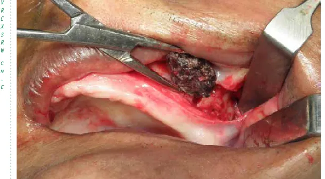
the treatment involved the surgical reduction and fixation of these fractures with the performance of rigid internal fixa-tion with 1.5 mm miniplates in the region of the fronto-zygomatic suture and a 2.0 mm system in the left zygomatic pillar. for removal of the foreign body, after fixation of the fractures, an antrostomy of the left maxillary sinus was performed through the caldwell-luc access without lower meatal antrostomy to promote the drainage of all sinus content (figure 2)1, 8, 9.
the removed material had a hardened consistency, was firm to palpation, and was 2.4 cm in diameter, with a darkened coloration. After removal of the material within the maxillary sinus, a total sinusec-tomy was performed to remove all infec-ted sinus mucosa10. After performing all
the necessary sutures, postoperative me-dications, consisting of antibiotics (cepha-Figure 1 – Axial ct scan indicating the
pre-sence of hyperdense foreign body and narrowing in the posterior region of the left maxillary sinus, as well as fracture lines in the anterior wall and lateral sinus, suggestive of bilateral le Fort ii fracture.
••
256
••
Bezerra AV Mello MJR Lira RC Silveira DX Andrade GS Silva BR Aguiar ASW Asymptomatic antrolith in maxillary sinus. Report of a case Rev. Odontol. Univ. Cid. São Paulo 2013; 25(3):254-60, set-dez
losporin 1 gram intravenous (iV) every 6 hours), anti-inflammatory drugs (ketopro-fen 100 mg, diluted in 100 ml of saline solution (ss) 0.9% 12/12 hours, dexame-thasone 4 mg iV every 12 hours) and anal-gesics (dipyrone 500 mg diluted in 18 ml of ss 0.9%), were prescribed.
the foreign body was removed and threaded. the presence of a tooth root was detected in its interior (figure 3). the clinical and surgical diagnosis of an an-trolith caused by a piece of tooth root was suggested; the root was possibly released within the maxillary sinus with a probable
evolution of 30 years. the material was sent for histopathological examination, which revealed amorphous eosinophilic material under basophilic lines of mate-rial deposition, which did not resemble cement or any dental or osteoid tissue (fi-gure 4), with the final diagnosis consistent with the diagnosis initially proposed.
DISCUSSION
the mucus produced within the ma-xillary sinus plays an important role as a protective colloid, and thus the presence of minerals are not found in high concen-Figure 2 – calcified and blackened foreign body removed from the left maxillary sinus via
caldwell luc access.
Figure 3 – Fragmentation of the calcified foreign body, 2.4 cm in diameter, revealing the
Bezerra AV Mello MJR Lira RC Silveira DX Andrade GS Silva BR Aguiar ASW Asymptomatic antrolith in maxillary sinus. Report of a case
••
257
••
Rev. Odontol. Univ. Cid. São Paulo2013; 25(3): 254-60, set-dez
trations, even in an environment rich in calcium. However, the inflammatory pro-cess impedes ciliary movement and the barrier ability of the sinusal mucosa as it proceeds. these conditions can cause sta-sis in the mucosal secretions to modify the environment and encourage the concen-tration of inorganic salts11. the antrolith
is formed from the successive deposition of minerals in the form of concentric rings around an fB into the sinuses7. the main
components are calcium phosphate, cal-cium carbonate, organic matter and wa-ter7,11,12.
The antroliths can be classified as true and false, depending on the aetiology. in the first case, their origin is endogenous and can be formed around blood, mucus, pus, red blood cells or leukocytes. exoge-nous antrolith injuries are defined as false, and can develop around a foreign body, such as teeth, tooth roots, gauze, rocks, glass, paper, vegetables, beans, seeds, and even pearls of osseointegrated implants6,11,
13,14,15,16. however, some authors consider
that teeth or dental roots are endogenous causes15,17,18 because they are constituent
parts of the body.
the injury may have a variable consis-tency and be covered with a granulation tissue that has a rich blood supply; it may also vary in colour from black to grey or white. some studies have shown that the-re is no gender or age pthe-redilection, and its symptoms can be variable. in some cases, patients may be asymptomatic, or they may report pain in the affected hemi-facial or the frontal regions7. facial pain,
nasal obstruction, epistaxis, accumulation of purulent or bloody secretions are some of the signs and symptoms commonly associated with these calcareous masses in symptomatic cases. in cases with no symptomatology, the discovery of antroli-ths often happens accidentally after routi-ne imaging exams8,15,19.
radiographically, antroliths are ob-served as radiopaque masses that vary in size and shape, with irregular borders, and they occasionally may be accompanied Figure 4 – Histopathology showing amorphous eosinophilic material lines in the
basophi-lic material deposition that do not resemble limestone, cement, or any dental or osteoid tissue.
••
258
••
Bezerra AV Mello MJR Lira RC Silveira DX Andrade GS Silva BR Aguiar ASW Asymptomatic antrolith in maxillary sinus. Report of a case Rev. Odontol. Univ. Cid. São Paulo 2013; 25(3):254-60, set-dez
by sinus opacification caused by mucosal oedema, polyps and fluid. For the asses-sment of quality and precise location, at least two radiographic projections of the lesion or a computed tomography (ct) of the sinuses are recommended18, 19.
in the differential diagnosis of radio-paque lesions of the maxillary sinus in imaging exams, antroliths may resemble osteomas, odontomas, ossifying fibromas, calcifying odontogenic cysts fibrosarco-mas, teeth, aspergillosis, manifestations of fungal diseases, exostoses, radiopaque foreign bodies and inflammatory cysts12, 20.
in a study conducted in 2003, 28 an-troliths were reported from 1927 to 2002, demonstrating that these occurrences are rare7. in this study, the authors reported
that 16 cases had a history of tooth extrac-tion, which suggests an important role for dental procedures in the aetiology of an-troliths.
oroantral communication is mainly a complication of tooth extraction, which contaminates the sinus cavity with the mi-crobiota of the oral cavity, resulting in the appearance of maxillary sinusitis5, 14.
seve-ral studies have demonstrated that odon-togenic sinusitis is polymicrobial21. in the
case presented, prophylactic antibacterial therapy was not performed, so the patient had no risk of sinus infection at surgery.
studies have shown that approximate-ly 91% of foreign bodies inside the ma-xillary sinus are the result of unsuccessful dental procedures, among which dental fragments, remnants of dental materials (amalgam dental fragments, extrusion of calcium hydroxide in the interior the root canal system) and osseointegrated im-plants can be found19, 22, 23. the objects
most commonly released into the maxilla-ry sinus are fractured roots or teeth24. the
palatal root of the maxillary first molar is most commonly displaced to the maxilla-ry sinus during an extraction procedure24.
the penetration of foreign bodies into the interior of the paranasal sinuses during dental procedures may result from poor surgical planning and surgical inexperien-ce.
in the case presented here, the pre-sence of oroantral communication over approximately 30 years, associated with
the tooth root within the maxillary sinus, allowed for the penetration of saliva, food residues and even probable accumulation of blood, which led to focal chronic ma-xillary sinusitis and the progressive growth of the calcareous mass. As the patient did not seek dental care to resolve his problem due to the lack of symptoms, the foreign body could only be identified and remo-ved after its visualisation with a ct scan for diagnosis of the facial trauma.
the caldwell-luc access is a well-esta-blished procedure that promotes access to the jaws and is indicated for the removal of cysts and intra-sinus tumours, foreign bo-dies, oroantral fistula, osteonecrosis of the jaw, epistaxis control, fungal mycoses and facial trauma10, 25. in standard
caldwell--luc access surgery, the lower meatal antrotomia (lmA) is typically held to pro-mote postoperative drainage of the bloody content of the maxillary sinus. however, this procedure has been criticised because it induces another surgical wound and po-ses an additional risk of injury to the naso-lacrimal duct. Although endoscopic sinus surgery is applied in some cases of sinu-sitis, there are still some cases in which the direct approach of the affected sinus is mandatory, as in cases of fistula, oroantral or intra-sinusal odontogenic lesions, or in the presence of large foreign bodies1.
surgical treatment of antroliths is indi-cated not only for the removal of the cal-careous mass but also to promote proper treatment of the coexisting sinus disease7,
26, 27, 28, 29. A retrospective study was
con-ducted with the results of caldwell-luc surgeries without lmA for the treatment of maxillary sinusitis of odontogenic origin during the period from 2004 to 2010. the authors concluded that this modification of the technique would provide a more comfortable postoperative period with a low probability of complications9.
Histopathological examination confir-med the clinical suspicion of an antrolith, as the tooth root was observed to be asso-ciated with a large amount of calcareous material.
CONCLUSIONS
Antroliths are rare pathological con-ditions and should be taken into
consi-Bezerra AV Mello MJR Lira RC Silveira DX Andrade GS Silva BR Aguiar ASW Asymptomatic antrolith in maxillary sinus. Report of a case
••
259
••
Rev. Odontol. Univ. Cid. São Paulo2013; 25(3): 254-60, set-dez
deration in the differential diagnosis of radiopaque paranasal injuries. the surgi-cal approach by surgi-caldwell-luc access is a safe and effective procedure for the remo-val of the calcareous mass, as well as for the removal of associated sinusal mucosa and the restoration of normal sinus drai-nage and ventilation. A careful analysis of
imaging tests may show changes that are not common, especially those found in the paranasal cavities, even if no clinical symptoms occur.
conflict of interest
the authors declare that they have no conflict of interest.
REFERÊNCIAS
1. han JK, smith tl, loehrl tA, fong KJ, hwang ph. surgical revision of the post-caldwell-luc maxillary sinus. Am
J rhinol 2005 sep-oct;19(5):478-82.
2. rodrigues mt, munhoz eD, cardo-so cl, de freitas cA, Damante Jh. chronic maxillary sinusitis associ-ated with dental impression material.
med oral Patol oral cir Bucal 2009
Apr;14(4):e163-6.
3. shenoy V, maller V. maxillary antro-lith: a rare cause of the recurrent si-nusitis. case rep otolaryngol 2013 2013(527152.
4. liston pn, Walters rf. foreign bodies in the maxillary antrum: a case report.
Aust dent J 2002 Dec;47(4):344-6.
5. felisati G, saibene Am, lenzi r, pipo-lo c. late recovery from foreign body sinusitis after maxillary sinus floor aug-mentation. BmJ case rep 2012 2012(
6. Abuabara A, cortez Al, passeri lA, de moraes m, moreira rW. evaluation of different treatments for oroantral/oro-nasal communications: experience of 112 cases. int J oral maxillofac surg 2006 feb;35(2):155-8.
7. nass Duce m, talas Du, ozer c, yildiz A, Apaydin fD, ozgur A. Antro-lithiasis: a retrospective study. J
laryn-gol otol 2003 Aug;117(8):637-40.
8. Al-Belasy fA. inferior meatal antros-tomy: is it necessary after radical sinus surgery through the caldwell-luc ap-proach? J oral maxillofac surg 2004 may;62(5):559-62.
9. huang yc, chen Wh. caldwell-luc operation without inferior meatal an-trostomy: a retrospective study of 50 cases. J oral maxillofac surg 2012 sep;70(9):2080-4.
10. matheny Ke, Duncavage JA. contem-porary indications for the caldwell-luc procedure. curr opin otolaryngol
Head neck surg 2003 feb;11(1):23-6.
11. ogata y, okinaka y, takahashi m. Antrolith associated with aspergil-losis of the maxillary sinus: report of a case. J oral maxillofac surg 1997 nov;55(11):1339-41.
12. henriques Jc, Kreich em, rosa rr, castilho Jc, de moraes lc, de mo-raes me. noninvasive aspergillo-sis as a maxillary antrolith: report of a rare case. Quintessence int 2012 feb;43(2):143-6.
13. irish le, Gray rp, sorenson fm. Antro-lith. oral surg oral med oral Pathol 1990 nov;70(5):682-3.
14. Guler n, Duygu G. progressive swell-ing and radiopaque mass in maxil-lary sinus: formation of stone. Kulak
Burun Bogaz ihtis derg 2012
may-Jun;22(3):181-5.
15. manzi f, tuji f, halter neto f, Al-meida s. Antrólito maxilar observado em paciente assintomático: revisão de literature e relato de caso clíni-co. rev odontol Brasil central 2001 10(29):17-9.
16. sofat Jr, Greval rs. maxillary antrolith around tooth root tip with oro-antral fistula--a case report. J indian dent
••
260
••
Bezerra AV Mello MJR Lira RC Silveira DX Andrade GS Silva BR Aguiar ASW Asymptomatic antrolith in maxillary sinus. Report of a case Rev. Odontol. Univ. Cid. São Paulo 2013; 25(3):254-60, set-dez
17. ishiyama t. maxillary antrolith: report of a case. Auris nasus larynx 1988 15(3):185-9.
18. pokorny A, tataryn r. clinical and radiologic findings in a case series of maxillary sinusitis of dental ori-gin. int Forum Allergy rhinol 2013 Dec;3(12):973-9.
19. Guneri p, Kaya A, caliskan mK. An-troliths: survey of the literature and report of a case. oral surg oral med
oral Pathol oral radiol endod 2005
Apr;99(4):517-21.
20. langerman A, sigari f, naclerio r. Calcified maxillary cyst secondary to a foreign-body reaction at the site of a remote tooth extraction. ear nose
throat J 2010 Jan;89(1):42-3.
21. mehra p, Jeong D. maxillary sinusitis of odontogenic origin. curr Allergy
Asthma rep 2009 may;9(3):238-43.
22. Agustí eB, puiggrós iV, figuerola cr, Vecina Vm. cuerpos extraños en seno maxilar. Acta otorrinolaringológica
española 2009 June;60(3):190-3.
23. tabrizi r, Amid r, taha ozkan B, Khorshidi h, langner nJ. effects of ex-posing dental implant to the maxillary sinus cavity. J craniofac surg 2012 may;23(3):767-9.
24. Aguiar rc, silva Júnior An, hernan-dez pAG, pinto JG, ciprandi mto, Gassen ht. remoção cirúrgica de um instrumento deslocado acidentalmen-te para oinacidentalmen-terior do seio maxilar du-rante a instalação de implantes. rFo
UPF 2007 set.-dez.;12(3):65-8.
25. Barzilai G, Greenberg e, uri n. indica-tions for the caldwell-luc approach in the endoscopic era. otolaryngol Head
neck surg 2005 feb;132(2):219-20.
26. cohen mA, packota GV, hall mJ, steinberg J. large asymptomatic antro-lith of the maxillary sinus. report of a case. oral surg oral med oral Pathol 1991 feb;71(2):155-7.
27. orhan K, Kocyigit D, turkoglu K, Kar-tal y, Arslan A. illosis of maxillary si-nus in immunocompromised patient. case report. n Y state dent J 2012 Jan;78(1):46-9.
28. Wu cW, tai cf, Wang lf, tsai KB, Kuo Wr. Aspergillosis: a nidus of max-illary antrolith. Am J otolaryngol 2005 nov-Dec;26(6):426-9.
29. nair s, James e, Dutta A, Goyal s. An-trolith in the maxillary sinus: an un-usual complication of endoscopic si-nus surgery. indian J otolaryngol Head
neck surg 2010
2010/01/01;62(1):81-3.
recebido em 19-11-2013 Aceito em 02-12-2013