2019
UNIVERSIDADE DE LISBOA FACULDADE DE CIÊNCIAS
Vulnerability of reef-building corals towards global change
“Documento Definitivo”
Doutoramento em Biologia
Especialidade de Biologia Marinha e Aquacultura
Marta Andreia Duarte Dias
Tese orientada por:
Professora Doutora Catarina Vinagre Professor Doutor Henrique Cabral
2019
UNIVERSIDADE DE LISBOA FACULDADE DE CIÊNCIAS
Vulnerability of reef-building corals towards global change
Doutoramento em Biologia
Especialidade de Biologia Marinha e Aquacultura
Marta Andreia Duarte Dias
Tese orientada por:
Professora Doutora Catarina Vinagre Professor Doutor Henrique Cabral
Júri: Presidente:
● Doutora Maria Manuela Gomes Coelho de Noronha Trancoso, Professora Catedrática e Presidente do Departamento de Biologia Animal da Faculdade de Ciências da Universidade de Lisboa
Vogais:
● Doutor Miguel Ângelo do Carmo Pardal, Professor Catedrático, Faculdade de Ciências e Tecnologia da Universidade de Coimbra
● Doutor Rui Jorge Miranda Rocha, Investigador Auxiliar, Centro de Estudos do Ambiente e do Mar da Universidade de Aveiro
● Doutora Ana Margarida da Silva Faria, Investigadora Auxiliar, ISPA – Instituto Universitário de Ciências Psicológicas, Sociais e da Vida
● Doutor José Pavão Mendes de Paula, Professor Associado com Agregação, Faculdade de Ciências da Universidade de Lisboa
● Doutora Catarina Maria Batista Vinagre, Professora Auxiliar Convidada, Faculdade de Ciências da Universidade de Lisboa (orientadora)
Documento especialmente elaborado para a obtenção do grau de doutor
Colaboração com Oceanário de Lisboa, financiada por Fundação para a Ciência e a Tecnologia (FCT)
i
This study was funded by Fundação para a Ciência e a Tecnologia, through a PhD scholarship
SFRH/BD/103047/2014 and the research project WarmingWebs
(PTDC/MAR-EST/2141/2012). Additional funding was provided by the strategic projects UID/MAR/04292/2019 granted to MARE and UID/Multi/04378/2019 granted to UCIBIO.
ii Nota prévia
A presente tese apresenta artigos científicos já publicados ou submetidos para publicação (ca-pítulos 2 a 6), de acordo com o previsto no n˚2 do artigo 25˚ do Regulamento de Estudos de Pós Graduação da Universidade de Lisboa, publicado em Diário da República 2ª série – N˚155 – 11 de Agosto de 2017. Uma vez que estes trabalhos foram realizados em colaboração, a can-didata esclarece que participou integralmente na concepção dos trabalhos, obtenção de dados, análise e discussão dos resultados, bem como na redacção dos manuscritos.
A presente tese, por ser uma compilação de publicações internacionais, está redigida em Inglês. Uma lista de referências é dada no final de cada capítulo em vez de no final da tese e devido a este formato poderão haver casos de duplicação entre capítulos. Cada capítulo contém também a informação de suporte associada ao mesmo. O formato diferente de alguns capítulos reflete os requerimentos específicos dos jornais científicos aos quais os manuscritos apresentados fo-ram submetidos.
Lisboa, setembro de 2019
iii
Agradecimentos
Gostaria de agradecer à Fundação para a Ciência e a Tecnologia por financiar a minha tese de doutoramento.
À minha orientadora Catarina Vinagre por me ter proposto este tema de tese, despertando o meu interesse pela ecologia marinha tropical. Queria também agradecer por todo o apoio, amizade, disponibilidade e confiança ao longo destes 4 anos de trabalho.
Ao meu orientador Henrique Cabral por me ter acolhido como doutoranda, pela disponibilidade e pelas sábias sugestões.
Ao Mário Diniz que me ensinou todas as técnicas necessárias de bioquímica para a realização do trabalho no âmbito desta tese, bem como pelo usufruto do laboratório para a realização dos procedimentos. Queria também agradecer pela sua total disponibilidade e amizade.
Agradeço à Núria Baylina por ter proporcionado a oportunidade de desenvolvimento das experiências no Oceanário de Lisboa.
Agradeço ao staff do Oceanário de Lisboa, em particular aos aquaristas Raúl Gouveia e Ana Ferreira que disponibilizaram o seu tempo para me auxiliar e por todo o conhecimento de aquariologia transmitido.
Às minhas amigas Sandra Ferreira, Filipa Belo, Joana Roma, Mafalda Pastaneira, Helga Silva e Joana Cruz pela paciência e amizade.
Às amigas e colegas do grupo ECCOWEBS: Carolina Madeira, Vanessa Mendonça e Diana Madeira pela amizade, sábios conselhos e momentos de relaxamento.
iv
À minha família que sempre me apoiou no percurso académico, pelo carinho, paciência e palavras de conforto.
Ao meu companheiro Rui Cereja e ao meu filho Miguel Cereja por fazerem parte da minha vida, pelo carinho e paciência.
v ABSTRACT
Global warming is leading to large-scale coral bleaching and mass mortality, but also to increases in tropical storms' frequency and intensity. Storms allow fragmentation of reef-building corals and can lead to near-shore salinity reduction which, combined with ocean warming, will aggravate coral distress. In order to assess the susceptibility of different coral species to these environmental stressors, small fragments of nine coral species of the Indo-Pacific region were exposed to different thermal (26°C, 30°C, 32°C) and hyposaline (26°C-33psu, 30°C-(26°C-33psu, 26°C-20psu, 30°C-20psu) experimental treatments for 60 days. Several parameters were assessed at different levels of biological organization: at the organism level (total and partial mortality, and coral condition based in bleaching levels), physiological level (growth rate and regeneration rate of artificially inflicted lesions), and molecular level (superoxide dismutase (SOD), catalase (CAT), glutathione S-transferase (GST), and lipid peroxidation (LPO)). Also, in order to test two different approaches to be applied in the monitoring of the effects of heat stress, some parameters were combined in integrated biomarker response indices, either in a molecular approach, approach A, using GST, CAT, LPO, and SOD, or in an approach that integrates the molecular, physiological and organism levels, approach B, using GST, CAT, LPO, SOD, partial mortality, and growth rate. Results indicate that Pocillopora damicornis and Stylophora pistillata were the most vulnerable at 30°C. Psammocora contigua, Turbinaria reniformis, and Galaxea fascicularis were the most tolerant species at 32°C. The species P. contigua and G. fascicularis were the most tolerant to low salinity (26°C-20psu). The species G. fascicularis was the only one capable of surviving the combined effect of high temperature and low salinity (30°C-20psu). Approach B, the most integrative approach, was considered the most adequate for evaluating the health of reef corals since it better discriminated the stress suffered by the tested species.
Keywords: global climate change, heath stress, hyposaline stress, coral bleaching, health assessment
vii RESUMO
Os corais construtores de recife são fundamentais para a geomorfologia, biodiversidade, produtividade e complexidade estrutural dos recifes tropicais. O sucesso ecológico dos corais construtores de recife deve-se à sua simbiose com algas dinoflageladas unicelulares (zooxantelas). Como os corais depositam carbonato de cálcio e normalmente suplementam o seu metabolismo através da provisão de compostos fotossintéticos provenientes das zooxantelas, a disrupção desta associação simbiótica sob stress, denominada lixiviação, resulta em reduções significativas na fotossíntese das zooxantelas acompanhadas por alterações drásticas na fisiologia resultantes na perda dos simbiontes. As respostas dos corais à lixiviação podem incluir aumento da taxa respiratória, diminuição da taxa de calcificação do esqueleto e da capacidade de regeneração do tecido, redução da taxa de crescimento e das capacidades reprodutivas, degradação do tecido e consequente morte do coral, podendo levar à perda rápida de biodiversidade do recife afetado. Os recifes de coral estão entre os ecossistemas mais vitais mas também entre os mais ameaçados globalmente. Para além das pressões locais, estes ecossistemas estão atualmente a sofrer uma pressão sem precedentes e risco de extinção devido às alterações climáticas globais que têm resultado no aumento das temperaturas à superfície e na acidificação dos oceanos. Os ecossistemas de recife de coral, embora localizados nas águas marinhas mais quentes do mundo, são considerados particularmente vulneráveis ao aquecimento global devido ao facto de muitos organismos tropicais, incluindo os corais construtores de recife e espécies associadas, viverem próximos do seu limite de tolerância térmica máximo. O aquecimento global é a principal causa da lixiviação de corais em larga escala e de mortalidade em massa em comunidades de recife de coral em todo o mundo, levando à rápida degradação dos recifes de coral. Embora atuem à escala local, outros fatores para além da temperatura elevada têm sido correlacionados com a lixiviação de corais (por exemplo, salinidade reduzida). As técnicas mais recentes de modelação do clima global indicam que as condições térmicas para a lixiviação de corais estão a tornar-se mais frequentes. O contínuo aumento das temperaturas nos oceanos tropicais irá exacerbar a severidade e a frequência do
stress térmico sofrido pelos recifes de coral, ameaçando a persistência de recifes dominados
por corais ao longo dos trópicos, havendo previsões de que locais presentemente considerados refúgios térmicos para estas espécies poderão desaparecer a meio do século. Oceanos mais quentes conduzirão a tempestades tropicais mais frequentes e intensas levando a períodos cada vez mais curtos para recuperação das comunidades de corais entre recorrências e à perspetiva
viii
de que o dano físico experienciado pelos recifes de coral irá aumentar. Além do dano físico, a forte precipitação que acompanha as tempestades tropicais irá baixar a salinidade em áreas superficiais próximas da costa. Os efeitos da salinidade reduzida podem ser exacerbados durante episódios de temperaturas elevadas, tornando-se importante perceber o efeito conjunto destas duas variáveis ambientais. Com o previsto aumento de intensidade e frequência das tempestades tropicais também aumentará a probabilidade de reprodução assexuada por fragmentação das colónias de corais construtores de recife, podendo estes fragmentos de coral dar origem a novas colónias nos recifes degradados e desta forma contribuir para a recuperação em pequena escala dos recifes afetados. No entanto, estes fragmentos também estarão sujeitos a pressões ambientais que potencialmente condicionarão a sua sobrevivência e crescimento. O primeiro nível de resposta de um organismo a uma perturbação ambiental verifica-se a nível molecular, passando posteriormente para níveis de organização mais elevados, até haver uma clara resposta do organismo como um todo. Deste modo, o estudo de parâmetros de resposta a diferentes níveis de organização biológica irá permitir uma análise mais completa. Diferentes espécies de corais construtores de recife apresentam diferente suscetibilidade ao stress térmico e salino, podendo levar a alterações na composição das espécies de coral após eventos de stress desta natureza e até mesmo à extinção local dos corais mais sensíveis. Os principais objetivos deste trabalho foram 1) determinar a suscetibilidade de várias espécies de corais construtores de recife à exposição prolongada a temperatura elevada e salinidade reduzida, e 2) testar e otimizar duas abordagens baseadas no uso de índices de resposta integrada de biomarcadores (sigla inglesa IBR – “Integrated Biomarker Response”) para avaliação da saúde dos corais construtores de recife face ao stress térmico, num contexto de reprodução assexuada por fragmentação. Desta forma, pequenos fragmentos de nove espécies de corais construtores de recife da região do Indo-Pacífico foram submetidos a vários tratamentos experimentais, durante 60 dias. As espécies utilizadas apresentaram diferente morfologia da colónia: ramificada (Acropora tenuis, Pocillopora damicornis, Stylophora pistillata, e Psammocora contigua), massiva (Galaxea fascicularis), placa (Montipora capricornis (morfotipo castanho),
Echinopora lamellosa, e Turbinaria reniformis), e incrustante (Montipora capricornis
(morfotipo verde)), esta característica inerente às espécies está ligada à sua diferente suscetibilidade relativamente às variáveis ambientais em estudo. Os tratamentos térmicos experimentais incluíram a temperatura controlo (26°C) e as temperaturas de stress (30°C e 32°C). Por outro lado, a experiência de efeito combinado de temperatura e salinidade incluiu os seguintes tratamentos experimentais: controlo (26°C-33psu), temperatura elevada (30°C-33psu), salinidade reduzida (26°C-20psu) e combinação de temperatura elevada com salinidade
ix
reduzida (30°C-20psu). De modo a obter uma visão mais generalista do efeito das variáveis em estudo nos organismos testados, parâmetros de diferentes níveis de organização biológica foram estudados: nível do organismo (mortalidade total, mortalidade parcial, e condição do coral que inclui os vários níveis de lixiviação), nível fisiológico (taxa de crescimento, taxa de regeneração de lesões infligidas artificialmente no tecido dos fragmentos), e nível molecular (determinação de biomarcadores de stress oxidativo: peroxidação lipídica (LPO), superóxido dismutase (SOD), catalase (CAT), e glutationa S-transferase (GST)). Também, de modo a testar duas diferentes abordagens para serem aplicadas na monitorização dos efeitos do stress térmico em corais construtores de recife dos oceanos Indo-Pacífico, alguns parâmetros pertencentes aos vários níveis de organização biológica foram selecionados e combinados em IBR’s, ou na categoria de resposta de biomarcadores de stress oxidativo (abordagem A: GST, CAT, LPO, e SOD) ou na categoria de resposta integrada-performance do organismo (abordagem B: GST, CAT, LPO, SOD, mortalidade parcial, e taxa de crescimento). No presente trabalho observou-se que a mortalidade aumentou com a temperatura, atingindo 100% para a maioria das espécies após os 60 dias de exposição a 32ºC, exceto para T. reniformis, G. fascicularis, e P. contigua. Estas espécies apresentaram a mortalidade parcial e lixiviação mais baixos ao longo dos tratamentos térmicos experimentais. Relativamente a T. reniformis e P. contigua, não foi observado dano oxidativo na exposição a 32ºC, embora ambas as espécies tenham apresentado uma aparência pálida. Galaxea fascicularis foi a única das três espécies onde fragmentos lixiviados e dano oxidativo foram observados, embora tenha apresentado alguns fragmentos com uma aparência saudável a 32ºC. A taxa de crescimento diminuiu com o aumento da temperatura, sendo mais elevada em espécies de morfologia ramificada, enquanto que a taxa de regeneração de lesões geralmente aumentou com a temperatura. Foi concluído que T.
reniformis, G. fascicularis e P. contigua foram as espécies mais resistentes ao stress térmico.
Relativamente aos resultados do efeito combinado de temperatura e salinidade, no tratamento de temperatura elevada (30°C-33psu) apenas duas espécies morreram (P. damicornis e S.
pistillata), enquanto que no tratamento de salinidade reduzida (26°C-20psu) todas as espécies
morreram com a exceção de duas (P. contigua e G. fascicularis) passados 60 dias. Estas duas últimas espécies apresentaram a mortalidade parcial mais reduzida, a melhor condição, e nenhuma evidência de dano oxidativo tendo-se, no entanto, verificado resposta antioxidativa. A mortalidade foi mais elevada a 30°C-20psu devido ao efeito combinado da elevada temperatura e da salinidade reduzida, atingindo 100% em oito das nove espécies de coral, com apenas G. fascicularis sobrevivendo a este tratamento experimental. Contudo, a mortalidade foi elevada nesta espécie, observando-se também um aumento na atividade da SOD revelador de
x
uma resposta antioxidativa, não tendo, no entanto, o dano oxidativo sido detetado. As taxas de crescimento diminuíram com o aumento da temperatura e a diminuição da salinidade, enquanto que as taxas de regeneração aumentaram com a temperatura atingindo um máximo a 30°C-33psu e um mínimo a 20psu. Foi concluído que P. damicornis e S. pistillata foram as espécies mais vulneráveis ao tratamento de temperatura elevada, G. fascicularis e P. contigua foram os mais tolerantes ao stress hipossalino, e G. fascicularis foi a única espécie que tolerou o efeito combinado da temperatura elevada e da salinidade reduzida. Os resultados deste trabalho também indicam que o IBR pode ser um método a ser aplicado na avaliação da saúde de corais de recife sob stress térmico. A abordagem B, integradora da resposta molecular, fisiológica e do organismo, foi considerada a mais adequada uma vez que refletiu melhor o stress diferencial sofrido pelas espécies testadas, enquanto que a abordagem A, integradora apenas de biomarcadores moleculares, não foi suficiente para discriminar a resposta da maioria das espécies testadas. Assim, a integração da resposta de parâmetros de diferentes níveis de organização será a abordagem mais adequada para a avaliação de qualidade ambiental.
Palavras-chave: alterações climáticas globais, stress térmico, stress hipossalino, lixiviação, efeitos sinergísticos
xi TABLE OF CONTENTS Agradecimentos……… iii ABSTRACT……….. v RESUMO………..vii LIST OF TABLES………...xvi
LIST OF FIGURES………. xix
LIST OF SYMBOLS AND ABBREVIATIONS... xxi
CHAPTER 1. General Introduction……….. 1
1.1. Coral reef ecosystems………. 2
1.1.1. Reef-building corals and zooxanthellae symbiosis……….. 2
1.1.2. Factors influencing coral reefs’ community structure and development………. 4
1.1.2.1. Coral reproductive strategies………. 4
1.1.2.2. Colony growth rates………... 4
1.1.2.3. Competitive dominance relationships……… 5
1.1.2.4. Predation……… 6
1.1.2.5. Physical disturbances: storms……… 7
1.2. Effects of global climate change in coral reef ecosystems………. 7
1.2.1. Climate induced changes………. 8
1.2.1.1. Heat stress impacts on reef-building corals………... 9
1.2.1.2. Hyposalinity impacts on reef-building corals……… 11
1.2.2. Bleaching consequences……….. 14
1.2.3. Predictions under future climate change scenarios……….. 16
1.2.4. Importance of fragmentation in corals’ propagation by tropical storms………..17
1.2.5. Recovery after disturbance……….. 19
1.2.6. Oxidative stress in reef corals……….. 21
1.2.7. Different susceptibility to environmental variables among reef-building corals…….25
1.2.8. Health assessment of reef corals……….. 27
1.3. Aims and thesis layout……… 28
1.4. References………... 29
CHAPTER 2. Mortality, growth and regeneration following fragmentation of reef-form-ing corals under thermal ………... 85
xii
2.1. Introduction and literature review.……… 87
2.2. Materials and methods……….………... 89
2.2.1. Study species……….. 89
2.2.2. Acclimation conditions and experimental setup………. 89
2.2.3. Analytical procedures……….. 91
2.2.3.1. Mortality assessments and bleaching level………... 91
2.2.3.2. Growth rate measurements………... 92
2.2.3.3. Regeneration rate of injuries………. 92
2.2.4. Statistical analyses……… 92
2.3. Results………….……… 93
2.3.1. Mortality and bleaching level………. 94
2.3.1.1. Mortality………... 94
2.3.1.2. Partial mortality……… 96
2.3.1.3. Bleaching level………..98
2.3.2. Growth rate……… 100
2.3.3. Regeneration rate………. 101
2.4. Discussion and conclusions……….……….. 102
2.5. References………... 108
2.6. Supplementary material……….. 117
CHAPTER 3. Synergistic effects of warming and lower salinity on the asexual reproduc-tion of reef-forming corals………...119
Abstract………….………... 120
3.1. Introduction and literature review …….……… 121
3.2. Materials and methods………….………... 125
3.2.1. Coral specimens and maintenance conditions……… 125
3.2.2. Experimental setup………... 129
3.2.3. Analytical procedures……….. 132
3.2.3.1. Mortality and coral condition assessments………... 132
3.2.3.2. Growth rate measurements………... 132
3.2.3.3. Regeneration rate of injuries………. 133
3.2.4. Statistical analyses……… 133
3.3. Results………. 134
3.3.1. Mortality and coral condition……… 134
xiii
3.3.1.2. Partial mortality……… 135
3.3.1.3. Coral condition………..137
3.3.2. Growth rate……… 141
3.3.3. Regeneration rate………. 142
3.4. Discussion and conclusions ………... 144
3.4.1. Mortality………. 144
3.4.2. Coral condition………. 146
3.4.3. Growth……… 147
3.4.4. Regeneration……….. 148
3.5. References………... 150
CHAPTER 4. Long-term exposure to increasing temperatures on scleractinian coral fragments reveals oxidative stress……….…………. 169
Abstract……….………... 170
4.1. Introduction and literature review …….……… 171
4.2. Materials and methods……….………... 174
4.2.1. Coral species………. 174
4.2.2. Experimental setup………... 174
4.2.3. Analytical procedures……….. 180
4.2.3.1. Survival, coral condition and samples storage……… 180
4.2.4. Protein extraction………. 182
4.2.5. Total Protein determination……… 182
4.2.6. Oxidative damage products – lipid peroxidation……… 182
4.2.7. Enzymatic assays……….. 183
4.2.7.1. Catalase activity……… 183
4.2.7.2. Glutathione S-transferase activity……… 184
4.2.8. Statistical analyses……… 184
4.3. Results……… 185
4.3.1. Survival………... 185
4.3.2. Coral general condition……….. 185
4.3.3. Lipid peroxidation (LPO)……… 186
4.3.4. Catalase (CAT)……….. 190
4.3.5. Glutathione S-transferase (GST)………... 190
4.4. Discussion and conclusions………... 191
xiv
4.6. Supplementary material……….. 218
CHAPTER 5. Oxidative stress on scleractinian coral fragments following exposure to high temperature and low salinity……….. 219
Abstract………….………... 220
5.1. Introduction and literature review ………... 221
5.2. Materials and methods……… 225
5.2.1. Study coral species………... 225
5.2.2. Experimental design………. 225
5.2.3. Survival, coral general condition and samples storage……… 228
5.2.4. Protein extraction………. 230
5.2.5. Total Protein determination……… 230
5.2.6. Oxidative damage products – lipid peroxidation……… 230
5.2.7. Enzymatic assays……….. 231
5.2.7.1. Superoxide dismutase activity……….. 231
5.2.7.2. Catalase activity……… 231
5.2.7.3. Glutathione S-transferase activity………. 232
5.2.8. Statistical analyses……… 232
5.3. Results………. 233
5.3.1. Survival………... 233
5.3.2. Coral general condition……….. 234
5.3.3. Lipid peroxidation (LPO)……… 235
5.3.4. Superoxide dismutase (SOD)……….. 237
5.3.5. Catalase (CAT)……….. 238
5.3.6. Glutathione S-transferase (GST)………... 239
5.4. Discussion and conclusions………... 240
5.5. References………... 245
5.6. Supplementary material……….. 260
CHAPTER 6. Integrative indices for health assessment in reef corals under thermal stress……….. 261
Abstract……….. 262
6.1. Introduction and literature review ………. 263
xv
6.2.1. Study species……….. 267
6.2.2. Experimental procedure……….. 267
6.2.3. Analytical procedures……….. 268
6.2.3.1. Partial mortality assessment……….. 268
6.2.3.2. Growth rate measurements………... 268
6.2.3.3. Samples collection and storage………. 268
6.2.4. Molecular biomarker assays……….. 269
6.2.5. Data analysis………. 269
6.3. Results………. 271
6.3.1. IBR approach A - biomarkers of oxidative stress response category……….. 271
6.3.2. IBR approach B - integrative stress response category (organism performance)….. 277
6.3.3. Fitness index……… 282
6.4. Discussion and conclusions………... 284
6.5. References………... 287
6.6. Supplementary material……….. 297
CHAPTER 7. Final remarks and future perspectives………. 299
7.1. Concluding remarks……… 300
7.2. Future perspectives………. 302
ANNEXES……… 305
xvi LIST OF TABLES
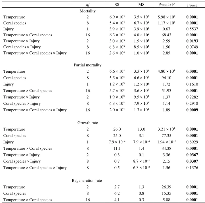
Table 2.1. Summary of results of PERMANOVA permutation tests applied to report the effects of temperature, coral species and presence/absence of injury in coral fragments’ mortality, par-tial mortality, growth and regeneration rates. Significant differences are marked a bold…. 93
Table 2.2. Coral fragments’ average growth rate ± standard deviation in the three temperature
experiments (% day-1). NA – not available………...101
Table 2.3. Coral fragments’ average regeneration rate ± standard deviation in the three temper-ature experiments (mm2 day-1) measured until complete regeneration or the last time point they
were still alive. For more information see Supplementary material……….. 102
Table 3.1. Characteristics of the coral species in study. Data taken from Veron, 1990, 2000; Richmond, 1997; Loya et al., 2001; LaJeunesse et al., 2004. ………. 126
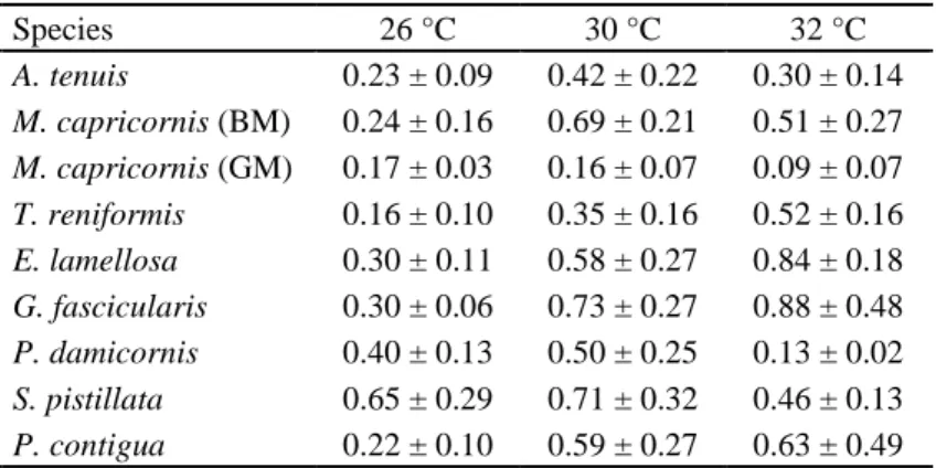
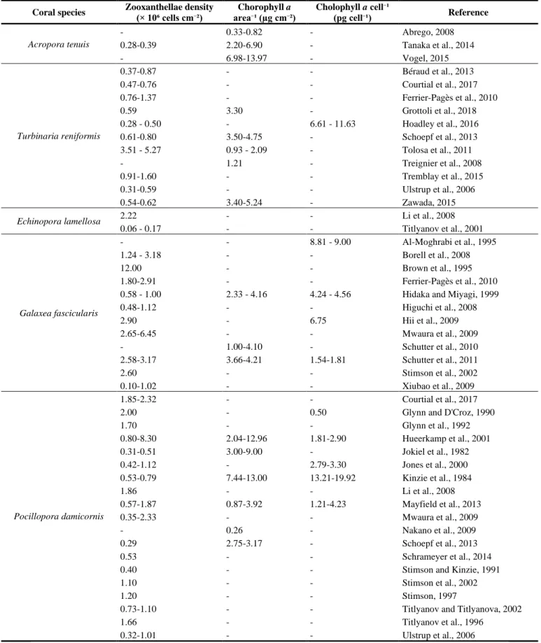
Table 3.2. Published data of zooxanthellae density and chlorophyll a concentration for the coral species in study. No data were found for Montipora capricornis (both morphotypes)……. 127
Table 3.3. Summary of results of PERMANOVA permutation tests applied to report the effects of coral species, temperature, salinity, and injury in coral fragments’ partial mortality, growth, and regeneration rates. Significant differences are marked a bold……… 134
Table 3.4. Mortality rate of the nine coral species studied on specific days of the four experi-ments. C - control, HT - high temperature, LS - low salinity……… 136
Table 3.5. Partial mortality rate of the nine coral species studied every 20 days of the four experiments (C - control, HT - high temperature, LS - low salinity) and separated in percentage classes [0,25%[, [25,50%[, [50,75%[, and [75,100%]………... 138
Table 3.6. Coral condition of the nine coral species studied every 20 days of the four experi-ments (C - control, HT - high temperature, LS - low salinity) and separated in four categories: normal, pale, bleached, and dead………... 140
xvii
Table 3.7. Coral species’ average growth rate ± standard deviation (% day-1) in the four
exper-iments. C - control, HT - high temperature, LS - low salinity. NA – Not Available………142
Table 3.8. Coral species’ average regeneration rate ± standard deviation (mm2 day-1) in the four experiments. C - control, HT - high temperature, LS - low salinity. *Only one individual. NA – Not Available………... 143
Table 4.1. Characteristics of the coral species in study………. 175
Table 4.2. Sea surface temperature (SST) range of the studied coral species according with their geographical distribution………... 177
Table 4.3. Number of surviving fragments of the nine-coral species tested on the relevant days of the three temperature treatments………181
Table 4.4. Summary of results of PERMANOVA permutation tests applied to report the effects of coral species and temperature in coral fragments’ lipid peroxidation (MDA) and both catalase (CAT) and glutathione S-transferase (GST) activities. Some combinations in the interaction temperature × species had no fragments as observed in Table 4.3. Significant differences are marked in bold………... 187
Table 4.5. Mean ± SD of MDA (µmol.mg−1 of total protein), catalase (CAT) activity (µmol.min−1.mg−1 of total protein), and glutathione S-transferase (GST) activity (µmol.min−1.mg−1 of total protein) for the nine-coral species tested in the three temperature treatments. NA – not available due to total mortality before the 60th day of experiment…. 189
Table 5.1. Number of surviving fragments of the nine-coral species tested on the relevant days of the four experimental treatments. C – control; HT – high temperature; LS – low salin-ity... 229
Table 5.2. Summary of results of PERMANOVA permutation tests applied to report the effects of coral species (Sp), temperature (Te), and salinity (Sa) in coral fragments’ lipid peroxidation (LPO); superoxide dismutase (SOD), catalase (CAT) and glutathione S-transferase (GST) ac-tivities. Significant differences are marked in bold………... 237
xviii
Table 6.1. Principal components analysis results of the four biomarkers of oxidative stress (GST – glutathione S-transferase; CAT – catalase; LPO – lipid peroxidation, SOD – superoxide dis-mutase) in the seven Indo-Pacific coral species. PC1 and PC2 stands for axes 1 and 2 of the PCA, respectively, and the values indicated for the variables are the factor loadings for PC1 and PC2……….. 273
Table 6.2. Sensibility of the seven Indo-Pacific coral species to heat stress calculated as rate of IBR variation and expressed as %. Negative percentages correspond to IBR values decrease with increase in temperature. NA – not available due to complete mortality at 32 °C……. 275
Table 6.3. Principal components analysis results of the six biomarkers relative to organism per-formance (GST – glutathione S-transferase; CAT – catalase; LPO – lipid peroxidation, SOD – superoxide dismutase, PM – partial mortality, GR – growth rate) in the seven Indo-Pacific coral species. PC1 and PC2 stands for axes 1 and 2 of the PCA, respectively, and the values indicated for the variables are the factor loadings for PC1 and PC2. NA – parameter with value = 0 in the three temperature treatments……….. 278
Table 6.4. Fitness index presented as a color hitmap: IBR scores were compared for biomarkers between control organisms and those exposed to both 30 °C and 32 °C. Red boxes denote del-eterious effects, green denotes positive effects, whereas yellow denotes no detected effect; val-ues are ± 0.5 (or higher) from control's score valval-ues. GST – glutathione S-transferase; CAT – catalase; LPO – lipid peroxidation; SOD – superoxide dismutase; PM – partial mortality; GR – growth rate. NA – not available due to total mortality……….. 283
Table 6.5. Biomarker scores (mean) for all species at different combinations of temperatures. GST – glutathione S-transferase; CAT – catalase; LPO – lipid peroxidation; SOD – superoxide dismutase; PM – partial mortality; GR – growth rate……… 283
xix LIST OF FIGURES
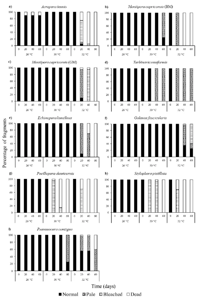
Figure 2.1. Mortality of the coral species’ fragments studied throughout the three temperature experiments……… 95
Figure 2.2. Partial mortality of the coral species’ fragments studied throughout the three tem-perature experiments separated in percentage classes [0,25%[, [25,50%[, [50,75%[ and [75,100%]………...97
Figure 2.3. Bleaching level of the coral species’ fragments studied throughout the three tem-perature experiments……….. 99
Figure 4.1. Coral condition of the nine-coral species studied in the three temperature treat-ments………... 186
Figure 4.2. Mean ± SD of (a) MDA (µmol.mg−1 of total protein), (b) catalase activity (µmol.min−1.mg−1 of total protein), and (c) glutathione S-transferase (GST) activity (µmol.min−1.mg−1 of total protein) for the nine-coral species studied in the three temperature treatments. Asterisks (*) mark significant differences (P < 0.05) in relation to control tempera-ture………. 188
Figure 5.1. Coral general condition of the nine-coral species studied in the four experimental treatments. C - control, HT - high temperature, LS - low salinity………. 234
Figure 5.2. Mean ± SD of (a) LPO (MDA concentration µmol.mg−1 of total protein), (b) super-oxide dismutase activity (% inhibition.mg-1 of total protein), (c) catalase activity (µmol.min−1.mg−1 of total protein), and (d) glutathione S-transferase (GST) activity (µmol.min−1.mg−1 of total protein) for the nine-coral species studied in the four experimental treatments. C - control, HT - high temperature, LS - low salinity. Asterisks (*) mark significant differences (p-value < 0.05) in relation to control treatment………. 236
Figure 6.1. Ordination plot of the first two axes of the principal components analysis carried out to assess the effect of the three temperature treatments in the biomarkers of oxidative stress
xx
response in seven coral species of the Indo-Pacific region. Variable’s factor loadings are repre-sented in blue and relative to GST - glutathione S-transferase; CAT – catalase; LPO – lipid peroxidation; SOD – superoxide dismutase……….. 274
Figure 6.2. Star plots with mean scores for the seven coral species exposed to 26 °C (control) and both 30 ºC and 32 °C (stress temperatures). GST – glutathione S-transferase; CAT – cata-lase; LPO – lipid peroxidation; SOD – superoxide dismutase……….. 276
Figure 6.3. Ordination plot of the first two axes of the principal components analysis carried out to assess the effect of the three temperature treatments in the biomarkers of oxidative stress, partial mortality and growth rate response in seven coral species of the Indo-Pacific region. Variable’s factor loadings are represented in blue and relative to GST - glutathione S-transfer-ase; CAT – catalS-transfer-ase; LPO – lipid peroxidation; SOD – superoxide dismutS-transfer-ase; partial mortality; and growth rate……….………. 279
Figure 6.4. Star plots with mean scores for the seven coral species exposed to 26 °C (control) and both 30 ºC and 32 °C (stress temperatures). GST – glutathione S-transferase; CAT – cata-lase; LPO – lipid peroxidation; SOD – superoxide dismutase; PM – partial mortality; GR – growth rate………. 281
xxi LIST OF SYMBOLS AND ABBREVIATIONS
ANOVA, analysis of variance
APX, ascorbate peroxidase
BSA, bovine serum albumin
CAT, catalase
cm, centimeter
CDNB, chloro-2,4-dinitrobenzene
CO2, carbon dioxide
DGAV, Direcção Geral de Veterinária
e.g., for example
EDTA, ethylenediaminetetraacetic acid
ENSO, El-Niño Southern Oscillation
Eq, equation
ɛ, extinction coefficient
FCT, Fundação para a Ciência e Tecnologia
FELASA, Federation of European Laboratory Animal Science Associations
Fig., figure g, grams GPx, glutathione peroxidase GSH, reduced glutathione GST, glutathione S-transferase h, hour
hsp, heat shock protein
H2O2, hydrogen peroxide
HO●, hydroxyl radical
xxii
IPCC, Intergovernmental Panel on Climate Change
K, kelvin
KCl, potassium chloride
KH2PO4, monopotassium phosphate
KIO4, potassium periodate
L, litre
Ln, natural logarithmic
LPO, lipid peroxidation
M, molar concentration
m, meter
MARE, Marine and Environmental Sciences Centre
MDA, malondialdehyde bis (dimethylacetal)
mg, milligrams
Min, minutes
mL, mililitre
mm, milimetres
mM, milimolar
NA, not available or not-applicable
Na2HPO4, disodium phosphate
NaCl, sodium chloride
NBT, nitroblue tetrazolium
nm, nanometers
1O
2, singlet oxygen
O2•-, superoxide anion radical
˚C, degree Celsius OH, hydroxide
xxiii
OTB, oxidative theory of coral bleaching
p, p-value
PAR, photosynthetically active radiation
PBS, phosphate buffered saline
PCA, principal component analysis
PERMANOVA, permutational multivariate analysis of variance
pg, picogram
psu, practical salinity unit
RGR, relative growth rate
RNS, reactive nitrogen species
ROS, reactive oxygen species
SD, standard deviation
SDS, sodium dodecyl sulphate
SOD, superoxide dismutase
SST, sea surface temperature
TBARS, thiobarbituric acid reactive substances
XOD, xanthine oxidase
%, percentage
~, approximately
±, more or less ∆, variation
µE m-2 s-1,µEinsteins per m2 per second µg, micrograms
µL, microlitres
1
CHAPTER 1
2 1.1. Coral reef ecosystems
Coral reefs are ecologically and economically important ecosystems found across the world’s tropical and sub-tropical oceans (Moberg and Folke, 1999; Wild et al., 2011). These ecosystems function as important spawning, nursery, breeding and feeding areas for a multitude of organisms, being among the most productive and biologically diverse marine ecosystems on Earth (e.g. Connell, 1978; Hughes et al., 2017a). Despite covering less than 0.1% of the ocean floor, reefs host 35% of all species living in the oceans (Ormond and Roberts, 1997; Spalding et al., 2001; Knowlton et al., 2010), with approximately 95,000 described coral reef species (Reaka-Kudla, 2005). Being one of the habitats most rich in species of the world, these ecosystems are important in preserving a vast biological diversity and genetic archive for future generations (Moberg and Folke, 1999). The remarkably high habitat heterogeneity of reef systems created by the complex three-dimensional structure enables niche diversification, making possible the evolution of new species (Birkeland, 1997; Paulay, 1997). Coral reefs’ complex three-dimensional structure also dissipates wave energy to protect coastlines from storms, flooding, and erosion (Heron et al., 2017; Harris et al., 2018). In terms of economic benefits, coral reefs supply many millions of people with goods and services such as seafood (e.g. fish, mussels, crustaceans, sea cucumbers, and seaweeds, Craik et al., 1990; Birkeland, 1997), recreational possibilities, coastal protection, as well as aesthetic and cultural benefits (e.g. Cesar, 1996; Done et al., 1996; Peterson and Lubchenco, 1997; Hughes et al., 2017a), with an estimated value of over USD $1 trillion globally (Costanza et al., 2014; Hoegh-Guldberg, 2015).
1.1.1. Reef-building corals and zooxanthellae symbiosis
Corals, like jellyfishes, hydras, and sea anemones, belong to the phylum Cnidaria. Corals begin life as ciliated planular larvae, which, after a period of growth, attach to a suitable substrate and remain sessile for the rest of their adult lives (Harrison and Wallace, 1990). Once attached, corals are called polyps, and can reproduce by asexual budding to form colonies. Corals can be divided into the broad categories of soft corals (e.g. sea fans and sea whips, Order Alcyonacea), and hard corals (Order Scleractinia), which lay down a calcium carbonate skeleton and are capable of building reefs (Meehan and Ostrander, 1997). Scleractinian corals are fundamental to the geomorphology, biodiversity, productivity, and structural complexity of tropical coral reef ecosystems (Hoegh-Guldberg, 2004; Idjadi and Edmunds, 2006; Hart and
3
Kench, 2007; Pratchett et al., 2008). The ecological success of shallow-water reef-building corals throughout tropical oligotrophic waters relies on the symbiosis between the coral animal and its unicellular dinoflagellate algae (zooxanthellae, family Symbiodiniaceae [LaJeunesse et al., 2018]; Hoegh-Guldberg et al., 2007a). Zooxanthellae live inside membrane-bound vacuoles (symbiosomes) within gastrodermal cells of the coral host in extremely high densities (106 cm-2) (Wakefield and Kempf, 2001; Wooldridge, 2014) and selectively transfer up to 90% of their photosynthetic products (mycosporine-like amino acids, glycerol, glucose or lipids; Wang and Douglas, 1999; Shick and Dunlap, 2002; Kopp et al., 2015) across the host-symbiont barrier (Muscatine, 1990; Hoegh-Guldberg, 1999; Stanley, 2006) with the remaining percentage of photosynthetic products being used for the respiration and growth of zooxanthellae (Edmunds and Davies, 1986). Zooxanthellae also cover 30% of the host’s nitrogen requirements for growth, reproduction and maintenance from dissolved nutrient uptake (Bythell, 1990). It is a crucial energy source in an otherwise clear and nutrient-poor tropical waters that powers the metabolically costly process of calcification, allowing corals to deposit huge amounts of calcium carbonate (Muscatine, 1990; Hoegh-Guldberg, 1999; Hart and Kench, 2007; Ganot et al., 2011) within the warm and sunlit subtropical and tropical waters. Furthermore, during photosynthesis, algae produce large amounts of molecular oxygen for the respiration of corals (Solayan, 2016). In return, the coral host ensures protection to the symbiont and provides a source of nutrients from its waste metabolism (carbon dioxide, ammonia, sulfates, and phosphates) essential for photosynthesis (Yellowlees et al., 2008; Shinzato et al., 2011; Davy et al., 2012). This symbiotic relationship is of extreme importance to the overall reef community. Reef-building corals provide habitat, shelter and food to many phyla of reef organisms (Jones et al., 2004; Cole et al., 2008; Rotjan and Lewis, 2008; Dubinsky and Stambler, 2010; Gibson et al., 2011), as well as mediate biological interactions among coral associated organisms (e.g., competition, Munday, 2001, Holbrook and Schmitt, 2002; predation, Coker et al., 2009), thus promoting coexistence of many species. However, due to this distinct symbiosis, stony corals are more sensitive to changes in the marine environment, and display systemic responses to environmental stress (Parkinson and Baums, 2014). Since the ability of reef-building corals to build reefs is dependent upon the presence of zooxanthellae (Muscatine and Weis, 1992; Berkelmans and Van Oppen, 2006), the disruption of this symbiotic association under stress, i.e. bleaching, can cause rapid loss of reef biodiversity (Glynn, 1993; Brown, 1997a; Guldberg, 1999; Hughes et al., 2003; Hoegh-Guldberg et al., 2007a) and general lowering of the skeletal calcification rate in the affected zones of the reef (Gattuso et al., 1999; Allemand et al., 2004; Colombo-Pallotta et al., 2010).
4
1.1.2. Factors influencing coral reefs’ community structure and development
Major factors influencing coral reefs’ community structure and development are: (1) coral reproductive strategies; (2) colony growth rates; (3) competitive dominance relationships; (4) predation; and (5) physical disturbance, such as storms (Maguire and Porter, 1977).
1.1.2.1. Coral reproductive strategies
Most reef-building corals reproduce both sexually and asexually (Wallace, 1985; Richmond and Hunter, 1990). Sexual reproduction is achieved by either free spawning eggs and sperm, or internally brooding larvae inside the coral polyp (Harrison, 2011). The resulting planula larvae then disperse, settle, and metamorphose to form a new coral polyp. Asexual reproduction is achieved by budding of individual coral polyps (Jackson, 1977) and vegetative fragmentation (Tunnicliffe, 1981; Highsmith, 1982; Lirman, 2000). While sexually produced larvae may establish highly dispersed and genetically diverse populations, asexual recruitment through fragmentation or fission may allow rapid expansion of a population locally and enable the propagation of well-adapted genotypes within an area (Miller and Ayre, 2004; Baums et al., 2006), or assist in the colonization of habitats unfavorable for larval settlement (Heyward and Collins, 1985). The balance between sexual and asexual reproduction within a species can be influenced by both biotic and abiotic factors. In marine environments, disturbance events (e.g. ENSO events, global warming, tropical storms, Baums et al., 2006) can dramatically alter the contribution of asexual reproduction to recruitment (Henry and Kenchington, 2004; Le Goff-Vitry et al., 2004) and affect the genotypic diversity of a species (Hunter, 1993).
1.1.2.2. Colony growth rates
Reef-building corals present a wide range of life-history strategies according with species traits (Darling et al., 2012). These life-history strategies include a) “competitive” fast-growing branching and plating species; b) “weedy” corals which are small and brood their larvae; c) “stress-tolerant” slow-growing, long-lived massive, sub-massive and encrusting species, and d) “generalist” species that display characteristics of the other three strategies (Darling et al., 2012). Competitive corals such as branching corals of both genus Acropora and the family Pocilloporidae dominate many Indo-Pacific reef communities in terms of substrate cover and species diversity (Cumming, 1999; Dalton and Carroll, 2011; Lenihan et al., 2011). These
5
corals channel resources into growth for space monopolization and reproduction rather than for maintenance of colony integrity (Wallace, 1999), being the major contributors to reef-building species richness and biomass (Aronson et al., 2002; Bellwood et al., 2004), provision of food (Pratchett, 2010), and shelter for reef dwellers (Munday, 2004; Wilson et al., 2008; Alvarez-Filip et al., 2009; Bonin, 2012). Published linear growth rates for Acropora and
Pocillopora are 2.09-18.5 cm a year and 1.22-4.50 cm a year, respectively (Harriott, 1999). On
the other hand, stress-tolerant corals are the long-lived and slow-growing persistent corals such as massive faviids and poritids with published linear growth rates of 0.17–1.60 cm a year and 0.13–2.21 cm a year, respectively (Babcock, 1991; Harriott, 1999; Lough et al., 1999), which allocate considerable energy towards colony maintenance (Wallace, 1999).
1.1.2.3. Competitive dominance relationships
Competition has been established as a major structuring force on coral reefs (Chadwick and Morrow, 2011), it alters demographic patterns on coral reefs in terms of population growth, decline, and turnover rates. Competition among sessile organisms is a major process on coral reefs (Done, 1992; Bak et al., 1996; Griffith, 1997; Hughes et al., 2007). Macroalgae, soft corals, ascidians, and corallimorpharians are major competitors with scleractinian corals on tropical reefs (Chadwick and Morrow, 2011). Reef-building corals can be overgrown and smothered by both soft corals (Alino et al., 1992) and corallimorpharians (Chadwick, 1991), whereas the mechanisms employed by macroalgae to compete with corals include diverse physical and chemical processes that impact all stages of the coral life cycle (Titlyanov et al., 2007). The effectiveness of competitive mechanisms in sessile organisms depends on environmental factors, especially levels of nutrients and seawater temperature, which alter their growth rates relative to scleractinian corals (Chadwick and Morrow, 2011). Intraspecific competition among stony corals is also very common on tropical reefs, it can be either direct (involving contact and aggression between adjacent colonies; Dai, 1990), or indirect. Interactions between colonies are mediated by the water column, whereas indirect competition mechanisms include allelopathy and overtopping (Baird and Hughes, 2000). Studies have revealed alteration of species diversity on reefs, and in some cases, decreased diversity, due to competitive interactions mainly among reef-building corals (e.g. Connell et al., 2004). Thus, intraspecific competition is highly important in defining coral reefs structure (Baird and Hughes, 2000).
6
1.1.2.4. Predation
Many observational and manipulative studies have shown the importance of predators in structuring the benthic communities and maintaining spatial heterogeneity (Glynn, 1976; McClanahan and Muthiga, 1988; Dulvy et al., 2004). Corallivorous fishes inhabiting tropical reefs include butterflyfishes (most common fish, browsers that cause damage to the tissue; Motta, 1989), and parrotfish, puffers, triggerfish, filefish, wrasses, and damselfish (excavators or scrapers with the ability to remove skeletal material along with coral tissue; Rotjan and Lewis, 2008). Damage by corallivores ranges from minor to lethal, even limited removal of tissue or skeletal structures has growth and/or fitness consequences for a scleractinian coral colony (Rotjan and Lewis, 2008). The intensity of predation can have major cascading effects on coral populations (Done et al., 2010). Population outbreaks of both crown-of-thorns starfish (Acanthaster planci (Linnaeus, 1758)) and muricid gastropods of the genus Drupella Thiele, 1925 represent one of the most significant biological disturbances on tropical coral reefs throughout the Indo-Pacific oceans (e.g. Lourey et al., 2000; Pratchett, 2005; Baine, 2006; Bruno and Selig, 2007; Pratchett et al., 2009, 2011; Osborne et al., 2011; De’ath et al., 2012). Outbreaks of these two major invertebrate corallivores have the potential to decimate coral communities, thereby altering the biological and physical structure of reef habitats (Hoeksema et al., 2013; Scott et al., 2015; Moerland et al., 2016). These two corallivores feed preferentially on fast-growing corals with high rates of recruitment, mainly Acropora spp., Montipora spp., and Pocilloporidae corals (Pratchett, 2001, 2010; Morton et al., 2002; Kayal et al., 2012; Baird et al., 2013; Tsang and Ang, 2015). Aside from being responsible for large declines in live coral cover, reduced reef resilience and recovery (e.g. Carpenter, 1997; Lam et al., 2007), selective feeding by these corallivores causes differential mortality among coral species, resulting in population regime shifts in dominance where the non-preferred coral species, usually less abundant, increase in relative abundance (e.g. De’ath and Moran 1998; Cumming, 1999, 2009; Nicolet et al., 2013). Studies have observed shifts from complex staghorn
Acropora-dominated habitats to relatively impoverished assemblages dominated by
alcyonacean soft corals at Lizard Island, Australia (e.g. Pratchett, 2010). Thus, localized population outbreaks of both A. planci and Drupella spp. can rapidly and severely reduce coral survival (e.g. Cumming, 2009).
7
1.1.2.5. Physical disturbances: storms
Coral reefs are routinely affected by structural disturbances (Tanner, 2017). The level at which such disasters occur may range from whole reefs during infrequent hurricanes (Stoddart, 1974) to local fragmentation of single colonies on a day to day basis (Tunnicliffe, 1981). Catastrophic events such as storms are one of the most dramatic stressors on reef communities. The effects of storms are well-documented, and can range from minimal impact to total destruction of the reef structure (Rogers, 1993; Connell, 1997; Nott and Hayne, 2001; Connell et al., 2004; Fabricius et al., 2008; De’ath et al., 2012). Storm damage is important in the dynamics of many reefs (Huston, 1985; Rogers, 1993). Effects of tropical storms may be direct, including removal of reef matrix, scouring and fragmentation (Van Woesik et al., 1991; Done, 1992), or indirect, through the near-shore salinity reduction following intense rainfall and flooding (e.g. Goreau, 1964). Survivorship among the fragments of a storm damaged coral may be high during moderate storms (Highsmith, 1982), with subsequent regeneration and growth of dispersed fragments increasing clonal biomass and spreading the risk of future mortality among independent units. Severe storm damage, however, may be followed by prolonged mortality among fragments because of their increased susceptibility to disease and predation (Knowlton et al., 1981).
1.2. Effects of global climate change in coral reef ecosystems
Coral reefs are among the most threatened global ecosystems, and among the most vital (Costanza et al., 1997; Bryant et al., 1998; Boesch et al., 2000; Reaser et al., 2000). In addition to the local stressors (e.g. pollution, dredging, overexploitation, and outbreaks of both A. planci and Drupella spp., Hughes et al., 2003; Hoegh-Guldberg et al., 2007a; Lough, 2008; Rotjan and Lewis, 2008), coral reefs are experiencing unprecedented pressure and extinction risk due to global climate change (Pandolfi et al., 2003; Hoegh-Guldberg et al., 2007b; Spalding and Brown, 2015; Hughes et al., 2017a). Rapid build-up of carbon dioxide (and other greenhouse gases) in the atmosphere has resulted in the increase in sea surface temperatures (SSTs, Parmesan, 2006; IPCC, 2014), and ocean acidification in tropical ecosystems (Parry et al., 2007; Crabbe, 2008; Doney et al., 2009). Nevertheless, recent studies have shown that the warming of tropical oceans is a much more imminent threat to coral reefs' survival than ocean acidification (e.g. Chua et al., 2013; Frieler et al., 2013). Global warming impacts on marine ecosystems have become an international concern (Wellington et al., 2001; Hughes et al.,
8
2018a; IPCC, 2018). Coral reef ecosystems, although located in the world’s naturally warmest marine waters, are considered to be particularly vulnerable to global warming (Hughes et al., 2003; Hoegh-Guldberg et al., 2007a) due to many tropical marine organisms, including reef-building corals and associated species, living near their upper thermal tolerance limits (Goreau, 1992; Hoegh-Guldberg, 1999; Randall and Szmant, 2009; Vinagre et al., 2018, 2019). Global warming is the primary cause of coral bleaching and mass mortality in coral reef communities around the world (Hoegh-Guldberg, 1999; Lesser, 2004; Hoegh-Guldberg et al., 2007b; Hughes et al., 2017a, 2018a; Langlais et al., 2017), leading to the rapid deterioration of the world’s tropical coral reefs. Although acting in a local scale, other environmental factors besides high temperature have also been correlated with coral bleaching. These include salinity fluctuations (Goreau, 1964; Nakano et al., 1997), low and high levels of illumination— especially ultraviolet radiation (Lesser and Shick, 1989a; Gleason and Wellington, 1993; Banaszakand and Trench, 1995), increased sedimentation (Stafford-Smith, 1993; Riegl and Bloomer, 1995), nutrient imbalance (Rosset et al., 2017), aerial exposure during low tide (Leggat et al., 2006; Teixeira et al., 2013), low temperatures (Steen and Muscatine, 1987; Kobluk and Lysenko, 1994), as well as combinations of these factors (e.g. Berkelmans and Oliver, 1999; Lesser and Farrell, 2004; Ainsworth et al., 2016), disease (Kushmaro et al., 1996; Rosenberg and Loya, 1999), and various anthropogenic toxicants (e.g. pesticides, Van Dam et al., 2015).
1.2.1. Climate induced changes
The natural environment of a coral consists of many physical, chemical and biological factors interacting to produce the ecological framework within which the organism must survive and reproduce (Coles and Jokiel, 1978). Coral reefs have long been considered stenotolerant ecosystems, confined by a relatively narrow range of environmental conditions (Kleypas et al., 1999). Seawater temperature and salinity are major environmental factors contributing to the survival, growth, and photosynthesis of corals (Ferrier-Pagès et al., 1999, 2007; Baird and Hughes, 2000; Chow et al., 2009). Large-scale bleaching due to increased temperature has been a major area of coral research as such events have become more frequent and are correlated with large-scale coral mortality (Hoegh-Guldberg et al., 2007b; Le Nohaïc et al., 2017; Frölicher and Laufkötter, 2018; Hughes et al., 2018b; Montefalcone et al., 2018; Stuart-Smith et al., 2018; Sully et al., 2019; Vargas-Ángel et al., 2019). However, understanding the effects of localized stressors leading to bleaching, such as reduced salinity, is critical for protection of
9
corals growing in marginal habitats (Mayfield and Gates, 2007). Field and laboratory evidence reveal that reef-building corals are highly sensitive to both thermal and salinity stress (Goreau, 1964; Van Woesik et al., 1995; Hoegh-Guldberg, 1999; Donner et al., 2005; Hoegh-Guldberg et al., 2007b). Corals and their symbiotic algae maintain pools of small, organic molecules within the coral cells (Gates and Edmunds, 1999). Rapid fluctuations in these biochemical pools due to environmental stress may lead to osmotic stress and ultimately the degradation of the symbiosis (Mayfield and Gates, 2007). Both heat and hyposaline conditions can change the osmotic capacity of corals (Mayfield and Gates, 2007; Wagner et al., 2010) and can, in extreme cases, lead to bleaching, although the causative agents differ (Gates et al., 1992). Coral bleaching in response to low salinity seawater after an increase in seawater temperature is the second most commonly cited cause of coral bleaching (Glynn, 1991).
1.2.1.1. Heat stress impacts on reef-building corals
Coral reefs thrive at water temperatures ranging from 17-18°C to 33-34°C (Guilcher, 1988; DeVantier et al., 2004; Chen et al., 2005; Faxneld et al., 2011; Foster et al., 2014; Tong et al., 2017; Ross et al., 2018), with the most optimum growth occurring between 25-29 °C (Jokiel and Coles, 1977; Abramovitch-Gottlib et al., 2003; Bhagooli and Hidaka, 2004; Marshall and Clode, 2004). Aside from the wide tolerance of the reefs to temperatures, the coral species composing a given reef are really stenothermal and are adapted to only the ambient water temperatures in which they grow (Wafar, 1990; Salvat and Salvat, 1992; Fan and Dai, 1999; Guest, 2005; Silverstein et al., 2011; Richards and Rosser, 2012; Turak and DeVantier, 2012; Zhao et al., 2013). Short-term exposure to heat stress leads to negative impacts in both coral host’s respiration rate and symbiont’s photosynthesis (Jokiel and Coles, 1990; Castillo and Helmuth, 2005). The photosynthetic machinery of the symbiotic zooxanthellae is susceptible to moderate increases in temperature, enhancing chronic photoinhibition through the degradation of photosystem II (Iglesias-Prieto et al., 1992). Long-term exposure to heat stress leads to reduced growth rates (Abramovitch-Gottlib et al., 2003; Cantin and Lough, 2014), decrease in both tissue regeneration and reproductive capacities (Hoegh-Guldberg, 2004; Diaz-Pulido et al., 2009; Albright and Mason, 2013; Levitan et al., 2014; Osborne et al., 2017), increased susceptibility to disease (Miller et al., 2009; Mydlarz et al., 2009; Burge et al., 2014) and can, in extreme cases, cause a breakdown in coral-zooxanthellae symbiosis leading to coral bleaching (Edwards et al., 2001; Liu et al., 2003; Mayfield and Gates, 2007; Adjeroud et al., 2009; Carroll et al., 2017), which may be followed by tissue degradation, and consequent death
10
of the affected tissue. One of the most dramatic impacts of ocean warming on coral reefs is mass coral bleaching (Glynn, 1993; Hoegh-Guldberg and Salvat, 1995; Hughes et al., 2017c; 2018a). Field and laboratory studies have shown unequivocally that sustained and anomalously high summer water temperatures are associated with mass coral bleaching events (Glynn and D’Croz, 1990; Hoegh-Guldberg, 1999, 2007b; Loya et al., 2001; Podestá and Glynn, 2001; McClanahan et al., 2009; Langlais et al., 2017; Hughes et al., 2018a). Warming-related mass bleaching events are among the greatest threats to coral reefs around the world today (Pandolfi et al., 2011; Heron et al., 2016; Donner et al., 2017; Hughes et al., 2017b,c, 2018a). Since these events can lead to mass mortality from regional to global scales, they impact both the diversity and functioning of coral reef ecosystems (Graham et al., 2007; Baker et al., 2008; Pratchett et al., 2011a). Corals are known to have adapted or acclimatized to local environmental conditions (Logan et al., 2014; Palumbi et al., 2014) with temperature thresholds for bleaching varying locally and being linked to local summertime conditions (Glynn and D’ Croz, 1990). Thermal bleaching can be induced by short-term exposure (i.e. 1-2 days) at temperature elevations of 3-4 °C above the average maximum summer temperature, or by long-term exposure (i.e. several weeks) at temperature elevations of only 1-2 °C above the average maximum summer temperature (Jokiel and Coles, 1990; Glynn, 1991), but the bleaching threshold also depends on the duration and magnitude of the heat stress and on the stress history of the corals (background climate conditions, Glynn, 1996; Baker et al., 2008; Carilli et al., 2012). For instance, impacts from thermal stress have been lower at sites where short-term pulses of low-level temperature stress preceded higher thermal stress later in summer (Ainsworth et al., 2016) or that have been affected by a prior but recent thermal stress event (e.g. Thompson and Van Woesik, 2009; Heron et al., 2016). For most low-latitude ‘tropical’ reef systems the observed temperature threshold for bleaching lies close to 30 °C (e.g. Hoegh-Guldberg and Smith, 1989; Glynn and D’Croz, 1990; Jokiel and Coles, 1990; Brown et al., 1996; Davies et al., 1997; Mumby et al., 2001; Adjeroud et al., 2009; Guest et al., 2012). A central role for the coral host in determining upper thermal bleaching thresholds supports observations that some genera, such as Acropora, Stylophora, Seriatopora, and Pocillopora, are highly susceptible to bleaching, whereas others such as Cyphastrea, Goniopora, and
Porites, are highly resistant, and this hierarchy of susceptibility is consistent over wide
geographic scales (Marshall and Baird, 2000; Loya et al., 2001; McClanahan, 2004).
As stenothermic organisms, corals are particularly sensitive to warming (Brown and Suharsono, 1990; Marshall and Baird, 2000). Since the early 1980s, large-scale mass coral
11
bleaching events have increased in geographic extent, intensity, and frequency in response to global warming (Hoegh-Guldberg, 1999; Aronson et al. 2000; Lough, 2000; Kleypas et al., 2001; Wellington et al., 2001; Heron et al., 2016; Hughes et al., 2018a) and have contributed to the rapid degradation of coral reefs (Eakin et al., 2016; Donner et al., 2017; Hughes et al., 2017b, 2018a). Warming of tropical seas has already pushed many scleractinian coral species close to their upper thermal limit (Hoegh-Guldberg, 1999; Langdon and Atkinson, 2005; De’ath et al., 2009; Tanzil et al., 2009; Manzello, 2010). Over the past four decades, a loss of > 40% of the world’s coral reefs has been observed (Burke et al., 2011). Three pan-tropical global mass bleaching events (1998, 2010, and 2015/16) associated with El Niño-Southern Oscillation (ENSO) driven warming events affected virtually all reefs in the world (Heron et al., 2016; Van Open et al., 2017; Hughes et al., 2018a). The 2015–16 El Niño event resulted in significant warming of large areas of the tropical oceans and continued bleaching of substantial areas of reef (Hughes et al., 2017b; L’Heureux et al., 2017), affecting 75% of the Indo-Pacific coral reefs (Hughes et al., 2018a). This event was unprecedented in duration and magnitude resulting in the longest and most severe global coral bleaching event on record (Eakin et al., 2016). Coral mortality was among the worst ever observed (Hughes et al., 2017b), and even remote and pristine reefs that experience minimal human degradation were severely affected (Hughes et al., 2017a).
1.2.1.2. Hyposalinity impacts on reef-building corals
Corals and other reef organisms can live in normal salinities as low as 25 psu and as high as 45 psu, even though most coral reefs occur in a more moderate salinity environment (Wolanski, 1981; Coles, 1988; Coles and Jokiel, 1992; Kleypas et al., 1999; Berkelmans et al., 2012). Osmotic stress represents a limiting physical parameter for marine organisms and especially for sessile scleractinian corals which are known to be basically stenohaline and osmoconformers, even with some species being euryhaline and withstanding significant changes in external osmolarity (Muthiga and Szmant, 1987; Coles, 1992; Manzello and Lirman, 2003; Mayfield and Gates, 2007). Since reef-building corals have a limited osmoregulation capability (Ferrier-Pagès et al., 1999; Kerswell and Jones, 2003; Manzello and Lirman, 2003), they do not possess a constant cellular osmolarity, but respond to dynamic changes in their environment by rapidly absorbing water to become iso-osmotic with their surroundings (Rankin and Davenport, 1981; Titlyanov et al., 2000; Mayfield and Gates, 2007). All cells require a stable environment to function properly. Thus, osmotic stress due to
12
fluctuations in cell volume and osmolyte, that is, failure of coral and algae to maintain a compatible osmotic environment, can compromise macromolecular structures and metabolic function (e.g. enzyme kinetics, Vernberg and Vernberg, 1972; Fabricius, 2005; Berkelmans et al., 2012) and lead to the production of reactive oxygen species (ROS) from increased metabolism in response to the osmotic stress (Freire et al., 2012). Therefore, major salinity changes (outside typical daily and seasonal fluctuations) can cause important cellular damage, since corals lack any developed physiological regulatory system, and can lead to the breakdown of the symbiosis (Kerswell and Jones, 2003) or even death (Hoegh-Guldberg and Smith, 1989; Mayfield and Gates, 2007; True, 2012).
The immediate reaction of corals subjected to low salinity include polyp retraction and tissue paling (Lirman and Manzelo, 2009). Short-term exposure (hours) to salinity stress can negatively influence the basal metabolic functions of the corals, inducing changes on the symbiotic algae photosynthetic efficiency (Muthiga and Szmant, 1987; Coles and Jokiel, 1992; Moberg et al., 1997; Ferrier-Pagès et al., 1999; Porter et al., 1999; Alutoin et al., 2001; Kerswell and Jones, 2003; Manzello and Lirman, 2003; Chartrand et al., 2009) and in the respiratory pathways of the coral host (Ferrier-Pagès et al., 1999; Porter et al., 1999; Alutoin et al., 2001; Faxneld et al., 2010), which in turn reduce energy levels and organic carbon (Moberg et al., 1997; Ferrier-Pagès et al., 1999; Chavanich et al., 2009). Long-term exposure to reduced salinity conditions can imply decreased growth potential (Coles, 1992) and also give rise to higher-order physiological diseases, such as gamete abnormalities and reduced viabilities, that affect fecundity, settlement success, and larval survivorship (Jokiel, 1985; Richmond, 1993; Humphrey et al., 2008; True, 2012; Scott et al., 2013) and can, in extreme cases, cause a breakdown in coral-zooxanthellae symbiosis leading to coral bleaching (Goreau, 1964; Egaña and DiSalvo, 1982; DeVantier et al., 1997; Mayfield and Gates, 2007). Acute stress during exposure to low salinity often leads to tissue damage (Van Woesik et al., 1995) and consequent coral death (Hoegh-Guldberg and Smith, 1989; Jokiel et al., 1993), followed by immediate coral’s tissue sloughing. An effect that probably underlies the mass mortalities of corals after severe rainy storms or flood events (Kerswell and Jones, 2003).
Coral reefs are frequently subject to fluctuations in seawater salinity, due to precipitation, storms, freshwater runoff, periods of prolonged drought or desalination processes (Coles and Jokiel, 1992, Leichter et al., 1996; Devlin et al., 2001). Several studies investigated the response of corals to low salinity bleaching, indicating that hyposalinity is a significant

![Figure 2.2. Partial mortality of the coral species’ fragments studied throughout the three temperature experiments separated in percentage classes [0,25%[, [25,50%[, [50,75%[ and [75,100%]](https://thumb-eu.123doks.com/thumbv2/123dok_br/15679794.1063388/124.892.111.762.98.1095/figure-partial-mortality-fragments-temperature-experiments-separated-percentage.webp)