UNIVERSIDADE DE LISBOA
Faculdade de Medicina
U
nraveling the
M
olecular
M
echanisms
U
nderlying
A
lpha-
S
ynuclein
O
ligomerization and
C
ytotoxicity
S
usana
A
lexandra
de
B
arros
G
onçalves
Orientador: Professor Doutor Tiago Fleming de Oliveira Outeiro
Tese especialmente elaborada para obtenção do grau de Doutoramento em
Ciências Biomédicas
,Especialidade em Neurociências
UNIVERSIDADE DE LISBOA Faculdade de Medicina
U
nraveling the
M
olecular
M
echanisms
U
nderlying
A
lpha-
S
ynuclein
O
ligomerization and
C
ytotoxicity
Susana Alexandrade Barros GonçalvesOrientador: Professor Doutor Tiago Fleming de Oliveira Outeiro
Tese especialmente elaborada para obtenção do grau de Doutoramento em Ciências Biomédicas, Especialidade em Neurociências
Júri
Presidente: Professor Doutor José Luís Bliebernicht Ducla Soares, Professor Catedrático em regime de
tenure e Vice-Presidente do Conselho Ciêntífico da Faculdade de Mecinina da Universidade de Lisboa.
Vogais:
– Doutor Duarte Custal Ferreira Barral, Professor Auxiliar Convidado da Faculdade de Ciências Médicas da Universidade nova de Lisboa;
– Doutora Patrícia Espinheira Sá Maciel, Professora Associada do Instituto de Investigação em Ciências da Vida e Saúde da Universidade do Minho;
– Doutora Luísa Maria Vaqueiro Lopes, Investigadora e Group Leader do Instituto de Medicina Molecular, unidade de investigação associada à Faculdade de Medicina da Universidade de Lisboa;
– Doutora Ana Maria Ferreira de Sousa Sebastião, Professora Catedrática da Faculdade de Medicina da Universidade de Lisboa;
– Doutor Joaquim José Coutinho Ferreira, Professor Associado Convidado da Faculdade de Medicina da Universidade de Lisboa;
– Doutor Tiago Fleming de Oliveira Outeiro, Professor Associado Convidado da Faculdade de Medicina da Universidade de Lisboa (orientador).
Instituições Financiadoras: Axa Research Fund e Fundação para a Ciência e Tecnologia (SFRH/BD/79337/2011)
O trabalho experimental relatado nesta tese foi realizado na Unidade de Neurociências Celular e Molecular, Instituto de Medicina Molecular, Faculdade de Medicina de Lisboa, Universidade de Lisboa.
As opiniões expressas nesta publicação são da exclusiva responsabilidade da autora.
A impressão desta tese foi aprovada pelo Conselho Científico da Faculdade
de Medicina de Lisboa em reunião de 23 de Novembro de 2016.
Aos meus pais.
“Como é fascinante escrever para saber o que é. (...) Mas o que se sabe é frágil e há que procurá-lo até à eternidade. Porque o que se encontra é ainda a procura, o além de todo o aquém. E é porque nunca se encontra, que a arte continua.”
Table of Contents
1 Table of Contents ... I 2 Acknowledgments ... III 3 Preface ... V 4 Publications ... V 5Communications in Scientific Meetings ... VI 6
Abstract ... IX 7
Resumo ... XIII 8
List of Abbreviations ... XVII 9
I. Introduction ... 21 10
1 Protein Misfolding Diseases ... 23 11
1.1 Loss of Neuronal Proteostasis and Neurodegeneration ... 25 12
1.2Synucleinopathies ... 26 13
2The Role of Alpha-Synuclein in Health and Disease ... 37 14
2.1Structure and Function of Alpha-Synuclein ... 37 15
2.2Genetic Association Between Alpha-Synuclein and Parkinson’s Disease ... 40 16
2.3Alpha-Synuclein post-Translational Modifications ... 41 17
2.4Alpha-Synuclein Aggregation and Cellular Dysfunction ... 46 18
2.5Alpha-Synuclein and Neuronal Trafficking ... 47 19
2.6Intercellular Propagation of Pathologic Alpha-Synuclein ... 50 20
2.7Cellular Models of Alpha-Synuclein Oligomerization and Aggregation ... 54 21
II. Aims ... 63 22
III. Results ... 67 23
Author Contributions... 69 24
A. Alpha-Synuclein Subcellular Dynamics in Living Cells ... 71 25
3.1. Assessing the Subcellular Dynamics of Alpha-Synuclein using Photoactivation Microscopy 26
... 71 27
B. Insights into the Mechanisms of Alpha-Synuclein Oligomerization and Aggregation 91 28
3.2. The Small GTPase Rab11 co-Localizes with Alpha-Synuclein in Intracellular Inclusions and 29
Modulates its Aggregation, Secretion and Toxicity ... 91 30
3.3. shRNA-Based Screen Identifies Endocytic Recycling Pathway Components that Act as 31
II
3.4 Antibodies Against Alpha-Synuclein Reduce Oligomerization in Living Cells ... 145 33
IV. Conclusions and Future Directions ... 161 34
V. Annexes ... 171 35
5.1. Assessing the Subcellular Dynamics of Alpha-Synuclein using Photoactivation Microscopy 36
... 173 37
5.2. shRNA-Based Screen Identifies Endocytic Recycling Pathway Components that Act as 38
Genetic Modifiers of Alpha-Synuclein Aggregation, Secretion and Toxicity ... 181 39
VI. References ... 211 40 41 42 43 44 45 46 47 48 49 50 51 52 53 54 55 56 57 58 59 60 61 62 63 64
III
Acknowledgments
6566
Besides answering questions, our duty as thinkers is to renew the right questions to 67
answer. Thus, a PhD never ends; in fact it is the beginning of knowledge consolidation and 68
an unfinished search of deeper understanding. 69
I am a privileged person as I am walking through this pathway surrounded with special, 70
unique and transcendent people around me, to whom I am deeply grateful: 71
O meu reconhecido agradecimento ao Prof. Dr. Tiago Fleming Outeiro. Este 72
doutoramento não seria uma realidade sem ele. Para além das capacidades de excelência 73
que detém, exerceu uma mentoria excepcional que me permitiu cimentar as minhas 74
capacidades de pensar, interpretar e trabalhar. No entanto, a humildade e transparência 75
são as capacidades que mais me orgulho de ter desenvolvido com ele. Manter-se-á a 76
honra que sinto de poder ter estado no laboratório de Lisboa desde o início. Considero 77
que tive toda a liberdade de movimentos e apoio necessários a toda a minha 78
investigação. 79
Agradeço também a todas as pessoas do laboratório e do Instituto de Medicina Molecular 80
com que me cruzei. De todos guardo actos, frases, interajuda, troca de ideias, ou simples 81
gestos como sorrisos. No seu conjunto constroem uma entidade de Ciência sólida cuja 82
excelência seria menor sem um desses elementos que fosse. 83
To Dr. Flav Giorgini for all the support and collaboration in this work. 84
Ao Dr. Duarte Barral por toda a disponibilidade em me ajudar e receber sempre que tive 85
dúvidas e pela preciosa colaboração neste doutoramento. 86
Ao Dr. José Rino e António Temudo pela formação contínua em Bioimaging, e pela 87
assistência sempre prontamente prestada nas minhas longas sessões de microscopia. 88
Ao Dr. Pedro Daniel Simões, Dra. Catarina Ferreira Moita, Dra. Helena Raquel, e Dr. Luís 89
Ferreira Moita, pela colaboração essencial na produção de vírus e na ajuda teórica sobre 90
screenings de RNAi. 91
I am deeply grateful to my current supervisor, Dr. Matthew Hoare, for his support and 92
ingenious mentorship. 93
IV
Desejo expressar a minha sentida gratidão aos meus pais, Maria Mercedes e Ricardo, e à 95
minha irmã, Paula. Pela ternura e protecção, pela confiança nas minhas escolhas; pelos 96
exemplos de humildade, carácter, generosidade, trabalho árduo e dedicação como 97
caminho único para o sucesso; pelo culto da simplicidade e genuinidade como a forma 98
mais feliz de se viver. 99
Ao Pedro Matos Soares, pelos inúmeros momentos substanciais, medulares. Por tão 100
peculiarmente ousar “Ser” e “estar” com uma inteligência ávida de sensações e 101
estímulos. Pela sua ânsia interior de mais humanidade e altruísmo, que admiro. Agradeço 102
também todos os momentos que partilhámos ao longo destes anos de amizade, em 103
poesia, em dança, em silêncios que tão bem se decifram e tão cheios de significado; por 104
ter sido essencial num processo de crescimento interior que me permitiu peneirar o que é 105
importante cultivar e manter. Agradeço por fim, a partilha de opiniões sempre de forma 106
justa, digna, e acima de tudo, evitando enviesamentos. 107
Ao António Bastos, pela descontraída amizade, e pelos abraços calorosos, templos de paz. 108
Ao António Cavaleiro, por todos os estados de alma partilhados, pela alegria intrínseca e 109
pela forma tão genuína de ser. Por caminhar comigo em todos os meus passos, e, acima 110
de tudo, por me ouvir. Pela generosidade, que se impõe de forma dominante, e que 111
revela alguém com um carisma muito forte, iluminado, que admiro e agradeço por ter 112
como amigo. 113
To my dear friend Prof. Dr. Volker Sommer, for sharing his intelligent, bright reflections 114
regarding humanity (and inhumanity), in the sense of its behavior, life and love. For being 115
the most substantial, interesting, funny and complex person I have met in Cambridge. I 116
am deeply grateful for the support and for sharing a life experience. That is awe-inspiring. 117
118
The financial support was given by AXA Research Fund and Fundação para a Ciência e 119
Tecnologia (SFRH/BD/79337/2011). 120 121 122 123 124 125
V
Preface
126127
All the results here presented were reported in the following scientific meetings, journals 128
and books: 129
130
Publications 131
132
Goncalves, S. A., J. E. Matos and T. F. Outeiro (2010). "Zooming into protein 133
oligomerization in neurodegeneration using BiFC." Trends Biochem Sci 35(11): 643-651. 134
Badiola, N., R. M. de Oliveira, F. Herrera, C. Guardia-Laguarta, S. A. Goncalves, M. Pera, M. 135
Suarez-Calvet, J. Clarimon, T. F. Outeiro and A. Lleo (2011). "Tau enhances alpha- 136
Synuclein aggregation and toxicity in cellular models of Synucleinopathy." PLoS One 6(10): 137
e26609. 138
Nasstrom, T., Goncalves S., C. Sahlin, E. Nordstrom, V. Screpanti Sundquist, L. Lannfelt, J. 139
Bergstrom, T. F. Outeiro and M. Ingelsson (2011). "Antibodies against alpha-Synuclein 140
reduce oligomerization in living cells." PLoS One 6(10): e27230. 141
Gonçalves, S., H. Vicente Miranda and T. F. Outeiro (2012). Novel molecular therapeutics 142
in Parkinson’s disease. Human Molecular Therapeutics. R. R. David Whitehouse. UK, John 143
Wiley & Sons. 1: 245-265. 144
Herrera, F., S. Goncalves and T. F. Outeiro (2012). "Imaging protein oligomerization in 145
neurodegeneration using bimolecular fluorescence complementation." Methods Enzymol 146
506: 157-174. 147
Goncalves, S. and T. F. Outeiro (2013). "Assessing the subcellular dynamics of alpha- 148
Synuclein using photoactivation microscopy." Mol Neurobiol 47(3): 1081-1092. 149
Basso, E., P. Antas, Z. Marijanovic, S. Goncalves, S. Tenreiro and T. F. Outeiro (2013). 150
"PLK2 modulates alpha-Synuclein aggregation in yeast and mammalian cells." Mol 151
VI
Chutna, O., S. Goncalves, A. Villar-Pique, P. Guerreiro, Z. Marijanovic, T. Mendes, J. 153
Ramalho, E. Emmanouilidou, S. Ventura, J. Klucken, D. C. Barral, F. Giorgini, K. Vekrellis 154
and T. F. Outeiro (2014). "The small GTPase Rab11 co-localizes with alpha-Synuclein in 155
intracellular inclusions and modulates its aggregation, secretion and toxicity." Hum Mol 156
Genet. 157
Goncalves, S. A., D. Macedo, H. Raquel, P. D. Simoes, F. Giorgini, J. S. Ramalho, D. C. 158
Barral, L. Ferreira Moita and T. F. Outeiro (2016). "shRNA-based screen identifies 159
endocytic recycling pathway components that act as genetic modifiers of alpha-Synuclein 160
aggregation, secretion and toxicity." PLoS Genet 12(4): e1005995. 161
Goncalves, S. A. and T. F. Outeiro (2016). "Traffic jams and the complex role of alpha- 162
Synuclein aggregation in Parkinson’s disease." Small GTPases: 1-7. 163
164
Communications in Scientific Meetings 165
166
Gonçalves, S. and Outeiro T.F (2009), “Insights into Parkinson’s disease pathophysiology”. 167
AXA Research Fund Meeting, Paris, invited oral communication. 168
Gonçalves, S. and Outeiro T.F (2009), “Novel insights into alpha-Synuclein intracellular 169
dynamics”. 11th Meeting of the Portuguese Society for Neurosciences, School of Health 170
Sciences, University of Minho, Braga, Portugal, poster presentation. 171
Gonçalves, S. Moita, L.F. and Outeiro T.F (2009), “Dangerous attractions: modifying alpha- 172
Synuclein dimerization in living cells”. AXA Talent Day on Longevity and Long-Term Care 173
meeting, AXA headquarters, Paris and III IMM PhD Student Meeting, Instituto de 174
Medicina Molecular, Lisbon, Portugal, poster presentation. 175
Gonçalves, S. Moita, L.F. and Outeiro (2009), “Genetic modifiers of alpha-Synuclein 176
oligomerization in living cells”, Society for Neurosciences, Chicago, Illinois, EUA, poster 177
VII
Gonçalves, S. Moita, L.F. and Outeiro (2010), “Genetic modifiers of alpha-Synuclein 179
oligomerization in living cells”. EMBO Workshop Proteolysis and Neurodegeneration, 180
Fundación Ramón Areces, Madrid, Spain, poster presentation. 181
Gonçalves, S. and Outeiro T.F (2010), “Monitoring alpha-Synuclein intracellular dynamics 182
using photoactivation”. IV IMM PhD Student Meeting, Instituto de Medicina Molecular, 183
Lisbon, Portugal, poster presentation. 184
Gonçalves, S. Moita, L.F. and Outeiro (2010), “Genetic modifiers of alpha-Synuclein 185
oligomerization”, George-August University of Göttingen, invited oral communication. 186
Gonçalves, S. Moita, L.F. and Outeiro (2011), “Modifying alpha-Synuclein dimerization in 187
living cells”. The 10th International Conference on Alzheimer’s & Parkinson’s Diseases, 188
Barcelona, and 9th Göttingen Meeting of the German Neuroscience Society, Göttingen, 189
poster presentation. 190
Gonçalves, S. Moita, L.F. and Outeiro (2011), “Modifying alpha-Synuclein dimerization in 191
living cells”, AXA Talent Day on Longevity and Life Risks meeting, Paris, poster 192
presentation. 193
Gonçalves, S. Moita, L.F. and Outeiro (2011), “Modifying alpha-Synuclein dimerization in 194
living cells”, V IMM PhD Student Meeting, Instituto de Medicina Molecular, Lisbon, 195
Portugal, oral presentation. 196
Gonçalves, S. and Outeiro T.F (2012), “Estudo dos mecanismos moleculares envolvidos na 197
patologia da doença de Parkinson, AXA Portugal Meeting, Lisbon, Portugal, invited oral 198
presentation:”. 199
Gonçalves, S. Barral D. C., Ramalho, J., Moita, L.F. and Outeiro (2013), “Elucidating the 200
effect of modulators of alfa-Synuclein aggregation in vesicular trafficking”, XIII reunião da 201
Sociedade Portuguesa de Neurociências, Luso, Portugal, oral and poster presentations. 202
Gonçalves, S. and Outeiro T.F (2014), “Neurodegenerative disorders and cognitive 203
VIII
Envelhecer na Contemporaneidade. Universidade de Passo Fundo, Brasil, invited oral 205
presentation. 206 207 208 209 210 211 212 213 214 215 216 217 218 219 220 221 222 223 224
IX
Abstract
225226
Neurodegenerative disorders (NDs) are proteinopathies characterized by the 227
accumulation of misfolded and aggregated proteins. Either through loss of normal protein 228
function and the generation of abnormal protein interactions, the protein network 229
deteriorates inside neurons and subsequently along the neuronal networks. Parkinson’s 230
disease (PD) is the second most frequent ND and is associated with the misfolding and 231
aggregation of alpha-Synuclein (aSyn), a pre-synaptic protein whose function is still 232
unclear. Importantly, aSyn dysregulation is also involved in other NDs, as Dementia with 233
Lewy Bodies and Multiple System Atrophy, jointly referred to as Synucleinopathies. Thus, 234
the study of aSyn became crucial for understanding the etiology of those pathologies. 235
There is ample debate as to what the toxic species of aSyn are, although it has been 236
postulated that misfolded oligomeric species of aSyn represent the toxic genus. 237
This thesis aimed to generate new insights into the role of aSyn in health and disease, at a 238
molecular level. To visualize aSyn in the biological orchestra of the cell, we first studied 239
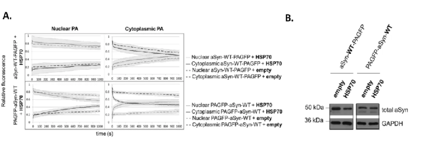
its intracellular dynamics in a cellular model through photoactivation microscopy. Using 240
photoactivatable green fluorescent protein as a reporter, we found that the availability of 241
the aSyn amino-terminus modulates its shuttling into the nucleus. This finding has 242
important implications regarding both the species of aSyn that enter the nucleus and also 243
the function of the protein within that compartment. aSyn was recently suggested to 244
exist naturally as a tetramer. Due to the nuclear pore size, only monomeric or dimeric 245
forms of aSyn can enter the nucleus, and this has been related to a deleterious effect and 246
neurotoxicity, due to transcription deregulation. Interestingly, intracellular dynamics of 247
aSyn was finely modulated by the HSP70 chaperone, PD-associated mutations and by the 248
phosphorylation state of the protein on S129 site. We found that the molecular 249
chaperone HSP70 accelerates the entry of aSyn into the nuclear compartment. Also, A30P 250
and A53T aSyn mutations increased the speed at which the protein moves between the 251
nucleus and cytoplasm, respectively. Finally, specific kinases potentiate the shuttling of 252
aSyn between nucleus and cytoplasm. Importantly, a mutant aSyn form that blocks S129 253
X
that phosphorylation modulates aggregation, and thus, alter the normal aSyn intracellular 255
dynamics. 256
To better understand the aggregation process in disease, we focused on the initial steps 257
of aSyn aggregation, thought to be the causative agents of pathology. We used cell-based 258
models of Synucleinopathy to investigate the molecular mechanisms underlying aSyn 259
oligomerization. In particular, we screened, in an unbiased manner, a subset of the 260
human genome-wide collection of lentiviral RNA-interference constructs, targeting genes 261
involved in signal transduction players, to identify modifiers of aSyn oligomerization, 262
using the bimolecular fluorescence complementation assay (BiFC) as readout. Through 263
this approach we identified 9 genetic modifiers of aSyn oligomerization. Interestingly, the 264
hits we identified were functionally related, and associated with neuronal trafficking 265
processes. We then characterized these hits with respect to their effects on aSyn 266
aggregation, toxicity and protein levels. After this first level of general characterization, 267
we further investigated the mechanism of action of the hits by assessing their effects on 268
aSyn secretion, a central aspect in the spreading of aSyn pathology. aSyn is secreted 269
under physiological conditions, via non-classical exocytosis, in association with exosomes, 270
and possibly via other less conventional mechanisms. However, it was demonstrated that 271
pathological and aggregated aSyn species can also be secreted, suggesting that 272
aggregated and misfolded aSyn may be the key agent for propagation of aSyn pathology, 273
possibly in a prion-like manner. Thus, in our study we selected four trafficking hits, based 274
on the literature and on their relevance to secretory pathways. Ras-related Protein in 275
Brain 8b (Rab8b), Rab11a, Rab13 and Synaptotagmin-Like Protein 5 were found to 276
promote the clearance of aSyn inclusions and reduce aSyn toxicity. Moreover, we found 277
that endocytic recycling and secretion of aSyn was enhanced upon expression of Rab11a 278
or Rab13 in cells accumulating aSyn inclusions. Importantly, in cells with inclusions, the 279
trafficking proteins co-localized with aSyn in inclusions. Altogether, our findings suggest 280
specific trafficking steps may prove beneficial as targets for therapeutic intervention in 281
Synucleinopathies, and should be further investigated in other models. 282
Here, we also studied the effects of monoclonal aSyn antibodies on the early stages of 283
aggregation using the BiFC assay. Our results support passive immunization against 284
Synucleinopathies by demonstrating that extracellular administration of monoclonal 285
XI
behave as a prion-like protein, immunization can be a mid-term strategy to delay the 287
progression of Synucleinopathies. 288
The present study uncovered novel aspects about the intracellular dynamics of aSyn and 289
allowed the identification of new genetic players involved in the aggregation, toxicity, 290
secretion and immunization of aSyn, opening novel avenues towards the understanding 291
of the molecular bases of Synucleinopathies. 292
293
Key-words: Alpha-Synuclein, Parkinson’s Disease, Oligomerization, Aggregation. 294
295 296 297 298 299 300 301 302 303 304 305 306 307 308 309 310 311 312 313 314 315 316 317 318 319
XII 320 321 322 323 324 325 326 327 328 329 330 331 332 333 334 335 336 337 338 339 340 341 342 343 344 345 346 347 348 349 350 351 352
XIII
Resumo
353354
As proteínas são os principais efectores biológicos na célula e regulam os processos vitais 355
na mesma. Assim, a desregulação funcional daquelas em regiões específicas do cérebro 356
pode culminar numa descontextualização espacial e temporal dos processos celulares. A 357
acumulação destas proteínas disfuncionais pode, por sua vez, originar agregados de 358
proteínas, que caracterizam as doenças neurodegenerativas (DNs). 359
A relação entre o misfolding de determinadas proteínas e a evolução para uma patologia 360
cerebral não é totalmente compreendida. A função alterada de uma proteína neuronal 361
pode culminar na formação de deposições proteicas no interior ou no exterior do 362
neurónio, levando à perturbação dos mecanismos de síntese e transporte de moléculas, 363
dos mecanismos de controlo de qualidade da célula e a uma perturbação na comunicação 364
interneuronal. No seu conjunto, estas doenças designam-se também de doenças 365
conformacionais, e representam grandes desafios para a Medicina actual, que tenta 366
encontrar terapias apropriadas que minimizem o impacto da deposição de agregados 367
proteicos nos neurónios. Em alguns casos, a deposição de agregados proteicos parece 368
perturbar fisicamente o funcionamento de alguns grupos de células específicos e 369
estender-se posteriormente para os respectivos tecidos e regiões adjacentes. Noutros 370
casos, a ausência de proteína funcional, devido ao seu recrutamento para os agregados 371
acumulados, resulta na falha de processos celulares cruciais. Segundo a hipótese 372
amilóide, a agregação de proteínas numa estrutura fibrilhar em DNs está relacionada com 373
interacções proteicas aberrantes que culminam na disfunção neuronal e, em última 374
instância, em neurodegeneração. Apesar de a célula possuir mecanismos de defesa e de 375
reparação que o próprio organismo acciona contra essas proteínas tóxicas, as DNs surgem 376
quando já nenhum mecanismo de defesa funciona na sua plenitude, e quando já há 377
saturação dessas proteínas disfuncionais nos neurónios. Assim, no contexto das doenças 378
neurodegenerativas, a hipótese amilóide postula que as proteínas podem ser convertidas, 379
sob certas circunstâncias, em estruturas não nativas com propensão para a instabilidade. 380
Nestas patologias, as proteínas podem apresentar estados conformacionais alternativos 381
ou misfolding que podem estar associados a disfunção celular, mas os mecanismos 382
XIV
inclusões nas DNs partilham vias de formação comuns, como seja perda de função das 384
proteínas envolvidas e formação de interações aberrantes. Assim, a comunicação entre as 385
proteínas deteriora-se intraneuronalmente e, por conseguinte, entre neurónios. Perante 386
este panorama, é essencial explicar a etiologia das DNs ao nível bioquímico e molecular, 387
para que haja impulso para o desenvolvimento de novas estratégias terapêuticas. 388
A doença de Parkinson (DP) é a segunda DN mais frequente e está associada ao 389
misfolding e agregação de alfa-Sinucleína (do inglês alpha-Synuclein, aSyn), uma proteína 390
neuronal cuja função não é totalmente conhecida. É de notar que a disfunção proteica de 391
aSyn também foi relacionada posteriormente com outras DNs, como sejam Demência 392
com Corpos de Lewy e Atrofia Sistémica Múltipla, sendo no seu conjunto designadas de 393
Sinucleinopatias. Assim, o estudo da aSyn tornou-se essencial para compreender a 394
etiologia e o denominador comum daquelas doenças. Existe uma forte controvérsia 395
relativamente à identificação das espécies tóxicas de aSyn; no entanto, as espécies 396
oligoméricas e misfolded têm sido postuladas nos últimos anos como as mais tóxicas. 397
Apesar de a função da aSyn ser pouco clara, existem várias implicações fisiológicas 398
propostas para a mesma, sendo uma das mais relevantes o seu envolvimento na 399
plasticidade sináptica, na medida em que ratinhos knockout para a aSyn possuem défices 400
de produção de vesículas celulares. Além disso, a aSyn parece actuar como um regulador 401
negativo da neurotransmissão de dopamina. Outros estudos sugerem o envolvimento da 402
aSyn no recrutamento de complexos necessários para o transporte entre o retículo 403
endoplasmático e o complexo de Golgi e para a fusão vesicular com a membrana 404
plasmática. Por outro lado, a disfunção de aSyn está associada a défices funcionais do 405
proteossoma, aumento da produção de espécies de oxigénio reactivas e disfunção 406
mitocondrial. 407
Assim, esta tese teve como principais objectivos entender a nível molecular e celular, a 408
função da aSyn na normalidade e na patologia. Para tal, estudou-se a dinâmica 409
intracelular da aSyn em modelos celulares, através de microscopia de fotoactivação. 410
Assim, usando uma forma fotoactivável da proteína verde fluorescente como repórter da 411
aSyn, verificou-se que a disponibilidade da sua extremidade amino-terminal determina a 412
sua deslocação para o núcleo. Esta evidência tem importantes implicações no que se 413
refere às espécies de aSyn que efectivamente entram no núcleo e à sua função no interior 414
XV
tetrâmero está certa, deve-se considerar que apenas monómeros ou dímeros de aSyn 416
podem entrar no núcleo, tendo em conta o tamanho descrito para o poro nuclear. Por 417
outro lado, a presença de aSyn no núcleo está associada a neurotoxicidade, porque 418
promove desregulação transcriptional. Curiosamente, verificamos que a dinâmica 419
intracelular de aSyn é elegantemente modulada pela chaperone HSP70; a presença desta 420
acelera a entrada de aSyn no compartimento nuclear. Para além disso, mutações 421
associadas a DP e o estado de fosforilação da proteína no local S129 alteraram o 422
comportamento dinâmico da aSyn na célula. Especificamente, as mutações A30P e A53T 423
aumentaram a velocidade a qual a proteína se desloca para o núcleo e para o citoplasma, 424
respectivamente. Por último, verificámos que certas cinases que fosforilam aSyn, também 425
têm um efeito na dinâmica intracelular da mesma. O resultado mais claro acerca do efeito 426
do estado de fosforilação na dinâmica da aSyn foi obtido com uma forma mutante que 427
bloqueia a fosforilação no local S129, designada por S129A. A expressão desta forma 428
mutante resultou na formação de inclusões citoplasmáticas, sugerindo que a fosforilação 429
modula a agregação e assim, altera a dinâmica intracelular de aSyn. 430
Para melhor compreender o processo de agregação e a sua evolução num contexto 431
patológico, estudou-se de seguida os passos iniciais de agregação de aSyn na célula. 432
Segundo a hipótese amilóide, o inicio da patologia reside na formação de espécies 433
diméricas e oligoméricas, diferentes da conformação nativa de aSyn. Assim, pensa-se que 434
são estas espécies, que se acumulam de forma aberrante, as causadoras de toxicidade e 435
de propagação da patologia. Para estudar os mecanismos moleculares por detrás da 436
formação de espécies oligoméricas tóxicas, usaram-se modelos celulares de 437
Sinucleinopatias. Em particular, efectuou-se um screening de interferência de RNA contra 438
genes envolvidos em vias de transdução de sinalização na célula. O objectivo foi 439
identificar de forma não enviesada, moduladores da oligomerização de aSyn, que foi 440
monitorizada através do método de complementação biomolecular por fluorescência. 441
Nove moduladores genéticos da oligomerização de aSyn, funcionalmente relacionados e 442
associados ao tráfego neuronal, foram identificados e validados. Assim, estes 443
moduladores foram caracterizados no que diz respeito aos seus efeitos na agregação de 444
aSyn, toxicidade e níveis proteicos. Após este primeiro nível de caracterização, investigou- 445
se o mecanismo de acção destes moduladores a nível da secreção de aSyn, um paradigma 446
XVI
condições fisiológicas está descrita como ocorrendo por exocitose não-convencional, em 448
associação com exossomas e possivelmente por outras vias menos convencionais. 449
Contudo, está também demonstrado que as espécies patológicas e agregadas de aSyn 450
podem ser secretadas, sugerindo-se que estas espécies podem ser um poderoso agente 451
de propagação da patologia de aSyn, possivelmente à semelhança dos agentes priónicos. 452
Assim, neste projecto, selecionaram-se quatro moduladores de tráfego, com base na 453
literatura e na sua relevância para as vias secretórias da célula: Ras-related protein in 454
Brain 8b (Rab8b, Rab11a, Rab13 e Synaptotagmin-Like Protein 5. Estas proteínas, quando 455
sobre-expressas, promoveram a remoção das inclusões proteicas de aSyn e reduziram a 456
toxicidade celular associada a aSyn. Para além disso, verificou-se um aumento do uso da 457
via endocítica e da secreção de aSyn quando Rab11a e Rab13 foram expressas num 458
modelo de agregação de aSyn. Por fim, verificou-se que no mesmo modelo, aqueles 459
quatro moduladores de tráfego co-localizaram com inclusões de aSyn. 460
Na sua totalidade, este trabalho sugere que certas vias específicas de tráfego celular são 461
benéficas para a intervenção terapêutica a nível das Sinucleinopatias, e devem ser 462
validadas noutros modelos. 463
Estudou-se também o efeito de anticorpos de aSyn em estados precoces de agregação da 464
mesma, através de complementação biomolecular por fluorescência. Os resultados aqui 465
descritos apoiam a imunização passiva contra Sinucleinopatias como sendo uma 466
estratégia eficaz a médio prazo para atrasar o progresso de Sinucleinopatias. 467
O presente estudo põe a descoberto a dinâmica intracellular da aSyn, uma vez que a 468
localização sob-celular dos muitos complexos proteicos que existem numa célula pode 469
ajudar a desvendar as suas funções e mecanismos de acção que culminam na patologia 470
de muitas DNs. Por outro lado, permitiu a identificação de novos moduladores genéticos 471
que envolvem oligomerização, agregação, toxicidade, secreção e imunização de aSyn, 472
contribuindo para o complemento do complexo esquema mecanístico que pode vir a 473
explicar as bases moleculares das Sinucleinopatias. 474
475
Palavras-chave: Alfa-Sinucleína, Doença de Parkinson, Agregação, Oligomerização 476
477 478
XVII
List of Abbreviations
479480 aβ Amyloid-beta peptide
AcbA acyl-CoA binding protein AD Alzheimer’s disease
AGE Advanced glycation end-products ALS Amyotrophic Lateral Sclerosis
aSyn alpha-Synuclein
aSynT Truncated aSyn-GFP fusion protein ATP Adenosine triphosphate
ATP6AP2 ATP hydrolase 6 lysosomal accessory protein 2 ATP13A2 ATP hydrolase 13A2
BAD Bcl-2-associated death protein Bax Bcl-2-associated X protein BBB Blood-brain barrier Bcl-2 B-cell lymphoma 2 BFA Brefeldin A
BRET Bioluminescence resonance energy transfer
bSyn beta-Synuclein
bZIP basic leucine zipper C-terminal Carboxy-terminal
CHIP Carboxyl terminus of Hsp70-interacting protein CI Confidence interval
CK Casein kinase
CM Conditioned media
CMA Chaperone-mediated autophagy co-IP co-Immunoprecipitation
COMT Catechol-O-methyltransferase CSF Cerebrospinal fluid
DBS Deep brain stimulation DLB Dementia with Lewy bodies ENS Enteric nervous system ER Endoplasmic reticulum
ERC Endosomal recycling compartment ERK Extracellular signal-regulated kinases FCS Fluorescence correlation spectroscopy FRET Fluorescence resonance energy transfer GBA Glucocerebrosidase
GFP Green fluorescent protein
GRK G-protein coupled receptor kinase
gSyn gamma-Synuclein
GTPase Guanosine triphosphate hydrolase
XVIII
HD Huntington’s disease hGH human growth hormone HNE 4-Hydroxynonenal
Hsc70 Heat shock cognate protein 70 HSP Heat shock protein
Htt Huntingtin
L-DOPA L-3,4-Dihydroxyphenylalanine
LAMP2 Lysosomal associated membrane protein 2
LB Lewy body
LDH Lactate dehydrogenase
LN Lewy neurite
LRRK2 Leucine-rich repeat kinase 2
MAO-B Monoamine oxidase B
MAPT Microtubule-associated protein Tau
miRNA Micro RNA
MPTP 1-Methyl-4-phenyl1,2,3,6-tetrahydropyridine MSA Multiple system atrophy
MVB Multivesicular body N-terminal Amino-terminal
N2 Notch2
NAC non-Amyloid-beta component
NADPH Nicotinamide adenine dinucleotide phosphate ND Neurodegenerative disorder
NF-kB Nuclear factor kappa-light-chain-enhancer of activated B cells P25 Tubulin polymerization-promoting protein
P62 Nucleosporin p62 PA Photoactivation PAGFP Photoactivatable GFP
PB Photobleaching
PD Parkinson's disease
PGC-1α Peroxisome proliferator-activated receptor gamma coactivator 1-alpha PINK1 PTEN-induced putative kinase 1
PLD2 Phospholipase D2
PMD Protein misfolding diseases PNS Peripheral nervous system
PPAR Peroxisome proliferator-activated receptor PPI Protein protein interactions
PTEN Phosphatase and tensin homolog PTM Post-translational modification RAB Ras-related proteins in brain
RanBP2 Ras-related nuclear binding protein 2 RE Recycling endosome
RNA Ribonucleic acid
RNAi Ribonucleic acid interference ROS Reactive oxidative species
XIX
S Seconds
shRNA Short-hairpin RNA
SIAH Seven in absentia homolog protein SNARE Soluble NSF attachment protein receptor
SNCA Synuclein alpha gene
SUMO Small Ubiquitin-like Modifier SUS Split ubiquitin system
Tf Transferrin
ThT Thioflavin-T
UPS Ubiquitin-proteasome system v-ATPase Vesicular adenosine triphosphatase VPS35 Vacuolar protein sorting 35
Y2H Yeast two-hybrid system
481 482 483 484 485 486 487 488 489 490 491 492 493 494 495 496 497 498 499 500 501 502 503 504
XX 505 506 507 508 509 510 511 512 513 514 515 516 517 518 519 520 521 522 523 524 525 526
I. I
ntroduction
______________________________This chapter contains parts of the following publications:
Gonçalves SA, Miranda HV Outeiro TF (2012), Novel molecular therapeutics in Parkinson’s disease. 1: 245-265. Molecular and Cellular Therapeutics, First Edition, David Whitehouse and Ralph Rapley. John Wiley & Sons.
Gonçalves SA, Matos JE and Outeiro TF (2010), Zooming into protein oligomerization in neurodegeneration using BiFC, Trends Biochem Sci 35(11): 643-651.
Goncalves, S. A. and T. F. Outeiro (2016). Traffic jams and the complex role of alpha-Synuclein aggregation in Parkinson’s disease. Small GTPases: 1-7.
I. Introduction | 23
1
P
rotein
M
isfolding
D
iseases
Cell viability depends on the maintenance of proteins integrity, which is directly dependent on the strict balance between protein synthesis, folding and degradation mechanisms. Protein folding and degradation are key quality control systems of the cell. The former is performed by molecular chaperones such as heat shock proteins (HSPs), and the later comprises the ubiquitin-proteasome and autophagy-lysosome pathways. Misfolded and damaged proteins can be targeted to the ubiquitin-proteasome system (UPS) to avoid accumulation and subsequent potentially toxic effects on cells, or can be processed by the autophagy-lysosome system (Figure 1A). The first is mainly involved in the nuclear/cytosolic protein degradation, and the latter in the clearance of cytosolic organelles and long-lived proteins. Three major types of autophagy are described (Figure 1B): Chaperone-mediated autophagy (CMA) selectively degrades proteins containing a pentapeptide motif (KFERQ) that is recognized by the heat shock cognate protein 70 (Hsc70); microautophagy implies the direct sequestration of cytoplasmic cargo by engulfment of the lysosomal membrane; finally, in macroautophagy, double-membrane vesicles termed autophagosomes are formed and sequester portions of cytosolic content or intact organelles (Juenemann & Reits 2012, Yang & Klionsky 2010) . Fine-tuned regulated macroautophagy is required for the survival of neurons, as lack of it leads to degeneration, but, if increased, it can turn into a cell death mechanism (Hara et al 2006, Komatsu et al 2006).
Although the native state of proteins is energetically favored, misfolding of proteins can arise when quality control systems fail their surveillance due to metabolic changes related to aging, cancer, stress conditions or genetic alterations (Hartl & Hayer-Hartl 2009). In those conditions, partially folded or misfolded proteins tend to aggregate and state for aberrant intra- or intermolecular interactions. Protein aggregation is driven by hydrophobic forces and can lead to the formation of amorphous or fibrillary structures. These highly ordered, cross-beta structures are called amyloid deposits and histopathologically define protein misfolding diseases (PMDs).
I. Introduction | 25
Figure 1. The role of protein quality control systems in PMDs. A. When destabilization of the
native state of nascent proteins occurs, molecular chaperones are able to reverse it. In aging and disease, protein quality control systems are more prone to fail and proteins form amorphous aggregates, toxic oligomers or amyloid fibrils as a consequence of misfolding of the native states. Different HSPs can modulate the oligomerization state. In several diseases, Hsp70 and HSP90 are known to contribute to the correct folding of misfolded proteins, preventing the formation of aggregated forms. Oligomers are toxic soluble aggregated species that can occur as off-pathway intermediates of amyloid fibril formation. These may be either directly targeted by Hsp chaperones or they may also be directed for proteasomal degradation. Oligomeric and/or aggregated species that are not degraded by the proteasome may be processed by chaperone-mediated autophagy. B. Three different types of autophagy are represented: in macroautophagy, specialized vacuoles called autophagosomes are formed for cargo transportation. These vacuoles deliver proteins, lipids and organelles to the lysosome. Through Hsc70 complex, Chaperone-mediated autophagy recognizes proteins with KFERQ-like motif and targets them to lysosomal degradation. Finally, in microautophagy, cargo is directed engulfed by lysosomal membrane. AA: aminoacids, FFA: free fatty acids. Adapted from (Gonçalves et al 2012, Wirawan et al 2012).
1.1 Loss of Neuronal Proteostasis and Neurodegeneration
Neurons are long-lived post-mitotic cells and thus particularly susceptible in PMDs, as they are not able to dilute toxic proteins through cell division. Thus, they require fine-tuned quality control mechanisms when proteostasis is compromised. Autophagy-lysosome system is the ubiquitous and most well characterized mechanism to guarantee proteostasis in neurons. Notwithstanding, a specific “sort-and-degrade” mechanism has also been postulated (Wang et al 2013). Neuron-specific proteins, such as the regulator of membrane trafficking V0a1, the subunit a1 of V0-ATPase (a multi-subunit protein-complex that regulates endosomal to lysosomal pH), seems to be a neuron-specific health sustainer, as mutations in v0a1 homolog in Drosophila cause neurodegeneration (Williamson et al 2010). Lysosomes, the terminal organelles on the endocytic pathway, digest macromolecules and make their components available to the cell as nutrients. Hydrolytic enzymes specific to a wide range of targets reside within the lysosome; these enzymes are activated by the highly acidic pH (Wang & Hiesinger 2012).
26 | I. Introduction
Similarly, mutations in the synaptic vesicle SNARE neuronal Synaptobrevin seems to cause cargo overload in neurons. SNAREs are the core regulation proteins in membrane fusion. Overall, the “sort and degrade” mechanism is active in parallel to ubiquitous endolysosomal and autophagosomal degradation and is essential for the normal neurotransmitter release (Haberman et al 2012).
The most prevalent PMDs in the brain are Alzheimer’s disease (AD), Parkinson’s disease (PD), Huntington’s disease (HD) and Amyotrophic Lateral Sclerosis (ALS). Clinically they result from the aggregation of different proteins in distinct neurons that can disrupt essential cellular functions. Aggregates can be extracellular, as plaques in AD, or intracellular, neurofibrillary tangles in AD or Lewy bodies in PD. The intracellular aggregates can be rapidly devastating to neurons as they can sequester essential cellular components as molecular chaperones or trafficking players. Moreover, they can promote oxidative stress, inhibit degradation systems and physically impair the release of neurotransmitters thus inhibiting the communication to the adjacent neurons (Gonçalves et al 2012).
It is not clear if fibrils are the toxic species in NDs, or if they arise from a defensive answer of cells aimed to protect themselves from toxic oligomeric species. Even if the mechanistic explanation of pathology in those disorders is variable, the common denominator among these NDs is the gain of toxic properties associated to misfolding. The aberrant conformation favors environmental stress which is aggravated by aging (associated with the decline in protein homeostasis capacity) and, in a long-term perspective, it promotes further protein accumulation and the potential self-propagation of aggregates to other neighboring neurons (Hartl et al 2011).
1.2 Synucleinopathies
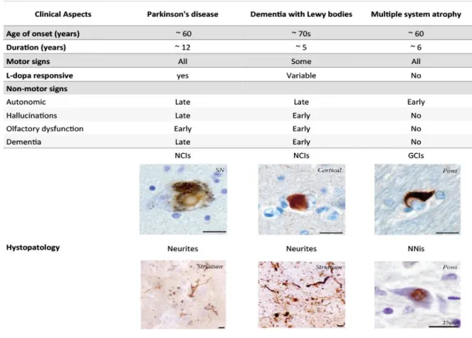
Synucleinopathies are neurodegenerative disorders (NDs) characterized by the abnormal neuronal accumulation of a small protein called alpha-Synuclein (aSyn). This protein is abundant in the central nervous system and its abnormal accumulation can occur in neurons, nerve fibers or glial cells. The major Synucleinopathies are PD, dementia with Lewy bodies (DLB) and multiple system atrophy (MSA) (Figure 2). AD and other neurodegenerative disorders related with iron accumulation in brain may also present
I. Introduction | 27 aSyn aggregation (Baba et al 1998, Irwin et al 2013, Spillantini et al 1997, Wakabayashi et al 1998).
Abnormal protein deposits were identified in brains from PD patients by Freiwdrich Lewy in the beginning of the twentieth century. However, only later aSyn was identified as the main component of Lewy bodies (LBs) (Lewy 1912, Spillantini et al 1997). Structurally, LBs are eosinophilic cytoplasmic large inclusions of 5-25 µm size compose of a halo of radial fibrils (Spillantini et al 1998b). The main component of LBs is phosphorylated (at S129), nytrosylated and also C-terminally truncated aSyn(Crowther et al 1998, Duda et al 2000, Fujiwara et al 2002, Giasson et al 2000, Spillantini et al 1997). However, the role of those post-translational modifications is not totally understood. In addition, molecular chaperones, proteasomal and lysosomal subunits were identified in LBs (Goedert et al 2013, Lowe et al 1988).
1.2.1 Parkinson’s Disease
1.2.1.1 Etiology and Pathophysiology of PD
PD was first described in 1817 by James Parkinson as “the shaking palsy” and is the second most common neurodegenerative disease affecting 1% of the world population over the age of 60. About 90% of PD cases are sporadic, while only a small proportion of the cases are known to have dominantly or recessively inherited familial forms caused by several mutations in specific genes (de Lau & Breteler 2006, Parkinson 2002). In addition, environmental factors as the exposure to pesticides (as rotenone or paraquat), heavy metals (iron, manganese, copper, zinc) and brain injury may cause sporadic PD (Critchley 1957, de Lau & Breteler 2006).
The neuropathological hallmarks of PD comprise the loss of dopaminergic neurons in the substantia nigra pars compacta and the presence of intracellular proteinaceous inclusions in the surviving neurons mainly composed of aSyn (Damier et al 1999, Lewy 1912, Spillantini et al 1997). The lack of dopamine results in abnormal neurotransmission and thus prevents appropriate information transfer from motor command centers in the cerebral cortex (Aosaki et al 2010). This leads to different severity degrees of motor
28 | I. Introduction
symptoms, including muscle rigidity, resting tremor, bradykinesia and postural instability (Marsden 1982, Parkinson 2002, Wu et al 2015). Non motor signs are believed to manifest prior to motor disabilities, starting with difficulty in problem-solving, attention capacities and decision making (Pfeiffer 2016). Autonomic dysfunction is a common non-motor sign that may precede clinical PD, comprising orthostatic hypotension, constipation, insomnia, abnormalities in olfactory and visual perception, urinary dysfunctions and sweating abnormalities (Liepelt-Scarfone et al 2015). Later disease stages include neuropsychiatric symptoms as depression, anxiety, apathy, and casually, dementia (Gelb et al 1999, Kulisevsky et al 2008).
Figure 2. Clinical and histopathological hallmarks of the three main Synucleinopathies.
Dementia with Lewy bodies (DLB) has the oldest age of onset. Multiple system atrophy (MSA) has the earliest autonomic features. Histologically, Both PD and DLB are characterized by neuronal cytoplasmic inclusions (NCIs) and neurites, while MSA has glial cytoplasmic inclusions (GCis) and neuronal intranuclear inclusions (NNis). SN, substantia nigra. Images show aSyn immunoreactive structures counterstained with cresyl violet. Scale bars: 25 µm. Adapted from (McCann et al 2014).
I. Introduction | 29 Histopathologically, surviving neurons often show protein inclusions, which develop as spindle-like Lewy neurites (LNs) (Braak et al 1994) or as globular LBs (Goedert et al 2013, Lewy 1912), both in sporadic and familial forms of PD (Spillantini et al 1997). As the disease progresses, aSyn aggregates can also be found in other areas of the brain as the olfactory bulb, neocortex and the limbic system (Braak et al 2003). Inclusions of aSyn can also occur in other regions of the central and peripheral nervous system as the enteric plexus of the gastrointestinal system (Dickson et al 2009).
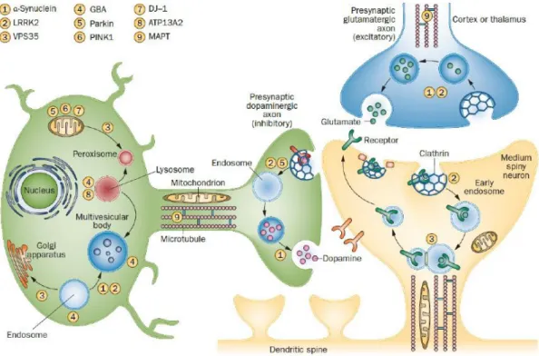
1.2.1.2 Familial PD Genes and their Convergent Role in Trafficking
Counting for 5-10% of the total cases, inherited PD has been correlated to autosomal dominant or recessive genetic mutations. Multiple genes have been implicated in PD through linkage analysis, genome sequencing and genetic association and the majority features mutations in cellular trafficking proteins (Table 1). The discovery of mutated genes associated with PD was elucidative on the cellular pathways that upon dysfunction triggers to pathology, not only in familial but also in sporadic forms of PD. Consequently, three main interconnected cellular processes may trigger PD upon dysfunction: first, synaptic transmission (exocytosis and endocytosis), lysosome-mediated autophagy; and third, mitochondrial quality control and stress response (Figure 3) (Trinh & Farrer 2013). The most common autosomal dominant inherited cases of PD present mutations in SNCA and LRRK2 genes encoding for aSyn (and discussed below in section 2) and Leucine-rich repeat kinase 2, respectively (Polymeropoulos et al 1997, Zimprich et al 2004). LRRK2 is a guanosine triphosphate hydrolase (GTPase) and kinase with defined roles in neuronal transmission, arborization, endocytosis, autophagy and immunity. Several PD-associated mutations in LRRK2 were identified, presenting a clinical phenotype that resembles idiopathic PD (Cookson 2012). G2019S mutation in LRRK2 has been shown to interfere with chaperone-mediated autophagy in neurons, and to enhance co-localization of aSyn with Lysosomal Associated Membrane Protein 2 (LAMP2) (Orenstein et al 2013). Interestingly, genome-wide association findings suggest that LRKK2 variability confers both significant risk or protection against PD (Trinh & Farrer 2013). Besides, mutations in
30 | I. Introduction
Vacuolar protein sorting 35 (VPS35), which mediates retrograde transport of endosomes to trans-Golgi network, cause late-onset PD (Vilarino-Guell et al 2011b).
Autosomal recessive cases of PD contributes for less than 4% of PD and involve genes for Parkin (PARK2), DJ1 (PARK7) and Pten-induced kinase 1 (PINK1), between others (Abbas et al 1999, Bonifati et al 2003, Kitada et al 1998, Valente et al 2004). PINK1 and PARK2 encode proteins involved in mitophagy. Specifically, Parkin, an E3 ubiquitin ligase, was described to facilitate the degradation of damaged mitochondria. PARK2 mutations are thought to result in insufficient protein clearance and subsequent protein accumulation and cellular damage (Kitada et al 1998). DJ1 is implicated in anti-oxidative stress responses, mainly through reactive oxidative species (ROS) scavenging (Ramsey & Giasson 2008). Mutations in the gene encoding for PINK1, a cytoplasmic but mitochondria-associated protein kinase, are thought to impair its kinase activity and contribute to disruption of mitochondrial trafficking, ROS formation, and protein aggregation (Liu et al 2009b, Valente et al 2004, Weihofen et al 2009). Moreover, mutant PINK1 is not able to translocate into the mitochondria, where it should stimulate mitophagy. Mutations in ATP13A2, a lysosomal ATPase, lead to impaired protein degradation (Park et al 2015, Ramirez et al 2006). Finally, mutations in ATP6AP2—a gene required for receptor-mediated endocytosis, membrane trafficking and lysosomal degradation—cause X‑ linked Parkinsonism (Korvatska et al 2013).
Recently, genome-wide studies have compelled the discovery of novel genes and polymorphisms associated with PD. One example is the established link between the genetic variability of microtubule-associated protein Tau (MAPT) loci and idiopathic PD. Tau is involved in microtubule stabilization, elongation and axonal transport (Lanktree et al 2011, Vandrovcova et al 2010). Moreover, carriers of a single Glucocerebrosidase (GBA) mutant allele have five times higher risk for PD. GBA is a housekeeping enzyme that helps to digest toxic molecules within lysosomes (Klein & Westenberger 2012). Remarkably all genetic forms present aSyn pathobiology with LBs, except for cases carrying PARK2 and LRRK2 mutations (Table 1 and Farrer et al 2001, van de Warrenburg et al 2001).
I. Introduction | 31
Table 1. Known genetic loci linked to Parkinson’s disease
Locus gene Protein Brain
accumulati on Age at onset Inheritance Genetic alterations
Loci implicated in late-onset Lewy body PD
Park 1 Park4
SNCA aSyn LBs 30-40s, fast
progression AD Missense/gene dosage Park 3 SPR Sepiapterin reductase LBs 60s AD DNA polymorphisms
Park 5 UCH-L1 Ubiquitin carboxy-terminal L1 LBs 50s AD Missense Park 8 LRRK2 Leucine-rich repeat kinase 2 LBs, not in all cases 40s, AD Missense
Park 10 PARK10 ? unknown 50s Risk factor DNA
polymorphisms
Park 11 GIGYF2 GRB10 interacting GYF protein 2
unknown late AD Missense
Park 12 PARK12 ATP6AP2 Taupathy juvenile and early onset
X-chromosome Synonymous
Park 13 HTRA2 HtrA serine peptidase 2
unknown 50s AD Missense
Park 16 PARK16 ? unknown ? Risk factor DNA
polymorphisms
Park 17 VPS35 Vacuolar protein sorting 35 Homolog
unknown Late onset AD Missense
Park 18 EIG4G1 Eukaryotic translation initiation factor 4 gamma, 1 LBs Late onset, mild AD Missense
Juvenile and early-onset recessively inherited parkinsonism
Park 2 PARK2 Parkin LBs, not in all cases
20s, slow progression
AR Missense
Park 6 PINK1 Pten-induced kinase 1
unknown 30s AR Missense/trun
cating/dosage
Park 7 PARK7 DJ1 unknown 30s, slow
progression
AR Missense/trun cating/dosage
Park 9 ATP13A2 ATPase type 13A2 Iron Juvenile, atypical
AR Truncating
Park 14 PLA2G6 Phospholipase A2, group VI
Iron Juvenile, atypical
AR Missense
Park 15 FBXO7 F-box protein 7 unknown Juvenile AR Truncating
Park 19 DNAJC6 HSP40 Auxilin unknown Juvenile, atypical
AR Splice site/ Truncating
Table 1. Known genetic loci linked to Parkinson’s disease. AD, autosomal dominant. AR, autosomal
32 | I. Introduction
Figure 3. Overview of cellular dysfunction and genes associated with PD. A glutamatergic cortical
neuron (blue), a dopaminergic substantia nigra neuron (green) and a dendritic spine of a medium spiny neuron (yellow) are represented. In presynaptic terminals, aSyn (1) promotes exocytosis and can play a part in endocytosis. Post-synaptically, LRRK2 (2) regulates the release of clathrin-coated endocytic vesicles through phosphorylation, neuronal polarity and arborization. LRRK2 also has roles in chaperone-mediated autophagy and microtubule stabilization. VPS35 (3) is an integral part of the retromer, a complex that mediates cargo endosomal-to-Golgi retrieval by forming a clathrin-independent carrier. Alternatively, cargoes may be destined for lysosomal degradation or exosome secretion. VPS35 mediates cargo recycling from endosomes to the Golgi apparatus or plasma membrane, and vesicle transport between mitochondria and peroxisomes. Lysosomal acid hydrolases, including GBA (4), also require the retromer for receptor recycling. Loss-of-function mutations in Parkin (5), PINK1 (6) and DJ1 (7) affect mitochondrial biogenesis and induction of autophagy. Parkin is involved in ubiquitination and proteasomal function, and PINK1 and Parkin are involved in mitochondrial maintenance. ATP13A2 (8) has a role in lysosome-mediated autophagy. MAPT (9) helps to regulate cargo trafficking and delivery, primarily in axons. Abbreviations: GBA, Glucocerebrosidase; LRRK2, Leucine-Rich Repeat Kinase 2; VPS35, Vacuolar Protein Sorting 35 (Trinh & Farrer 2013).
I. Introduction | 33 1.2.1.3 Current Therapies of PD
Most, if not all, currently available therapies for PD are just symptomatic. While they improve motor dysfunction symptoms, they do not modify disease progression nor prevent disease onset. These therapies include pharmacological modulation of the dopamine system, neurosurgery and physical therapy.
Since shortage of dopamine is one of the major deficits in the PD brain, current pharmacologic interventions are aimed either at replenishing dopamine levels in the brain or at modulating the dopamine system with specific agonists and antagonists. More specifically, the strategies are the immediate or controlled uptake of the stable dopamine precursor levodopa and the inhibition of monoamine oxidase B (MAO-B) or catechol-O-methyltransferase (COMT), which are enzymes that catabolize dopamine (Goetz et al 2005, Horstink et al 2006). Levodopa and dopamine agonists are the most widely used drugs, as they readily cross the blood-brain barrier (BBB) to exert their anti-Parkinsonian effects. However, long-term use of levodopa improves motor symptoms but does not slow disease progression and is associated with adverse effects such as motor fluctuations and dyskinesias (Fahn 2000, Olanow et al 2004). MAO-B inhibitors, such as Selegiline or Rasagiline, are thought to be neuroprotective as they can inhibit dopamine catabolism. COMT inhibitors also act on the dopamine pathway by inhibiting levodopa catabolism and by extending its half-life. For example, Tolcapone and Entacapone are effective in alleviating the motor impairments, but they are associated with hepatotoxicity (Williams et al 2010).
Peroxisome proliferator-activated receptors (PPARs) are also attractive targets to treat mitochondrial damage and oxidative stress associated with PD. They belong to a nuclear receptor superfamily involved in major biological processes such as inflammation, mitochondrial function, tissue differentiation, and lipid and glucose metabolism. Pioglitazone is a PPAR- agonist which, when administrated to mice before 1-methyl-4-phenyl1,2,3,6-tetrahydropyridine (MPTP, a prodrug to the neurotoxin MPP+ that causes symptoms of PD by destroying dopaminergic neurons in the substantia nigra of the brain) injection, prevents dopaminergic neuronal loss and glial cell activation, by inhibiting the conversion of MPTP into MPP+. Concordantly, in a rat model of PD, pioglitazone improved
34 | I. Introduction
Rosiglitazone, another PPAR- agonist, protected human neuroblastoma cells from acetaldehyde-induced ROS and apoptosis, through the induction of antioxidant enzymes. In in vitro models, ibuprofen and acetaminophen were also shown to impair neurotoxicity by binding to PPAR- and PPAR-α. PPAR agonists are thus promising therapeutic targets, but further studies are needed to prove their safety and efficacy in PD patients. Moreover, although PD is a multifactorial disorder, the widespread involvement of PPAR in cell biology must be carefully regarded to avoid putative severe side effects (Chaturvedi & Beal 2008).
Surgical approaches such as deep brain stimulation (DBS) are presently used, where a neurostimulator delivers electric stimuli to targeted brain areas that are responsible for motor control. This strategy constitutes an alternative treatment in patients who meet specific criteria. A clinical trial comparing drug therapy with a combined drug therapy and DBS showed that patients of the latter group have an improved quality of life, regarding motor impairment and dyskinesias although this is only a symptomatic treatment (Lozano et al 2010).
PD is a progressive ND and treatment is only efficient for a limited stage of the disease (Tambasco et al 2012). In order to develop novel therapeutic strategies for PD it is crucial to gain a detailed understanding of the molecular mechanisms involved in the disease. Since aSyn-induced cytotoxicity seems to be mainly associated with its misfolding and aggregation, it is important to understand how cells respond to the accumulation of these protein species.
Notwithstanding, regular body exercising and healthy nutrition are associated with the delay of disease progression. Moreover, coffee consumption seems to reduce the risk of PD as caffeine is an inhibitor of adenosine A2 receptors, that are responsible for
decreased dopaminergic activity and inhibition of neuronal excitation. Thus, by inhibiting A2 receptors, caffeine increases brain functions such cognition, learning, and memory and
improves motor deficits in a mouse model of PD (Ribeiro & Sebastiao 2010). Resveratrol, a non-flavonoid polyphenol found in red wine and grapes also protects dopamine neurons through its antioxidant and anti-inflammatory properties. Resveratrol-mediated neuroprotection seems to act by inhibiting both lipopolysaccharide-induced neurotoxicity and microglia activation (Zhang et al 2010).
I. Introduction | 35
1.2.2 Dementia with Lewy bodies
DLB is a ND characterized by dementia, cognitive impairment, visual hallucination and Parkinsonian motor symptoms. Patients with DLB also present LBs in midbrain but mainly in neocortical areas and brainstem. It is thought to account for up to 30% of dementia cases (Zaccai et al 2005). The most prominent difference between PD and DLB is that dementia can affect PD patients after more than one year with motor symptoms of parkinsonism while DLB patients suffer from it before or during the parkinsonism manifestation (Aarsland & Kurz 2010). Moreover, although most cases of DLB are sporadic, a genetic association is described whose profile overlaps with AD and PD ones. Thus, SNCA and LRKK2 mutations are found in DLB cases (Hyun et al 2013, Nervi et al 2011).
APOE Ɛ4 allele is a strong risk factor for DLB, while APOE Ɛ2 is protective. Moreover, mutations in GBA are a risk factor for DLB (Berge et al 2014, Bras et al 2014, Tsuang et al 2013).
The realization that patients with Parkinson’s disease often develop cognitive deficits and dementia has led to extensive research efforts and new diagnostic criteria for PD and DLB. Improving diagnosis by developing new biomarkers, clarifying terminology and criteria, and determining protective and risk factors are crucial for an accurate diagnosis.
1.2.3 Multiple System Atrophy
MSA is a sporadic progressive disease with mid-age onset. Clinically, patients can have a variable combination of autonomic and cognitive dysfunction, cerebellar ataxia or Parkinsonism. Histopathologically, MSA is characterized by the loss of neurons in the cerebellum, pons, basal ganglia and spinal cord. Genetic factors may play a role in the etiology of the disease, as SNCA variations were associated with MSA risk, as well as MAPT gene, encoding for Tau protein (Ross et al 2010, Vilarino-Guell et al 2011a). In addition, analysis of familial MSA has identified mutations in COQ2, a protein involved in the synthesis of coenzyme Q10 (Multiple-System Atrophy Research 2013). However, until now no gene was associated to MSA.
36 | I. Introduction
The neuropathological hallmark of MSA is the presence of filamentous glial cytoplasmic inclusions of aSyn, called glial cytoplasmic inclusions (GCIs) (Trojanowski et al 2007). Actually, this aspect is sufficient to diagnose the disease. Although aSyn is the main component of GCIs, other proteins as ubiquitin, Nucleosporin p62 (p62) and tubulin polymerization-promoting protein (TPPP or p25) are also found. GCIs are located surrounding the nucleus randomly arranged with packed filaments (Papp et al 1989). Interestingly, aSyn can also form glial nuclear inclusions (GNIs), or be aggregated in neurons (Papp & Lantos 1992). While the presence of aSyn in oligodendrocytes is still not well understood given the fact that those cells do not express aSyn mRNA, it was suggested that a neuron-to-oligodendrocyte transfer of aSyn may occur (Reyes et al 2014). GCIs are associated with myelin degeneration, microglia activation and ultimately to cell death. Once this happens, aSyn inclusions can be uptake by surrounding neurons and the process of inflammation and neuronal and oligodendrial dysfunction perpetuates to other brain regions (Brundin et al 2008, Streit et al 2004).
Patients with MSA usually do not respond well to dopamine replacement, probably because other populations than dopamine-producing cells are affected, including spiny neurons in the striatum (Sato et al 2007).