1
Drug-like properties and ADME of xanthone derivatives: the antechamber of clinical trialsAna Sara Gomes, Pedro Brandão, Carla Fernandes, Marta Correia-da-Silva, Emília Sousa*, Madalena Pinto#
# all authors contributed equally to this work *corresponding author
Abstract
Xanthone derivatives have been described as compounds with privileged scaffolds that exhibited diverse interesting biological activities, such as antitumor activity, directing the interest to pursue the development of these derivatives into drug candidates. Nevertheless, to achieve this purpose it is crucial to study their pharmacokinetics and toxicity (PK/tox) as decision endpoints to continue or interrupt the development investment. This review aims to expose the most relevant analytical methods used in physicochemical and PK/tox studies in order to detect, quantify and identify different bioactive xanthones. Also the methodologies used in the mentioned studies, and the main obtained results, are referred to understand the drugability of xanthones derivatives through in vitro and in vivo systems towards ADME/tox properties, such as physicochemical and metabolic stability and biovailability. The last section of this review focus on a case-study of the development of the drug candidate DMXAA, which has reached clinical trials, to understand the paths and the importance of PK/tox studies. In the end, the data assembled in this review intends to facilitate the design of potential drug candidates with a xanthonic scaffold.
Xanthone; analytical method; drug-like; pharmacokinetics; toxicity; drug development; preclinical; metabolism; chromatography.
1. Introduction
The xanthone nucleus or 9H-xanthen-9-one (dibenzo-γ-pirone 1, Fig. 1) comprises an important class of oxygenated heterocycles and is considered a privileged structure 1. It includes relevant secondary metabolites which are present in commercialized extracts with human health promotion properties 2. Considering synthetic derivatives, one compound emerged as drug candidate, 5,6-dimethylxanthenone-4-acetic acid (DMXAA, Vadimezan, ASA404, 2, Fig. 1) and reached phase III clinical trials towards antitumor activity 3. Due to a great number of studies in isolation, synthesis and biological/pharmacological properties of xanthone derivatives, several reviews 1,4-16 have gathered important information to guide the design of these compounds as potential drug candidates. Nevertheless, little focus was given to drug pharmacokinetics (PK), toxicity studies, and structure-properties relationships of xanthone derivatives.
2
Fig. 1: Xanthone (1) and DMXAA (2).Nowadays it is recognized that in addition to pharmacological properties, PK and toxicological properties are crucial determinants of the ultimate clinical success of a drug. This recognition has led to the early introduction of absorption, distribution, metabolism, excretion and toxicity (ADME/Tox) screening during the drug discovery process, in an effort to filter drugs with problematic ADME/Tox profiles. Prior to the early 1980s, for example, PK studies in the pharmaceutical industry were largely detailed and were primarily focused on clinical development efforts in support of product registration, with little attention being devoted to the PK characteristics of new chemical entities and to mechanistic insight into the fate of candidate drugs in biological systems. The fact that the primary reason for failure of drug candidates in clinical development during the 1960s and 1970s was judged to be due to inappropriate human PK led to the progressive integration of PK studies as a key component of the overall drug discovery process 17. This shift was relatively easily accomplished using existing methodologies and, currently, development attrition due to PK properties is no longer the bottle-neck in drug discovery and development 18.
The optimization of a large number of variables is required early in the design and evaluation of the chemical entities to increase the probability of finding clinical candidates not only of complex parameters related to toxicity and bioavailability but also the most favorable physicochemical properties (i.e., molecular weight, solubility, polar surface area, lipophilicity, among others). The importance of these properties in drug design has always been recognized in an implicit manner in structure-activity relationship studies; nevertheless, it is established that analyzing the data of these properties makes more probable the success in drug discovery 19.
The physicochemical properties of some investigated xanthone derivatives suggest that they might exhibit high bioavailability, an important factor contributing to their efficacy and success as drug candidates. However, studies involving drug-like properties of xanthone derivatives are sparse. This review provides an opportunity to organize the emerging data concerning the methods used to investigate PK and physicochemical properties of bioactive xanthone derivatives. Firstly, the most relevant analytical methods and conditions are described for xanthones from different sources (natural, metabolic, or synthetic) and classes (prenylated, glycosylated, carboxylated). Following, methodologies and results involved in drug-like properties characterization are revised. At the end of this review, a case-study of the drug DMXAA (2) highlights the rational of PK studies in the drug discovery and development pipeline. The methodologies and data gathered in this review may pave the way for the design of potential drug candidates with a xanthonic scaffold.
3
2. Chromatographic methods applied to xanthones in preclinical and clinical studiesChromatographic methods have been used to study drugs and their PK behavior, namely gas chromatography – mass spectrometry (GC-MS) and liquid chromatography (LC) with ultraviolet (UV), fluorescence, radioactivity and MS detection systems, being the late one probably the most applied nowadays, both in in vitro and in vivo studies 20,21. In the case of LC-MS, the LC (usually HPLC – High Performance Liquid Chromatography - or UPLC – Ultra Performance Liquid Chromatography) allows the separation of the different components present in the sample and the MS enables the identification of the compounds previously separated in the chromatographic method 22.
The establishment of quantitative structure-property relationships for xanthone derivatives is a major focus of interest for PK studies. The use of reversed-phase HPLC coupled with a UV detector has been developed in order to describe the behavior of xanthone derivatives according to a model in which both the molecular structure parameters and mobile phase properties are taken into consideration. Appling this method, the chromatographic behavior of xanthone derivatives are dependent of the solvent properties (polarity/polarizability parameter, hydrogen-bond basicity) and solute properties (most positive local charge, the sum of positive charges on hydrogen atoms contributing in hydrogen interaction and lipophilicity index (Log P)) 23. Another chromatographic technique that can be applied in the evaluation of lipophilicity of xanthone derivatives is micellar electrokinetic chromatography, since the separation selectivity of xanthones can be modulated by changing the micellar phase or aqueous phase between which the analytes are partitioned, being this behavior closely associated with the hydrophobicity of the compounds 24. Techniques such as capillary zone electrophoresis (CZE), measuring the mobility of a given solute as a function of pH, is a reliable method to determine dissociation constants (pKa) for pharmacologically active xanthones 25.
2.1. Natural xanthones
Due to the wide distribution of xanthonic compounds in many natural products used in folk medicine, in the last decade the study of methods to purify, isolate, identify and analyze these derivatives has become an important goal in Medicinal Chemistry 5,6,24. Several natural xanthones are currently undergoing preclinical studies.
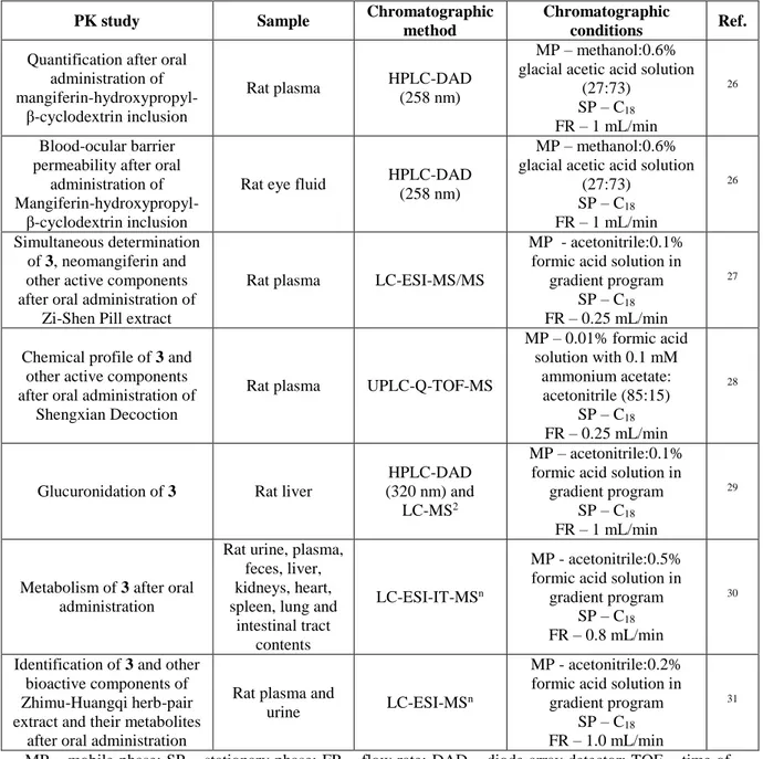
One example of these compounds is mangiferin (3, Fig. 2) an hydroxylated and glycosylated xanthone that was originally isolated from Mangifera indica L. and has already been reported to have several pharmacological activities, such as antitumor, antiviral, antidiabetic, among others 1. For all these reasons, many research groups have found great interest in studying the PK behavior of this compound, through chromatographic methodologies (Table 1).
4
Fig. 2: Chemical structure of mangiferin (3).Table 1: Chromatographic methods applied to PK studies of mangiferin (3).
PK study Sample Chromatographic
method
Chromatographic
conditions Ref. Quantification after oral
administration of
mangiferin-hydroxypropyl-β-cyclodextrin inclusion
Rat plasma HPLC-DAD (258 nm)
MP – methanol:0.6% glacial acetic acid solution
(27:73) SP – C18 FR – 1 mL/min
26
Blood-ocular barrier permeability after oral
administration of
Mangiferin-hydroxypropyl-β-cyclodextrin inclusion
Rat eye fluid HPLC-DAD (258 nm)
MP – methanol:0.6% glacial acetic acid solution
(27:73) SP – C18 FR – 1 mL/min 26 Simultaneous determination of 3, neomangiferin and other active components after oral administration of
Zi-Shen Pill extract
Rat plasma LC-ESI-MS/MS
MP - acetonitrile:0.1% formic acid solution in
gradient program SP – C18 FR – 0.25 mL/min
27
Chemical profile of 3 and other active components after oral administration of
Shengxian Decoction
Rat plasma UPLC-Q-TOF-MS
MP – 0.01% formic acid solution with 0.1 mM ammonium acetate: acetonitrile (85:15) SP – C18 FR – 0.25 mL/min 28
Glucuronidation of 3 Rat liver
HPLC-DAD (320 nm) and
LC-MS2
MP – acetonitrile:0.1% formic acid solution in
gradient program SP – C18 FR – 1 mL/min
29
Metabolism of 3 after oral administration
Rat urine, plasma, feces, liver, kidneys, heart, spleen, lung and
intestinal tract contents
LC-ESI-IT-MSn
MP - acetonitrile:0.5% formic acid solution in
gradient program SP – C18 FR – 0.8 mL/min
30
Identification of 3 and other bioactive components of Zhimu-Huangqi herb-pair extract and their metabolites
after oral administration
Rat plasma and
urine LC-ESI-MS
n
MP - acetonitrile:0.2% formic acid solution in
gradient program SP – C18 FR – 1.0 mL/min
31
MP – mobile phase; SP – stationary phase; FR – flow rate; DAD – diode array detector; TOF – time of flight; Q – quadrupole; LC – liquid chromatography; ESI – electrospray ionization; IT – ion trap; MS – mass spectrometry.
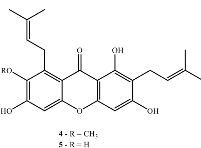
Other important representatives of natural xanthones with biological interest are α-mangostin (4, Fig. 3) and γ-mangostin (5, Fig. 3), prenylated xanthones present in high concentrations in the pericarp of
Garcinia mangostana L. These compounds have shown antioxidant, anti-inflammatory and antitumor
activities, among others 1,14,32-34. Due to the increasing interest and knowledge of the mechanisms behind the biological activities of mangosteens, several studies have been carried out over the last years to understand the PK proprieties of these compounds, using chromatographic methods (Table 2).
5
Fig. 3: Chemical structures of α-mangostin (4) and γ-mangostin (5).Table 2: Chromatographic methods applied to PK studies of α-mangostin (4) and γ-mangostin (5).
PK study Sample/ Matrix Chromatographic method Chromatographic conditions Ref. Quantification of 4 and 5 after oral and
intravenous administration of Mangosteen extract Mouse plasma HPLC-MS/MS MP – acetonitrile containing 0.1% formic
acid:0.1% formic acid solution (70:30)
SP – C18 FR – 0.35 mL/min
35
Tissue distribution of xanthones and their phase II metabolites
Mouse liver, tumor, feces and
serum
HPLC-DAD and HPLC-MS
MP – acetonitrile containing 2% acetic acid solution: n-butanol
in a gradient program SP – C18 FR – 0.5 mL/min
32,36
In vitro study of the
intestinal uptake, retention, transport and
metabolism of 4 Caco-2 human intestinal cells HPLC-DAD (254 nm) MP – acetonitrile: 2% acetic acid solution in a
gradient program SP – C18 FR – 0.8 mL/min
37
Study of absoption, tissue distributon and tissue metabolism of 4
after oral and intravenous administration
Mouse plasma, liver stomach, small and large intestines, lung, heart, kidney, fat, mesentery, muscle and brain HPLC-MS/MS MP – acetonitrile containing 0.1% formic
acid:0.1% formic acid solution (70:30)
SP – C18 FR – 0.35 mL/min
38
PKs of 4 after oral and intravenous administration
Rat plasma LC-MS/MS
MP – acetonitrile containing 0.05% formic
acid:0.05% formic acid solution (80:20)
SP – C18 FR – 0.5 mL/min
39
MP – mobile phase; SP – stationary phase; FR – flow rate; DAD – diode array detector; LC – liquid chromatography; MS – mass spectrometry.
Several other xanthones, isolated from natural sources have been investigated by chromatographic methods to obtain information about their PK proprieties (Table 3): the cytotoxic gambogic acid (6, Fig.
6
4), a polyprenylated xanthone isolated from the resin of several Garcinia species 40; the vasorelaxing and antihypertensive 1-hydroxyl-2,3,5-trimethoxy-xanthone (7, Fig. 4) isolated from the a Tibetan medicinal herb Halenia elliptica D. Don 41, the xanthone glycoside sibiricaxanthone F (8, Fig. 4), isolated fromPolygala sibirica L., which has shown in vitro effect on peroxisome proliferator-activated receptors
(PPARs) and in accelerating the differentiation of 3T3-L1 preadipocytes cells 42; the promising antitumor compound cudratricusxanthone B (9, Fig. 4), an isoprenylated xanthone isolated from Cudrania
tricuspidata (Carr.) Bur. 43.
Fig. 4: Chemical structures of gambogic acid (6), 1-hydroxy-2,3,5-trimethoxy-xanthone (7), sibiricaxanthone F (8), and cudratricusxanthone B (9).
Table 3: Chromatographic methods applied to PK studies of other natural xanthones (6-9).
PK study Sample Chromatographic
method Chromatographic conditions Ref. Quantification of 6
after intravenous administration
Dog plasma HPLC-UV detector (360 nm)
MP – methanol:0.05% phosphoric acid solution (94:6)
SP – C18 FR – 1.0 mL/min 44 Identification of metabolites of 6 after intravenous
Rat bile LC-TOF-IT-MS
MP – acetonitrile:0.05% acetic acid solution in gradient
program
7
administration SP – C18 FR – 0.2 mL/min Structure elucidation of metabolites of 6 after intravenous administration Rat bile HPLC-UV (280 nm), LC-ESI-IT-MSn and LC-NMR MP – methanol:water (85:15) SP – C18 FR – 1.0 mL/min 46 In vitro identification of cytochrome P450 isoforms responsible for the metabolism of7
Human liver
microsomes LC-IT-TOF-MS n
MP – acetonitrile:0.5%acetic acid solution in gradient
program SP – C18 FR – 0.8 mL/min 47 In vitro metabolite characterization of 7 and other bioactive
xanthones
Rat liver
microsomes LC-IT-TOF-MS n
MP – acetonitrile:0.5%acetic acid solution in gradient
program SP – C18 FR – 0.8 mL/min
41
Quantification of 8 after oral and
intravenous administration
Rat plasma LC-ESI-MS/MS
MP – acetonitrile containing 0.01% formic acid:0.01% formic acid solution in gradient
program SP – C18 FR – 0.4 mL/min 48 In vitro metabolite characterization of 8 Human and rat liver microsomes HPLC-EMS-IDA-EPI MP – acetonitrile containing 0.1% formic acid:0.1% formic
acid solution in gradient program SP – C18 FR – 0.3 mL/min 42 Quantification of 9 after intravenous administration
Rat plasma HPLC-ESI-MS/MS
MP – 0.5% formic acid in methanol
SP – C18 FR – 0.3 mL/min
43
MP – mobile phase; SP – stationary phase; FR – flow rate; TOF – time of flight; LC – liquid chromatography; NMR – nuclear magnetic resonance; ESI – electrospray ionization; IT – ion trap; EMS - enhanced mass spectrum; IDA - information dependent acquisition; EPI - enhanced product ion ; MS – mass spectrometry.
2.2. Synthetic xanthone derivatives
Due to the pharmacological interest in xanthone derivatives, several research groups work in the synthesis of xanthonic compounds in an attempt to find new drug candidates with better pharmacodinamic and PK properties. Table 4 shows different classes of synthetic derivatives, namely thioxanthones, in preclinical studies, and the applications of chromatographic methods in their PKs proprieties studies. It also displays the PK study of the 9H-xanthen-9-one (1) - in an in vivo model 49.
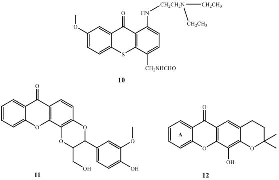
The development of an HPLC methodology to determine the amount of a thioxanthone drug candidate (10, Fig. 5) and its metabolites in plasma was applied to determine effective and toxic doses, as well as to predict and quantify the metabolic profile of the drug candidate in in vivo tests 50.
The intestinal absorption of a xanthonolignoid (11, Fig. 5), a protein kinase C inhibitor, was studied in our group, using Caco-2 cell lines as a model to predict the absorption of the drug candidate, using a HPLC to quantify the cell permeability 51.
A pyranoxanthone derivative (12, Fig. 5) with potential antitumor activity intestinal absorption was also studied applying the same chromatographic technique. Another HPLC methodology was developed
8
to evaluate the improvement of absorption and therefore enhancement of biological activity at lower doses of compound 12 by inclusion into drug delivery systems, namely PLGA nanoparticles 51,52.Fig. 5: Chemical structures of a thioxanthone derivative 10, a xanthonolignoid 11 and a pyranoxanthone 12.
Table 4: Chromatographic methods applied to PK studies of synthetic xanthones (1, 10-12).
PK study Sample Chromatographic
method Chromatographic conditions Ref. Determination of 1
after oral and intravenous administration
Rat plasma HPLC-DAD (254 nm)
MP – acetonitrile containing 0.1% trifluoroacetic acid:0.1% trifluoroacetic acid solution in a
gradient program SP – C18 FR – mL/min 49 Quantification of 10 and its metabolites
Mouse plasma HPLC-DAD (266 nm) MP - methanol:10 mM phosphate buffer pH 3.5 (45:55) SP - C18 FR - 0.8 mL/min (10'), 1.4mL/min (15') 50 Prediction of intestinal
absorption of 11 Caco-2 cell monolayers as an intestinal model HPLC-DAD (237 nm) MP - methanol:water (85:15) SP - C18 FR - 1 mL/min 51 Prediction of intestinal absorption of 12 HPLC-DAD (254 nm) MP - methanol:water (80:20) SP - C18 FR - 1 mL/min Study of the intracellular delivery of 12 entrapped in PLGA nanoparticles MCF-7 culture cell lines exposed
to the xanthone derivative included in nanocapsule formulation HPLC-DAD (254 nm) MP - methanol:water (85:15) SP - C18 FR - 1 mL/min 52,53
9
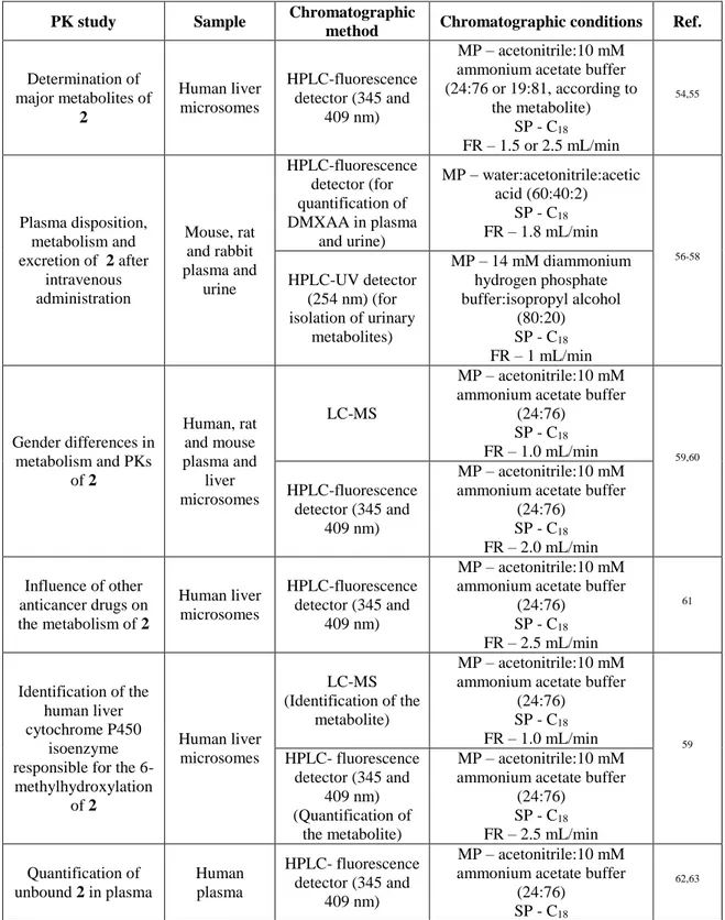
MP – mobile phase; SP – stationary phase; FR – flow rate; DAD – diode array detector.The introduction of a drug candidate in human trials is a highly demanding and expensive process. Therefore, only the drug candidates with best results in preclinical studies achieve this level in the pipeline of drug development 22. DMXAA (2, Fig.1) is a drug candidate that reached clinical trials and the chromatographic methods and conditions applied to PK studies are highlighted in Table 5.
Table 5: Chromatographic methods applied to PK studies of DMXAA (2).
PK study Sample Chromatographic
method Chromatographic conditions Ref.
Determination of major metabolites of 2 Human liver microsomes HPLC-fluorescence detector (345 and 409 nm) MP – acetonitrile:10 mM ammonium acetate buffer (24:76 or 19:81, according to the metabolite) SP - C18 FR – 1.5 or 2.5 mL/min 54,55 Plasma disposition, metabolism and excretion of 2 after intravenous administration Mouse, rat and rabbit plasma and urine HPLC-fluorescence detector (for quantification of DMXAA in plasma and urine) MP – water:acetonitrile:acetic acid (60:40:2) SP - C18 FR – 1.8 mL/min 56-58 HPLC-UV detector (254 nm) (for isolation of urinary metabolites) MP – 14 mM diammonium hydrogen phosphate buffer:isopropyl alcohol (80:20) SP - C18 FR – 1 mL/min Gender differences in metabolism and PKs of 2 Human, rat and mouse plasma and liver microsomes LC-MS MP – acetonitrile:10 mM ammonium acetate buffer
(24:76) SP - C18 FR – 1.0 mL/min 59,60 HPLC-fluorescence detector (345 and 409 nm) MP – acetonitrile:10 mM ammonium acetate buffer
(24:76) SP - C18 FR – 2.0 mL/min Influence of other anticancer drugs on the metabolism of 2 Human liver microsomes HPLC-fluorescence detector (345 and 409 nm) MP – acetonitrile:10 mM ammonium acetate buffer
(24:76) SP - C18 FR – 2.5 mL/min 61 Identification of the human liver cytochrome P450 isoenzyme responsible for the 6-methylhydroxylation of 2 Human liver microsomes LC-MS (Identification of the metabolite) MP – acetonitrile:10 mM ammonium acetate buffer
(24:76) SP - C18 FR – 1.0 mL/min 59 HPLC- fluorescence detector (345 and 409 nm) (Quantification of the metabolite) MP – acetonitrile:10 mM ammonium acetate buffer
(24:76) SP - C18 FR – 2.5 mL/min Quantification of unbound 2 in plasma Human plasma HPLC- fluorescence detector (345 and 409 nm) MP – acetonitrile:10 mM ammonium acetate buffer
(24:76)
SP - C18 62,63
10
FR – 2.5 mL/min Identification and reactivity of the major metabolite of 2 Rat, mouse, rabbit and human plasma and human urine HPLC-fluorescence detector (345 and 409 nm) MP – acetonitrile:10 mM ammonium acetate buffer (24:76 and 19:81, dependingon the metabolite)
SP - C18 FR – 2.5 mL/min and 2.0
mL/min (depending on the metabolite) 54,56,59,64 HPLC-fluorescence detector (345 and 409) MP – water:acetonitrile:acetic acid (60:40:2) SP - C18 FR – 1.8 mL/min Interspecies differences in the metabolism of 2 Rat, mouse, rabbit and human liver microsomes LC-MS (Identification of the metabolite) MP – acetonitrile:10 mM ammonium acetate buffer
(24:76) SP - C18 FR – 1.0 mL/min 54,59,65 HPLC- fluorescence detector (345 and 409 nm) (Quantification of the metabolite) MP – acetonitrile:10 mM ammonium acetate buffer
(24:76) SP - C18 FR – 2.5 mL/min Interspecies differences in the plasma protein binding and blood cells distribution of 2 Rat, mouse, rabbit and human plasma HPLC- fluorescence detector (345 and 409 nm) MP – acetonitrile:10 mM ammonium acetate buffer
(24:76) SP - C18 FR – 2.5 mL/min 56,66 Strain differences in the metabolism of 2 and its correlation with the maximum
tolerated dose Five different mouse strains liver microsomes HPLC-MS-APCI/ESP (Identification of the metabolite) MP – acetonitrile:10 mM ammonium acetate buffer
(24:76)
SP - C18 FR – 1.0 mL/min (for
HPLC-MS-APCI) or 0.5 mL/min (for HPLC-MS-ESP) 54,59,64,67 HPLC- fluorescence detector (345 and 409 nm) (Quantification of the metabolite) MP – acetonitrile:10 mM ammonium acetate buffer
(24:76) SP - C18 FR – 2.5 mL/min Impact of preclinical factors in the clearance of 2 Human plasma and liver microsomes LC-MS (Identification of the metabolite) MP – acetonitrile:10 mM ammonium acetate buffer
(24:76) SP - C18 FR – 1.0 mL/min 68 HPLC- fluorescence detector (345 and 409 nm) (Quantification of the metabolite) MP – acetonitrile:10 mM ammonium acetate buffer
(24:76) SP - C18 FR – 2.5 mL/min Quantification of unbound 2 in plasma Human plasma LC-MS/MS MP – acetonitrile containing 0.1% formic acid:0.1% formic
acid solution in gradient program
SP - C18 FR – 0.5 mL/min
11
Determination ofplasma and urine concentration of 2 and its metabolites after intravenous administration Human plasma, red cells, urine and tumor samples HPLC- fluorescence detector (345 and 409 nm) MP – acetonitrile:10 mM ammonium acetate buffer
(25:75)
SP - C18 FR – 2.0 mL/min
70,71
Determination of [14C] ASA404 and its
metabolites after intravenous administration Human plasma and feces HPLC - off-line radioactivity detector MP – gradient elution consisting of solvent A (5 mM ammonium formate with 0.1%
formic acid, pH~3) and B (acetonitrile with 0.1% formic
acid)
SP - Waters Xbridge column FR – 1.0 mL/min
72
Determination of 2 and its acyl glucuronide Caco-2 cell monolayers for transport studies Caco-2 cell monolayers HPLC- fluorescence detector (345 and 409 nm) MP – acetonitrile:10 mM ammonium acetate buffer, pH
5.0 (24:76)
SP - C18 FR – 1.2 mL/min
73
MP – mobile phase; SP – stationary phase; FR – flow rate; LC- liquid chromatography; MS – mass spectrometry; APCI/ESP - Atmospheric pressure chemical ionization/electrostatic precipitator.
According to all these data, it is possible to verify that the most commonly applied chromatographic method is LC, especially HPLC, using a C18 stationary phase in reversed phase, using mixtures of water and an organic solvent, most commonly acetonitrile or methanol. The detection method applied depends on the nature of the analyte and the aim of the PK work, being fluorescence and diode array detectors widely used for quantitative analysis and identification of compounds, especially when standards are available. In metabolism studies and identification of xanthone derivatives and its metabolites, mass spectrometers detectors appear to be the most applied, due to the sensitivity, reliability, specificity and information that can be assessed.
3. Physicochemical properties and preformulation studies of xanthone derivatives
The discovery of new drugs needs a thorough investigation for its safety and efficacy before their release into the market. The old paradigm of drug discovery process has changed due to technology innovation. Rational drug discovery requires an early appraisal of all factors impacting on the likely success of a drug candidate in the subsequent preclinical, clinical and commercial phases of drug development. Important preformulation considerations include solubility, stability, pka, all properties that affect drug permeability and distribution. The next sections highlight the main structure-properties relationships established from the analysis of xanthone derivatives.
3.1. Solubility
Solubility is emerging as one of the major issues in drug discovery and development of new chemical entities 74. Compounds with low solubility not only cause problems for in vitro and in vivo assays, but also have a higher risk of attrition in the drug discovery pipeline 75.
Lipinski has classified the solubility ranges (high is > 60 g/mL, moderate is 10-60 g/mL, low is < 10 g/mL) to provide a general guideline for achieving acceptable human absorption for compounds with average potency and permeability. This classification is different from the Biopharmaceutics
12
Classification Systems whereas, for example, for a 1 mg/kg intravenous (I.V.) dose in rat with an ideal dosing volume of 1-5 mL/kg, the required concentration is 0.2 – 1 mg/mL 76.Different analytical tools (i.e., light scattering/turbidity, UV plate reader, LC-UV and LC–MS/LC-CLND - chemiluminescence nitrogen) can be used for detection of the maximum amount of a compound that can remain in solution at a certain volume of solvent, temperature, and pressure, under equilibrium conditions. The pH of the buffer in solubility methods is typically 6.5 (intestine) or 7.4 (blood).
α-Mangostin (4) showed poor aqueous solubility and low oral bioavailability, hindering its
therapeutic application. The aqueous solubility of free α-mangostin (4) was found to be 0.2 ± 0.2 µg/mL 77. Briefly, an excess amount from the samples was added to phosphate-buffered saline (PBS) at pH 7.4 (Scheme 1). The mixtures were mixed and then centrifuged. The supernatant was collected, filtered and diluted in methanol. The samples were then analyzed using a spectrophotometer at 243.4 nmto determine
α-mangostin (4) concentration 77.
Scheme 1: Schematic representation of the solubility assay.
Other study investigated the thermodynamic solubility in water at pH 7.4 HEPES (N-2-hydroxyethylpiperazine-N-2-ethane sulfonic acid) buffer of 12-hydroxy-2,2-dimethyl-3,4-dihydropyran[3,2b]xanthene-6(2H)-one (12) and five of its analogues bearing substituents on ring A 75. All compounds showed low solubility (0.1-0.6 µM) which was explained by the high planarity and rigidity of these compounds. Each compound was added to 2 mL of HEPES buffer until a saturated solution was obtained (Scheme 2). The suspension of each compound was agitated at 37 °C for 24 h. Each sample was filtered through a 0.1 µm filter and 1485 µL of each filtrate was added to another eppendorf containing 15 µL of DMSO. The UV/Vis spectrum was traced in a double-beam spectrophotometer and the concentration was determined according to the standard calibration curve for each compound.
13
Oxo-9H-xanthene-3,6-diyl bis(3-chlorobenzoate), 9-oxo-9H-xanthene-3,6-diyl bis(4-tert-butylbenzoate) and 9-oxo-9H-xanthene-3,6-diyl bis(4-methoxybenzoate) also showed poor water solubility. This limitation was successfully overcome with the formulation of these derivatives into cyclodextrins 78.The intrinsic aqueous solubility of 40 structurally diverse Garcinia natural-product-like xanthones based on gambogic acid (6) was determined. All the synthetic compounds displayed aqueous solubility that was improved over that of gambogic acid. Among all the tested compounds, the most soluble exhibited an intrinsic solubility of 10.37 mM, almost 1000 times more soluble than gambogic acid. Some structure-properties were drawn: hydrophilic amine substitution, a hydroxyl group at position 1, and removal of the gem-dimethyl group provides improved water solubility, while bulky hydrophobic groups such as a pyran ring or prenyl, and hydroxyl in B ring decrease water solubility 79.
3.2. Lipophilicity
Lipophilicity is one of the first physicochemical properties to be evaluated in the early phases of a drug discovery program and has been correlated with several other physicochemical and PK properties, for example, solubility, permeability, plasma protein binding, metabolism, blood-brain barrier (BBB) penetration, volume of distribution and clearance. A compound with moderate lipophilicity has a good balance between solubility and permeability and is optimal for oral absorption, cell membrane permeation in cell-based assays, and is generally good for BBB penetration (optimal distribution coefficient logarithm -log D- ~2). Consequently, it may extensively influence the success or the failure of a drug discovery program. In fact, compounds with high lipophilicity have shown an increased risk of attrition during the clinical trials.
Lipophilicity is commonly evaluated by the logarithm of the partition coefficient (log P) of an un-ionized solute in a biphasic system using the so-called shake-flask method. Different solvent systems have been used for estimating this property, but the octanol/phosphate buffer pH 7.4 system remains the most common, and mimics, respectively, the biomembranes and the blood in vivo. The determination of the log P of a solute in a biphasic octanol–water system by the shake-flask method is described in the OECD (Organization for Economic Co-operation and Development) guidelines for testing chemicals 80. The solute is simply partitioned between the two liquid phases of the proposed solvent system in a test vessel. Both solvents should be mutually saturated at room temperature (20 to 25 ºC) before the log P determination. After equilibrium and separation, usually achieved by centrifugation, the relative solute concentration in each layer is determined using a variety of analytical techniques. These include spectroscopic methods, HPLC, GC, thin-layer chromatography (TLC), among others.
The octanol–water distribution of several hydroxylated and/or methoxylated xanthones was systematically investigated in a biphasic octanol–water system and their log P values lie in the range 2.90–3.80 81. Optical density of the aqueous phase and of the organic phase was measured on a spectrophotometer. Stock solutions of compounds (concentration 10–2 M) were prepared in octanol and shaken with a known volume of extractant (doubly distilled water saturated with octanol). The aqueous phase was acidified with HCl (1 N) to pH 2-3 in order to avoid dissociation for the study of the
14
distribution of compounds containing free phenolic groups. Experiments were conducted with varying phase (octanol–water) ratios (1:20, 1:30, 1:40, 1:50, 1:60) to cover a wide range of lipophilicity values, at 20–22°C 81.The octanol–water system has some limitations since it fails to create the anisotropic media that is found on biomembranes and encode some important interactions that take place between the solute and the membranes. Therefore, other models have been developed such as liposomes and micelles which have proved to be advantageous when compared to octanol–water. The lipophilicity of several prenylated and pyranoxanthones was determined as the partition coefficient (P) of the solute between buffer (pH 7.4) and micelles or liposomes, and calculated without a phase separation 75,82. The tested compounds showed a moderate to high lipophilicity. A good correlation between the two models, micelles and liposomes was observed, which implies that there is no clear tendency for the tested compounds to increase the affinity for either of the membrane models with an increase in hydrophobicity. The results obtained led to the observation that micelles can be used as a surrogate for liposomes for the studied compounds with the advantage of not having the light scattering limitation (wavelengths below 300 nm) as well as a higher applicability and easier preparation 75,82.
All the pyranoxanthones showed a Log Kp in liposomes and micelles superior to 3 but below the critical 5 (upper limit referred in the Lipinski “rule of five”). The presence of a hydroxyl ortho to the carbonyl leads to a dramatic increase in the lipophilicity. Considering the relationship between the hydroxyl or methoxyl group with the oxygen of the fused pyran ring, the compounds with an ortho relationship seem in most of the cases to have a lower partition coefficient than in meta position. Comparing the linear with the angular arrangement of the molecule of pyranoxanthones, it can be observed that in general, the latter have a less partition coefficient than the former. Regarding the presence or absence of the double bond in the fused ring, there is not a clear tendency among the different set of compounds 82.
Considering the effect of ring A (see Fig. 5) substituents on the Kp of compound 12, it was observed that chlorine and methyl were associated with an increase in Kp. In the case of the methoxyl, the introduction of this group in position 8 did not interfere with the Kp while the introduction in position 10 was associated with an increase of the Kp. For the hydroxyl group an unexpected result was observed and no decrease in Kp was determined. In fact, for assays with micelles, an increase in Kp was observed. Considering the analogues of compound 12 with a methoxyl in position 12, it can be observed that this substituent was associated with an increase in the Kp. Comparing with the analogues with a different ring D orientation, it can be observed that most of the compounds had a Kp near or higher than 4 with the chlorine and the diethylamine derivatives representing the compounds that showed a higher Kp 75.
15
Fig. 6: Pyranoxanthone (13) with the higher Kp.HPLC method is also described in the OECD guidelines for testing chemicals 83. With the development of reversed phase HPLC in many laboratories, readily accessible retention factor of a new compound, compared to the standard but time consuming shake-flask method, made the method extremely popular and widely used 84. Reversed phase HPLC is performed on analytical columns packed with a solid phase containing long hydrocarbon chains chemically bound onto silica. The chemicals are retained in the column in proportion to their hydrocarbon-water partition coefficient, with hydrophilic chemicals eluted first and lipophilic chemicals last 83.
Using HPLC methods, the log P values of a series of xanthone derivatives - obtained by cationic modification of the free C3 and C6 hydroxyl groups of α-mangostin with amine groups 85 - ranged between 5.42 and 15.01. In this study the log P and molecular hydrophobicity parameters were found less predictive than pKa for improving the antimicrobial properties of xanthone analogues.
Most solutes are weak bases or acids which become partially ionized when dissolved on water. This has raised the need to develop the distribution coefficient (D) which is used to determine the ratio of the sum of the concentrations of all forms of the solute (pH dependent mixture of ionized plus un-ionized forms) in each of the two phases. The logarithm to the base 10 of the distribution coefficient (log D) is normally used to express results 86.
The log D at pH 7.4 of a set of 40 compounds obtained by propargylation of hydroxyxanthone substrates were determined according to the method of Avdeef and Tsinman on a Gemini Profiler instrument (pION) by the “gold standard” Avdeef−Bucher potentiometric titration method. Log D values were between 0.44-6.0 79.
3.3. Acid dissociation constant (pKa)
It is well known that ionized compounds are more soluble in water than the neutral forms, but less permeable. Ionization is determined by the pKa and aqueous pH; thus, pKa has a major effect on PK properties. Buffer solutions with different pH values have to be prepared by mixing appropriate volumes of the stock solutions and then diluting to ionic strength I = 0.03, which were controlled by calculation and by acid–base equilibrium theory. The pH should be measured at 25 °C by a pH meter calibrated in advance with standard buffers at the pH range.
Mangiferin (3) pKa values were determined in aqueous solution by UV–Vis spectroscopic studies coupled with nuclear magnetic resonance (NMR) detector. The NMR study complemented the pKa
16
values assignment and evidenced a hydrogen interaction at C-1 with the carbonyl group. Results showed that the proton at hydroxyl in position 1 is the less acidic, assigning the last pKa to this proton. This result contrasted with the predicted by the ACD/pKa DB version 5 (Advanced Chemistry Development, Inc., Toronto, Ont., Canada, www.acdlabs.com, 2003) that estimated this proton as the most acidic one 87.Capillary zone electrophoresis (CZE) method, which relies on measuring the mobility of the solute as a function of pH, has been proved to be an effective and convenient technique for determining the pKa values being considered as an universal technique for determining pKa values in a wide pH range. The accuracy of pKa values obtained from CZE has been confirmed to be in agreement with those from potentiometric methods and, in general, better than those from single chromatographic or spectrophotometric methodologies. 25. Compared to spectroscopy, potentiometry, conductivity or other techniques, CZE has advantages concerning sensitivity (only small amounts of sample are required), analysis time (relatively short), the result is independent of the solute purity (high purity is not necessary), it is applicable to samples that are weakly soluble in water (can be performed in aqueous or nonaqueous solvents), and the procedure does not require solute measurement or titrant concentrations, but only the migration times of solutes 25. Another important advantage of CZE method is that the pKa values of solutes based on both their migration times and UV absorbance measured from online spectra can determine, when online diode array detection (DAD) is used 25.The pKa values of 10 hydroxylated and/or methoxylated xanthones, isolated from herbal medicine Securidaca inappendiculata, have been determined by CZE based on linear and nonlinear regression models and the results were confirmed by UV absorbance from online DAD 25. The results showed that the precision of the two methods, expressed in terms of the acceptable repeatability and reproducibility of the migration time, mobility and pKa values, is acceptable 25.
In another study, prediction of pKa values was highlighted as a new way to improve “hits” for the development of membrane-active antibiotics that target drug-resistant pathogens 85. Results showed that the antimicrobial activities of cationic xanthone derivatives can be generally predicted based on the pKa values of the corresponding amines. Pyrazole and triazol moieties which led to low pKa values reduced the antimicrobial activity of the derivatives when compared with the parent compound 85.
3.4. Permeability
With the development of drug discovery, methods that can screen in vitro permeability of compounds rapidly received significant attention. The Caco-2 cells represent a well-known and interesting cell culture-based model for enterocytes which has been used in several in vitro bioavailability assays to evaluate the intestinal absorption rate of a broad range of drugs. The first report on xanthones bioavailability and metabolism was performed using the coupled in vitro digestion/Caco-2 human intestinal cell model 88. Optimal bioavailability of α- (4) and γ-mangostin (5) were dependent on incorporation into bile salt mixed micelles. In addition, α-mangostin (4) was transported across the apical surface of enterocyte-like Caco-2 cells and partially converted to phase II metabolites 37. Both unconjugated α-mangostin (4) and its phase II metabolites were effluxed across the basolateral membrane suggesting that they were absorbed. Transepithelial transport was enhanced by addition of products of lipid digestion in the apical compartment, suggesting that absorption was dependent on the assembly and
17
secretion of chylomicrons. α-Mangostin metabolites also were retro-transported across the apical membrane into the simulated gut luminal compartment 37. An HPLC-UV analysis from the in vitro permeation assay with Caco-2 cells monolayer was performed to predict the intestinal permeability of different xanthonic derivatives (oxygenated, prenylated, xanthonolignoids) 89 which revealed high apparent permeability coefficients (Papp) similar to other well absorbed drugs 51.Parallel artificial membrane permeability assay (PAMPA) has been applied early in drug development. Most drugs are absorbed through passive diffusion across the lipid bilayer membrane of the epithelial cells into systemic circulation. The process is driven by concentration gradient. The PAMPA assay is designed to measure passive diffusion through an artificial lipid membrane. Permeability coefficients of a set of 40 propargylxanthone derivatives were determined using a standard PAMPA 79. The results suggested that the trends of permeability of propargylxanthone derivatives are similar to their behavior in solubilities (see section 3.1): compounds with smaller frameworks and hydrophilic groups are also likely to have better permeability.
3.5. Modulation of ABC Transporters
ATP-binding cassette (ABC) transporters are present in all cells of all organisms and use the energy of ATP binding/hydrolysis to transport substrates across cell membranes. Several membrane proteins belonging to the ABC family of proteins act by preventing the absorption of orally ingested or airborne xenobiotics or drugs. Among these proteins, P-glycoprotein (P-gp) and multidrug-resistance protein 1 (MRP1) are the most extensively studied 90,91. These ABC transporters are attractive therapeutic targets since their inhibition could overcome multidrug resistance to anticancer drugs while its induction/activation could limit the toxicity of substrates such as pesticides. Thus, investigation on ABC transporters modulation by xanthone derivatives is not limited to permeability studies and aims mainly these pharmacological perspectives.
3.5.1. MRP1 modulation
MRP1 promotes drug efflux in which is considered phase III metabolism in two possible ways: conjugation with glutathione (GSH) or by co-transport with free GSH 92. In the search of compounds with cytotoxicity towards cancer cells overexpressing MRP1, Genoux-Bastide et al. 93 studied a library of xanthones and compared the results with a well-known activator of massive GSH efflux by MRP1, the calcium channel blocker verapamil 94,95.
The total cellular GSH determination was measured using the enzymatic method described by Tiezte 96, as modified by Anderson 97 to the microtiter plate. From the library of xanthones evaluated, derivatives with more than 40% of GSH efflux scores were investigated to determine their cytotoxicity against a sensitive tumor cell line (NCI-H69) and a MRP1-overexpressing human resistant cell line (H69AR/MRP1 derived from NCI-H69). In order to evaluate if the most active compounds for selective killing of MRP1-overexpressing resistant cells activity was correlated with MRP1 expression, cytotoxicity of the two best compounds was tested on three HEK-293 cell lines, (one with wild-type Pgp, one with ABCG2 and one with pcDNA3.3) and on two MDCK2 cell lines (one with wild-type MRP2 and one without it). Since the achieved IC50 value was greater than 100 µM to the five cell lines, it allowed to conclude that the
18
analyzed compounds were selective killers of resistant cells overexpressing MRP1 93. Structures of the most promising compounds are shown on Fig. 7.Fig. 7: Chemical structures of
xanthones with GSH efflux of more than 40%.
The results allowed inferring that cell death was not dependent just by the GSH efflux, since some xanthones induced efflux of GSH by more than 50% and show no significant cytotoxicity towards the tested cell line. Therefore, the selective cytotoxicity presented by compound 17, Fig. 7 is more likely due to a MRP1 activation followed by apoptosis through an acceleration of GSH efflux from the cell 93.
3.5.2. P-gp modulation
P-gp is responsible for the transmembrane transport of neutral or positively-charged hydrophobic substrates, namely anticancer agents. Over the past decades, this target has drawn a lot of interest in several research areas, such as Medicinal Chemistry and Toxicology. First, because its inhibition can lead to increase cytotoxicity and therefore increase therapeutic efficacy of anticancer agents and, on the other hand, due to its broad substrate specificity and ubiquitous presence in excretory and barrier tissues, its activation can play a crucial role in limiting the absorption and distribution of toxic xenobiotics 98-100.
In our group, to improve the efficacy in sensitizing resistant P-gp overexpressing cell lines, (thio)xanthones P-gp modulators were obtained based on structure-based design approaches 101. The methods applied to validate a library of dual inhibitors of P-gp and tumor cell lines are summarized in Table 6 101.
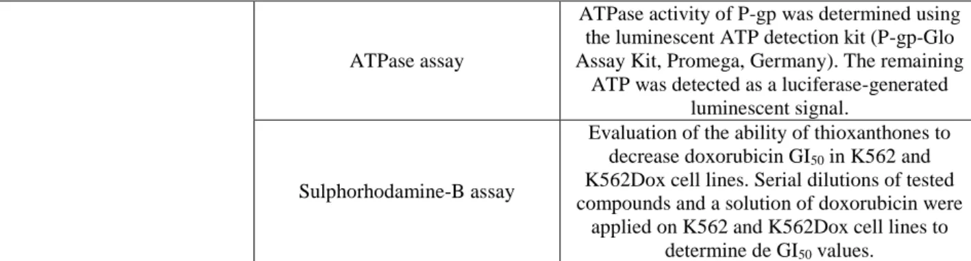
Table 6: Methods applied in biological studies of a library of thioxanthones.
Aim Methods Description
Cell growth study
Sulphorhodamine-B assay
Serial dilutions of tested compounds were applied on K562 and K562Dox cell lines to
determine de GI50 values. Trypan blue exclusion assay
Investigation of cellular viability in MRC-5 cell line, using the compounds in their GI50 concentration achieved to the K562 cell line. P-gp modulation study Rhodamine-123 accumulation
assay
K562 and K562Dox cell lines were incubated with the thioxanthones and rhodamine-123,
followed by flow cytometry analysis.
Compound R1 R2 R3 R4 14 OH CH3 OCH3 H 15 OH OH OCH3 H 16 OH OH H H 17 OH OH H OCH3 18 OH OH H CH3 19 OH OH H OH 20 OH OCH3 H OCH3
21 OCH3 O-Bz H OCH3
22 OH OH H NH-COCF3
19
ATPase assayATPase activity of P-gp was determined using the luminescent ATP detection kit (P-gp-Glo Assay Kit, Promega, Germany). The remaining
ATP was detected as a luciferase-generated luminescent signal.
Sulphorhodamine-B assay
Evaluation of the ability of thioxanthones to decrease doxorubicin GI50 in K562 and K562Dox cell lines. Serial dilutions of tested compounds and a solution of doxorubicin were
applied on K562 and K562Dox cell lines to determine de GI50 values.
K562 and K562Dox cell lines – human chronic myelogenous leukemia cell line and doxorubicin-induced P-gp overexpression K562 cell line; GI50 – drug concentration that exerts a growth inhibition rate to 50% of the maximum rate; MRC-5 cell line – human fetal lung fibroblast-like cell line; ATPase – adenosine triphosphatase; P-gp – permeability glycoprotein.
In this study, both inhibitors and activators of P-gp were found and in Fig. 8 are highlighted the hit compounds concerning P-gp noncompetitive and competitive inhibitors 101.
Fig. 8: Thioxanthone noncompetitive 24 and competitive 25 inhibitors of P-gp.
Further studies to evaluate the selectivity profile of thioxanthones as inhibitors of MDR were performed, by studying their interaction with other ABC transporters, namely MRP1, MRP2, MRP3 and BCRP. Another important assessment was the evaluation of the interaction of thioxanthones with P450 3A4 (CYP3A4), as well as the prediction of their binding conformations and metabolism sites 102. The results allowed to conclude that compound 25 (Fig. 8) is a promising multi-target inhibitor of P-gp, BCRP and MRP1. On the other hand, 1-[2-(diethylamino)ethyl]amino-4-propoxy-9H-thioxanthen-9-one (26, Fig. 9) is a multi-target inhibitor of P-gp, BCRP, MRP1 and MRP3.
20
Fig. 9: Multi-target inhibitor of P-gp, BCRP, MRP1 and MRP3 thioxanthone 26.Nowadays, strategies to increase P-gp expression and/or activity are considered as a potential detoxification pathway to prevent the harmfulness mediated by toxic P-gp substrates, by reducing their intracellular accumulation 100,103,104. Thioxanthones that revealed an effect compatible with P-gp activation were further validated as P-gp activators for their effect in protecting against Caco-2 cells from PQ-induced toxicity 105. A library of dihydroxylated xanthones (27-30, Fig. 10) was also evaluated on their effect towards P-gp activation and induction. Table 7 shows the methods applied in these investigations 100.
Table 7: Methods applied for the study of the effect of (thio)xanthones on P-gp.
Aim Method
Cytotoxicity evaluation
MTT reduction assay Neutral red uptake assay P-gp expression evaluation Flow cytometry
P-gp transport activity evaluation
Rhodamine-123 efflux assay in Caco-2 cells pre-exposed to xanthones for 24h
(Flow cytometry)
Rhodamine-123 efflux assay in Caco-2 cells in the presence of xanthones
(Flow cytometry) P-gp ATPase activity
evaluation ATPase screening assay
Paraquat cytotoxicity
evaluation Neutral red uptake assay
P-gp – permeability glycoprotein; ATPase – adenosine triphosphatase; Caco-2 cells – human epithelial colorectal adenocarcinoma cell line.
Fig. 10 shows structures of the tested dihydroxylated xanthones. The most active compound was 30, followed by 27, 29, 16, and 28 was the least active. The overall results allowed to conclude that a
21
hydroxyl group on position 3 of the xanthonic scaffold seems to increase the P-gp activation, while when in position 1, the activation was decreased 100.Fig. 10: Dihydroxylated xanthones tested for their P-gp activation/induction activity.
3.6. Plasma protein binding
For studying the interaction between small molecules as drugs and proteins, several techniques have been applied, such as electrochemistry, chromatography, NMR and spectral analysis. Spectral analysis is applied widely because of the easy operation, low cost, and abundant theory foundation 106.
A simple HPLC method with fluorimetric detection for the determination of the free DMXAA (2, Fig. 1) concentration in human plasma was reported 62. Sample preparation involves the ultrafiltration of plasma and extraction with an acetonitrile:methanol mixture. The HPLC method has been used for the analysis of preclinical studies 62. DMXAA (2) exhibited high plasma protein binding which was concentration-dependent and with significant variation between animal species 62.
Binding experiments with BSA were performed for mangiferin (3) 106. BSA exhibits an intrinsic fluorescence when excited at 280 nm, which gives valuable information regarding both the macromolecule structure and dynamic complex state 107. In the fluorescence assays is possible to observe a continuous decrease in tryptophan quenching of BSA with the binding. Mangiferin (3, Fig. 2) strongly absorbs wavelengths in the UV range due to its glycosidic residue at position 2 107. Complex formation is evident by both the hypochromic and bathochromic effects in the presence of BSA, where the bathochromic effects can be attributed to structural changes that are induced by the bound ligand 107. Synchronous fluorescence spectra indicated that there are conformational changes in the polypeptide backbone of BSA upon a ligand binding 106. In a different study performed by cyclic voltammetry, results indicated that there is an irreversible charge transfer between mangiferin (3) and BSA that is modulated by diffusion on the electrode surface, where two electrons are transferred 107.
3.7. Stability
Stability of drug conjugates - as metabolic and chemical stability- is a matter of great concern to be investigated during the drug development process, once it affects the safety and efficacy of the final drug product 108, being compounds containing highly reactive functional groups less stable 109. The methods generally used to assess the stability result of guidelines published by ICH (International Conference on Harmonization). This guidance state the requirement of stability testing data to understand how the quality of a drug substance and drug product changes with time under the influence of environmental
Compound R1 R2 R3 R4 R5 27 H H OH OH H 28 OH OH H H H 16 OH H OH H H 29 H OH OH H H 30 H H OH H OH
22
factors and specific experimental conditions 108. Investigations on stability include titration, spectrophotometric and chromatography methods. They are relatively inexpensive and easy techniques, being HPLC the most commonly used. To study stability it is necessary to analyze the structure and its physicochemical properties to understand which compounds can derive from the degradation of the drug candidate and identify them by a simple and reproducible chromatographic method 22,110.Studies performed with xanthone derivatives in biological and in non-biological samples considered different stress factors as highlighted in the following sections.
3.7.1. Stability in biological samples
3.7.1.1. Extreme temperatures stability
The study of stability in extreme temperatures involve cycles of freezing at -20ºC for hours to days and thawing at room temperature for hours, repeating three times. These kind of studies were developed for some xanthone derivatives, namely mangiferin (3) 27,111,112, neomangiferin (31, Fig. 11) 27, gambogic acid (6) 44, and its major metabolite 10-hydroxygambogic acid (10-OHGA) 113, polygalaxanthone (32, Fig. 11) 114, sibiricaxanthone F (8) 48, α- (4) and γ-mangostins (5) 35, and DMXAA (2) 69.. Analysis were conducted with comparison with QC (Quality Control) samples replicates, where a standard solution was added to a blank matrix sample submitted to the same conditions 27,44,112. Afterwards, the samples were analyzed in LC-ESI-MS/MS 27,35,69, UFLC-MS/MS 112 or HPLC-UV 44. Results obtained revealed that these compounds are stable 27,44,69,111,113,114 or did not exhibited significant degradation 7,35,112 under the studied conditions.
Fig. 11: Strucures of neomangiferin (31) and polygaloxanthone (32).
23
The short- term stability usually corresponds to the storage time of samples before analysis from a few hours (i.e. 4h) to 24h at room temperatures 10,35,44,111,113-115. Analysis were conducted with comparison with QC samples and analyzed in LC-ESI-MS/MS or HPLC-UV as mentioned in freeze/thaw studies.A polysulfated derivative of mangiferin (33, Fig. 12) was submitted to stability evaluation in human plasma for 3h at 37ºC and showed no significant difference from the standard solution (Scheme 3) 116. Also, DMXAA (2) showed stability in human plasma at 37 ◦C (in water bath) for at least 2 h through LC-MS/MS analysis 69.
Fig. 12: Structure of the polysulfated derivative of mangiferin (33).
For assessing the stability of biological samples there is necessary to a pre-treatment process before analysis. To fulfill such purpose, some stability studies were performed by leaving prepared samples at 4ºC for 8 h 112,114. Other studies were conducted storing pre-treated samples in the auto-sampler for 4h 111, 12h 27,35 at room temperature or 6 days at 2-8º C before re-injection in order to control reproducibility 69. No significant degradation occurred when the extracted samples were kept in the autosampler at room temperature for 12 h 27 or 6 days at 2-8º C 69.
3.7.1.3. Long-term stability
For the assessment of long-term stability, samples of α-mangostin (4) and γ-mangostin (5) were subjected to a storage time of 21 days 35, for one month to samples of sibiricaxanthone F (8) 7 and for two months to samples of 10-OHGA (hydroxylated derivative of gambogic acid in position 10) 113 at -20º C and no significant alterations after HPLC MS/MS 7,35 or LC-ESI-MS 108 analysis were observed.
3.7.2. Stability in non-biological samples
3.7.2.1. Short-term stability
Gambogic acid (6, Fig. 4) was dissolved in different extraction solvents and the sample solutions were stored for a week before being tested and analyzed by HPLC. The stability of gambogic acid (6) was dependent of the solvent used, once when extracted and stored in methanol appeared another peak in the chromatogram, further identified corresponding to10-OHGA 117.
3.7.2.2. Long-term stability
Polysulfated xanthone 33, analyzed by HPLC-DAD, was found to be chemically stable for 15 days under different temperature storage conditions, namely room temperature, 4º C and -20º C 116.
24
3.7.2.3. Accelerated stabilityForced degradation of α-mangostin (4) (Fig. 3) was carried out under thermolytic, photolytic, acid/base hydrolytic, and oxidative stress conditions and the analysis was performed in HPLC-UV 118. Thermal (in a controlled-temperature oven at 80º C for 3 h) and photo-degradation (under UV at 254 and 366 nm for 6 h) were performed in solid state. For hydrolytic and oxidative degradation, solutions were prepared by dissolving α-mangostin (4) extract in a small volume of methanol, and later dropped with 3% hydrogen peroxide, 3N HCl, or 3N NaOH solution and heated at 80º C for 3 h. All sample solutions used for for acid/base hydrolysis and oxidative stress were kept in a dark to prevent the effect of light. It was found that α-mangostin (4) was stable under light, heat, and basic hydrolytic conditions. Nevertheless, the
α-mangostin (4) demonstrated decomposition in acidic and oxidative conditions.
3.7.2.4. pH stability
To assess pH stability of polysulfated xanthone 33 the following buffers were used: HCl (pH 1.0), sodium acetate (0.05 M, pH 5.0), potassium phosphate (0.1 M, pH 6.8), PBS (pH 7.4), and sodium boric acid (0.05 M, pH 9.1). Solutions were left at 37º C for further analysis in HPLC-DAD at 0, 1, 2 and 3 h. This compound did not degrade at the range of pH values selected 116.
Regarding mangiferin (3), different solutions were prepared at acid, neutral and basic pH and analyzed after 24 h, revealing that a small variation within the experimental interval through UV/Vis spectroscopy occurred. These data leaded to the decision to not use mangiferin (3) solutions for more than 12 h after preparation in order to assure they will remain stable 119.
3.8. Clearance
The lack of data concerning bioavailability of interesting xanthone derivatives impelled the study of PK parameters including clearance which evaluates the rate of a compound that is cleared from the blood after administration. For mangiferin (3, Fig. 2) 27,120,121, neomangiferin (31) 27, cudratricusxanthone B (9) 43 and α-mangostin (4) 37 some in vivo studies have been carried out in rats after intravenous and/or oral administration. Blood samples were collected in different time periods to achieve a concentration versus time relationship. Samples were centrifuged to obtain plasma and after that pre-treated to eliminate proteins for chromatographic analysis; further PK parameters were calculated using a software 27,39,120. Regarding to mangiferin (3) 27,120,121 and cudratricusxanthone B (9) 43 these compounds have a low rate of clearance but neomangiferin (31) being a more hydrophilic compound has a higher clearance 27. Also differences in clearance may be found after an oral or intravenous treatment with mangiferin (3) 120.
4. Xanthone derivatives metabolism
Genetic aspects of both drug metabolism and susceptibility to drug toxicity have become widely assessed in an early stage of drug development aiming to diminish the attrition rate during development phases due to poor PK 17,122,123. Further, the study of the metabolic fate of a compound is interesting not only in a bioavailability and in a toxicity perspective, but also to discover possible active metabolites and lead compounds for optimization 124,125.
25
There are different approaches to assess drug metabolic profile that could be categorized in three major models: in silico, in vitro and in vivo 122. Regarding to in vitro models, beyond other methodologies, it is possible to assess drug metabolism (phase I and II) with rat and human liver microsomes and with recombinant isoforms of CYP450 enzyme (taking into consideration genetic polymorphisms). Also, it is important to search for glutathione conjugates allowing to search for electrophilic metabolites 122. In the presence of an inhibitor of CYP450 it is also possible to understand potential drug-drug interactions 17,122. Additionally, it is important to understand the metabolism by the intestinal flora that could be assessed in vitro, regarding a future oral bioavailability 30,42,126.The study of drug metabolism with in vivo models usually is conducted by analyzing plasma, urine and feces. The investigation of bile appears to be interesting in order to complement the study of the main metabolic and disposition pathways in animals. It allows metabolic profiling across species and offers insight into the extent of in vivo formation of metabolites 122. Usually the analysis, metabolites detection and identification, is made through LC-MS/MS 122.
Besides diverse and potential activities of xanthone derivatives have been well documented 1,4-6,15,16 the understanding of their metabolism is an area in exploration to realize the possible metabolic pathways and characterize their metabolites 30,127. Pre-clinical metabolism studies of xanthones have been carried out through in vitro (Table 8) and in vivo models (Table 9) in a quantitative and comparative way. Further, it is valuable to take into account the knowledge of the major CYP450 enzymes involved in the metabolism of potential drug candidates to predict possible drug-drug interactions, as well as genetically based individual variation in drug metabolism. In vitro models (Table 8) with rat and human liver microsomes and CYP450 isoforms allowed to discover and to identify metabolites of DMXAA (2), mangiferin (3), α-mangostin (4), gambogic acid (6), and other natural xanthone derivatives.
Table 8: In vitro models carried out for xanthones metabolism studies.
PK study Model Sample Pre-preparation Chromato-graphic method Ref. To investigate intestinal flora metabolism of mangiferin (3)
Rats intestinal flora homogeneized with anaerobic cultural solution. Filtration. Incubation with mangiferin
(37 oC. Reaction terminated by cooling them at 4º C, after 2, 6, 10
and 24 h). SPE column. Dryness by nitrogen gas. Dissolution in methanol. Centrifugation. LC-ESI-IT-MS 30 To investigate phase II conjugation of 3
Subcellular rat liver fractions (microsomal and cytosolic). Glucuronidation –microsomes (with
UDPGA, 37 oC, 60 min. Reaction terminated with ice-cold methanol). Sulphation – cytosolic fraction (with PAPS, same conditions as above).
Enzymatic hydrolysis of glucuronidated metabolites (37º C, 120 min). Centrifugation. Extraction with methylene chloride and centrifugation. Dryness by nitrogen gas. Dissolution in water followed by SPE column. HPLC-UV/Vis 29 To study the metabolism of α-mangostin (4) by different human
Human cell cultures: THP-1, HepG2, Caco-2 HTB-37, HT-29 and MDM (37 o C, 95% humidity and 5% CO2,
24 h). Medium and cells were